

Perfusion magnetic resonance imaging correlates with the duration of stages and lateral pillar class in Legg-Calvé-Perthes disease
- PMID: 40433248
- PMCID: PMC12088216
- DOI: 10.1016/j.jposna.2024.100019
Perfusion magnetic resonance imaging correlates with the duration of stages and lateral pillar class in Legg-Calvé-Perthes disease
Abstract
Background: Legg-Calvé-Perthes disease (LCPD) progresses through 4 stages characterized by unique radiographic features, and stage duration is recognized as an important prognostic factor. Newer perfusion magnetic resonance imaging (pMRI) allows for the evaluation of vascularity early in the disease process. This study aims to describe the relationship between global and regional perfusion patterns on early pMRI and the duration of Waldenström stages. A secondary aim was to verify the relationship between hypoperfusion and subsequent lateral pillar class.
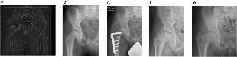
Methods: Through a prospectively collected multicenter international cohort, patients with early LCPD (Waldenström Stage I) and pMRI were followed with serial radiographs at 3-month intervals for a minimum of 2 years. Epiphyseal hypoperfusion was quantified by HipVasc Software for the entire epiphysis and regional thirds of the femoral head. Waldenström stages and lateral pillar class were determined by mode assessments from 3 pediatric orthopedic surgeons. Duration of the stage was defined as the interval between the first radiograph demonstrating features of stage IIa and stage IIIa for fragmentation and between IIIa and IV for reossification.
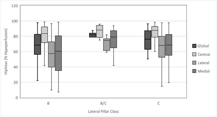
Results: One-hundred and seven patients (88.8% male, median age 8.0 years) met the study criteria. The average global hypoperfusion was 73.7%. Poorer global perfusion was predictive of a longer duration of fragmentation (rho = 0.34, P < .001) and of reossification (rho = 0.38, P = .003). The average regional hypoperfusion of the medial, central, and lateral third of the femoral head was 65.3%, 83.7%, and 61.3% respectively, and was similarly related to the duration of fragmentation (rho = 0.26, 0.24, and 0.31, respectively) and of reossification (rho = 0.31, 0.43, and 0.39, respectively) (P < .05 for all). Similar to previous studies, we found a significant positive association between hypoperfusion in the lateral third of the femoral head and lateral pillar class (P = .037).
Conclusions: The degree of both global and regional hypoperfusion correlated with the duration of fragmentation and reossification stages in LCPD. Lateral epiphyseal hypoperfusion is predictive of lateral pillar class. Taken together, the information provided by perfusion magnetic resonance imaging can provide crucial prognostic information for children with LCPD.
Key concepts: 1.Amount of hypoperfusion both globally and regionally on perfusion MRI correlates with the duration of fragmentation and reossification stages in LCPD.2.Lateral epiphyseal hypoperfusion correlates with lateral pillar class.3.Perfusion information offered by contrast MRI can offer crucial prognostic information in children with LCP.
Level of evidence: II Prognostic Study.
Keywords: Duration; Legg-Calve-Perthes; Perfusion MRI; Perthes.
© 2024 The Authors.
Conflict of interest statement
The authors declare the following financial interests/personal relationships which may be considered as potential competing interests: Royalties from Wolters Kluwer Health for edited textbook, consulting OrthoPediatrics, consulting Siemens—W.S. If there are other authors, they declare that they have no known competing financial interests or personal relationships that could have appeared to influence the work reported in this paper.
Figures

Similar articles
-
Perfusion MRI in Early Stage of Legg-Calvé-Perthes Disease to Predict Lateral Pillar Involvement: A Preliminary Study.J Bone Joint Surg Am. 2014 Jul 16;96(14):1152-1160. doi: 10.2106/JBJS.M.01221. J Bone Joint Surg Am. 2014. PMID: 25031369
-
Does Early Proximal Femoral Varus Osteotomy Shorten the Duration of Fragmentation in Perthes Disease? Lessons From a Prospective Multicenter Cohort.J Pediatr Orthop. 2020 May/Jun;40(5):e322-e328. doi: 10.1097/BPO.0000000000001451. J Pediatr Orthop. 2020. PMID: 31524767
-
The Role of the Artery of Ligamentum Teres in Revascularization in Legg-Calve-Perthes Disease.J Pediatr Orthop. 2022 Apr 1;42(4):175-178. doi: 10.1097/BPO.0000000000002061. J Pediatr Orthop. 2022. PMID: 35089880
-
Evolution in diagnosis and treatment of Legg-Calve-Perthes disease.Arch Bone Jt Surg. 2014 Jun;2(2):86-92. Epub 2014 Jun 15. Arch Bone Jt Surg. 2014. PMID: 25207324 Free PMC article. Review.
-
Evolution of Legg-Calvé-Perthes disease following proximal femoral varus osteotomy performed in the avascular necrosis stage:a prospective study.J Child Orthop. 2020 Feb 1;14(1):58-67. doi: 10.1302/1863-2548.14.190153. J Child Orthop. 2020. PMID: 32165982 Free PMC article. Review.
References
LinkOut - more resources
Full Text Sources
Research Materials
Miscellaneous
