

Common upper extremity gymnastics injuries and gymnastic specific return to play protocols
- PMID: 40433250
- PMCID: PMC12088353
- DOI: 10.1016/j.jposna.2024.100016
Common upper extremity gymnastics injuries and gymnastic specific return to play protocols
Abstract
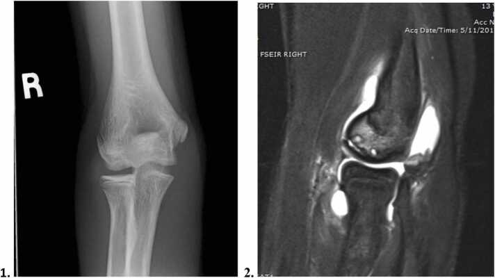
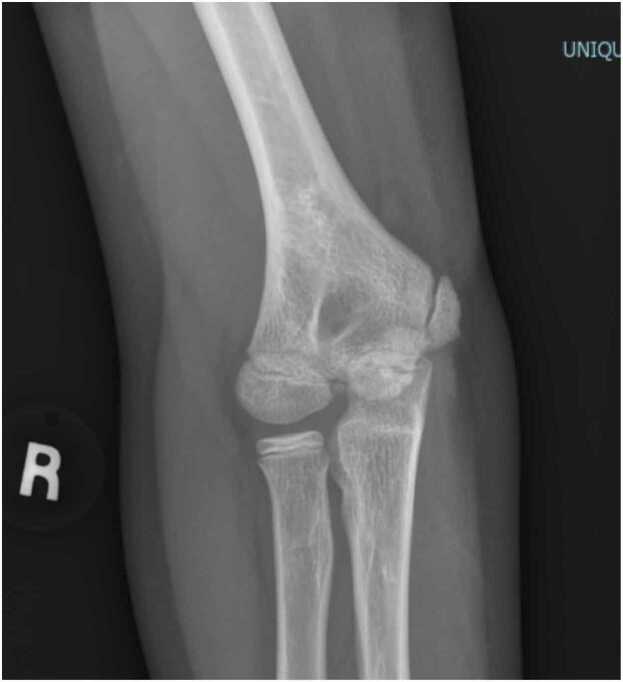
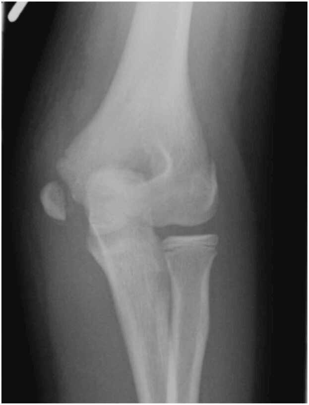
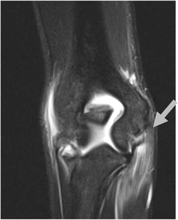
Young gymnasts use their upper extremities as weight-bearing joints, imparting high repetitive loads onto the growing upper limb. The purpose of this review is to provide orthopaedic and sports medicine clinicians practical information on the etiology, presentation, and treatment of 5 common upper extremity injuries in the young gymnast: (1) "gymnast wrist" (distal radial physeal injury); (2) grip lock (acute radius and ulna fracture); (3) osteochondritis dissecans of the capitellum; (4) medial tensile injuries of the elbow (medial epicondylar apophysitis, medial epicondyle fractures, and partial or full ulnar collateral ligament tears); and (5) glenohumeral instability (including labrum tears). Specific return to gymnastics protocols are provided to guide providers and athletes through safe return to participation following these injuries.
Key concepts: 1)Gymnastics is a unique sport in which the arms are used as weight-bearing limbs resulting in distinct injuries.2)Orthopaedic and sports medicine providers should understand these five diagnoses: Gymnast Wrist (distal radial physeal injury and the sequela), Grip Lock (acute radius and ulna fracture), elbow osteochondritis dissecans (OCD), Medial tensile injuries (medial epicondylar apophysitis, medial epicondyle fractures, and partial or full UCL tears), and shoulder instability (including labrum tears) if they will be evaluating gymnasts in their clinic.3)Pre-determined "return-to-gymnastics" protocols may aid successful progression back to training and competition after upper limb injury.
Keywords: Gymnastics; Gymnastics injuries; Return to gymnastics protocols; Return to play protocols; Upper extremity; Upper extremity injuries.
© 2024 Published by Elsevier Inc. on behalf of Pediatric Orthopaedic Society of North America.
Conflict of interest statement
The authors declare the following financial interests/personal relationships which may be considered as potential competing interests. Dr. Donald Bae reports Royalties, Lippincott Williams and Wilkins. Board member, Foundation for Advancing Pediatric Orthopaedic. Board member, Pediatric Orthopaedic Society of North America. Elspeth Hart is the founder of the nonprofit Gymnastics Medicine: Education and Research (GymnasticsMedicine.org) and also works as medical staff for USA Gymnastics. Dr. Andrea Bauer has no known competing financial interests or personal relationships that could have appeared to influence the work reported in this paper.
Figures
Similar articles
-
Gymnastics Medicine: A New Subspecialty in Sports Medicine.Curr Sports Med Rep. 2025 May 1;24(5):126-134. doi: 10.1249/JSR.0000000000001249. Curr Sports Med Rep. 2025. PMID: 40323057 Review.
-
Upper Extremity Injuries in Gymnasts.Hand Clin. 2017 Feb;33(1):187-197. doi: 10.1016/j.hcl.2016.08.010. Hand Clin. 2017. PMID: 27886834 Review.
-
Overuse Elbow Injuries in Youth Gymnasts.Am J Sports Med. 2022 Feb;50(2):576-585. doi: 10.1177/03635465211000776. Epub 2021 Mar 29. Am J Sports Med. 2022. PMID: 33780632 Review.
-
Artistic Gymnastics Injuries; Epidemiology, Evaluation, and Treatment.J Am Acad Orthop Surg. 2019 Jul 1;27(13):459-467. doi: 10.5435/JAAOS-D-18-00147. J Am Acad Orthop Surg. 2019. PMID: 31232791 Review.
-
Five-Year Follow-Up of Adolescent Gymnasts After Surgical Treatment of Osteochondritis Dissecans of the Elbow.J Hand Surg Am. 2024 Sep;49(9):934.e1-934.e6. doi: 10.1016/j.jhsa.2022.12.009. Epub 2023 Apr 28. J Hand Surg Am. 2024. PMID: 37115144
References
-
- Gymnastics participation in the U.S. 2010–2021. June 9, 2023. https://www.statista.com/statistics/191908/participants-in-gymnastics-in... (accessed December 14, 2023).
-
- Must-Know Gymnastics Statistics. November 14, 2023. https://blog.gitnux.com/gymnastics-statistics/ (accessed December 14, 2023).
-
- DiFiori J.P., Puffer J.C., Aish B., Dorey F. Wrist pain, distal radial physeal injury, and ulnar variance in young gymnasts: does a relationship exist? Am J Sports Med. 2002;(6):879–885. - PubMed
-
- DiFiori J.P., Caine D.J., Malina R. Wrist pain, distal radial physeal injury, and ulnar variance in the young gymnast. Am J Sports Med. 2006;(5):840–849. - PubMed
Publication types
LinkOut - more resources
Full Text Sources
