Review
. 2025 May 13:16:1543702.
doi: 10.3389/fimmu.2025.1543702. eCollection 2025.

"Remodeling the intestinal immune microenvironment": immune regulation and tissue regeneration by mesenchymal stem/stromal cells in the repair microenvironment of inflammatory bowel disease

Affiliations
Review

"Remodeling the intestinal immune microenvironment": immune regulation and tissue regeneration by mesenchymal stem/stromal cells in the repair microenvironment of inflammatory bowel disease

Hongkun Li et al. Front Immunol. .

Abstract

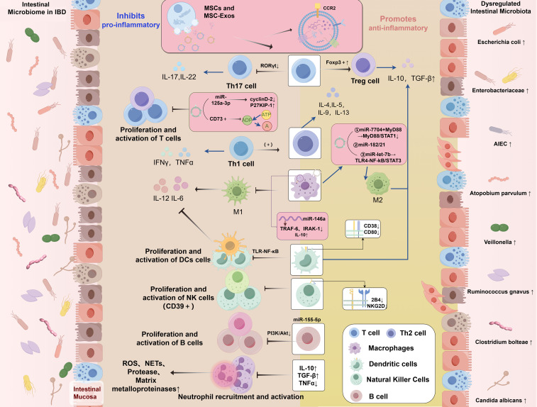
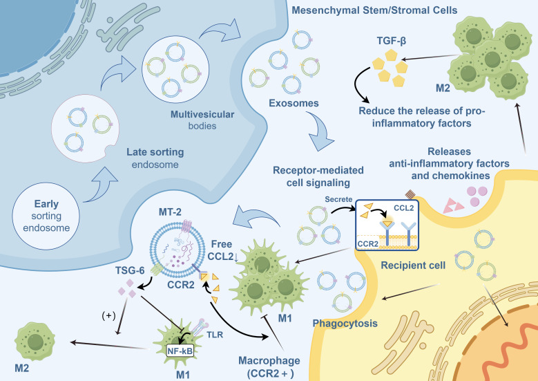
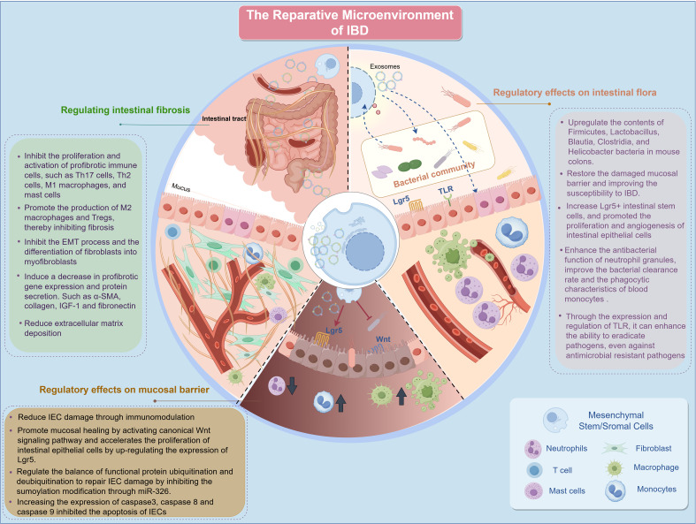
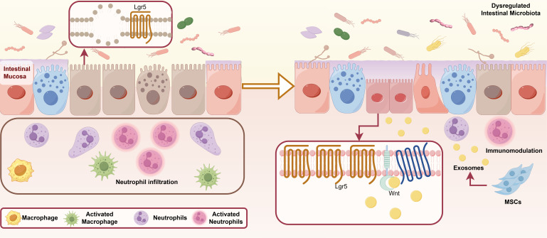
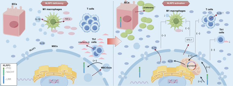
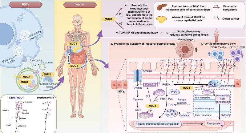
The global prevalence of inflammatory bowel disease (IBD) has significantly increased in recent decades. IBD is a long-term, recurring, gastrointestinal inflammatory condition that mainly comprises two primary clinical types: ulcerative colitis and Crohn's disease. The current treatment paradigm for IBD primarily focuses on symptom management. However, this approach does not support mucosal epithelial repair, maintenance of barrier homeostasis, or regulation of biological functions in the gut. Conventional therapies rely on the frequent use of high-dose medications, including antibiotics, nonsteroidal anti-inflammatory drugs, biological agents, and immunomodulators. Recently, mesenchymal stem/stromal cells (MSCs) have gained interest in tissue regeneration owing to their unique ability to differentiate and secrete regulatory factors, including extracellular vesicles (EVs), which play crucial roles in abnormal organization. Various routes of administration have been explored in preclinical and clinical studies to deliver MSCs from diverse tissue sources. The routes include intraperitoneal, intravenous, and local (intracolonic or rectal) delivery. The MSCs employed were obtained from various tissues, including bone marrow, umbilical cord, and adipose tissue. This article reviews the research framework for the application of MSCs and EVs secretion in the treatment of IBD, emphasizing key immunological effects, such as immune microenvironment regulation, intestinal barrier stabilization, and therapeutic approaches targeting intestinal barrier disorders. The discussion primarily focuses on the advantages of MSCs over other biologics, impairment of gut mucosal tissue-resident mesenchymal stem cells in IBD development, immune targets (at the cellular and molecular levels) within the framework of IBD, and the reparative effects of MSCs in the microenvironment of IBD. We aimed to present an overview of the current trends in MSC research and therapy, as well as to identify the challenges and future directions that must be addressed to advance research on MSC-mediated therapeutic strategies for IBD.

Keywords: biological therapies; inflammatory bowel disease; intestinal immune microenvironment; mesenchymal stem/stromal cells; tissue regeneration.


Conflict of interest statement

The authors declare that the research was conducted in the absence of any commercial or financial relationships that could be construed as a potential conflict of interest.

Figures

Figure 1
Figure 1
Dysregulation of the intestinal microbiome in IBD and the regulatory role of MSCs in modulating the immune microenvironment of IBD. IBD, inflammatory bowel disease; MSCs, mesenchymal stromal/stem cells; MSC-Exos, mesenchymal stromal/stem cell-derived exosomes; miRNAs, microRNAs; IL, interleukin; TNF-α, tumor necrosis factor alpha; IFN-γ, interferon-gamma; TGF-β, transforming growth factor-beta; ROS, reactive oxygen species; NETs, neutrophil extracellular traps; CCR2, Chemokine (C-C motif) Receptor 2; P27KIP-1, Cyclin-dependent Kinase Inhibitor 1B; CD, Cluster of Differentiation; RORγt, Retinoic acid-related Orphan Receptor gamma t; Foxp3, Forkhead box P3; A, Adenosine; ADP, Adenosine Diphosphate; ATP, Adenosine Triphosphate; MYD88, Myeloid Differentiation Primary Response Gene 88; STAT, Signal Transducer and Activator of Transcription; NF-κB, Nuclear Factor kappa-B; TRAF6, Tumor necrosis factor Receptor Associated Factor 6; IRAK1, Interleukin 1 Receptor Associated Kinase 1; TLR, Toll-like receptor; NKG2D, NK group 2 member D; PI3K, Phosphatidylinositol 3-kinase; AKT, protein kinase B; M1, Classically activated macrophages; M2, Alternatively activated macrophages; Tregs, regulatory T cells; Th, T Helper; DCs, dendritic cells; AIEC, Adherent Invasive Escherichia coli.
Figure 2
Figure 2
Regulation of macrophage polarization by MSC-Exos.MSC-Exos, mesenchymal stromal/stem cell-derived exosomes; M1, Classically activated macrophages; M2, Alternatively activated macrophages; TGF-β, transforming growth factor-beta; CCL2, C-C motif ligand 2; CCR2, C-C motif chemokine receptor 2; TSG-6, tumor necrosis factor-stimulated gene 6; NF-κB, nuclear factor kappa B; MT, metallothionein; TLR, Toll-like receptor.
Figure 3
Figure 3
Repair microenvironment of IBD. IBD, inflammatory bowel disease; IEC, intestinal epithelial cells; M1, Classically activated macrophages; M2, Alternatively activated macrophages; EMT, epithelial-to-mesenchymal transition; α-SMA, alpha-smooth muscle actin Lgr5, leucine-rich repeat-containing G-protein-coupled receptor 5; IGF-1, insulin-like growth factor 1; Th, T Helper; TLR, Toll-like receptor; miRNAs, microRNAs.
Figure 4
Figure 4
Restoration of the damaged mucosal barrier by MSCs. The fundamental causes of pathological changes in IBD include imbalances and disorders of mucosal immunity, surface mucus, intestinal bacteria, and other factors, which disrupt the IEB, leading to IEC necrosis, apoptosis, and increased intestinal wall permeability. Additionally, MSC-Exos can significantly reduce IEC damage through immune regulation. MSC-Exos activate the canonical Wnt signaling pathway to promote mucosal healing, which is characterized by reduced histopathological damage, decreased neutrophil infiltration, and upregulation of Lgr5, thereby accelerating the proliferation of IECs. IL-25 enhances the ability of MSCs to induce intestinal epithelial cell regeneration. MSCs alter the composition and diversity of the gut microbiota, thereby restoring gut microbiota balance and metabolism. IBD, inflammatory bowel disease; MSCs, mesenchymal stromal/stem cells; MSC-Exos, mesenchymal stromal/stem cell-derived exosomes; IEB, intestinal epithelial barrier; IEC, intestinal epithelial cells; Lgr5, leucine-rich repeat-containing G-protein-coupled receptor 5; Th, T Helper; IL, interleukin.
Figure 5
Figure 5
Role of NLRP3 inflammasome activation in MSCs and their immunomodulatory effects. In the intestinal microenvironment of IBD: Activated NLRP3 can stimulate the production of intestinal defensins. Enhanced immunoregulatory ability of MSCs: 1. Up-regulation of IL-10 production by Glut1; 2. Inhibit T cell proliferation, Th1 cell differentiation, Th1 cell function, and promote Th2 cell function; 3, inhibit the activation of M1 macrophages. MSC-Exos also regulate the activity of NLRP3 inflammasome. IBD, inflammatory bowel disease; MSCs, mesenchymal stromal/stem cells; MSC-Exos,mesenchymal stromal/stem cell-derived exosomes; M1, Classically activated macrophages; IEC, intestinal epithelial cells; IL, interleukin; NLRP3, NOD-like receptor protein 3; Th, T Helper; Leucine-Rich Repeat, LRR; Nucleotide-binding and oligomerization domain, NACHT; Pyrin domain, PYD; GLUT1, glucose transporter protein 1; STC-1, stanniocalcin-1.
Figure 6
Figure 6
Role of the apoptosis-related gene/tumor-associated antigen MUC1 in mesenchymal stem cell-mediated treatment of IBD. MSCs significantly upregulate the expression of MUC1, which is closely associated with the pathological processes of IBD, by preventing disruption of antioxidant defenses, regulating the sensitivity of cell ferroptosis, and improving inflammation and extraintestinal manifestations of IBD: (A) MUC1 is a tumor-associated antigen that is downregulated in IBD. The presence of abnormal forms of MUC1 has a profound impact on the timing and severity of inflammation, as well as the progression of certain extraintestinal manifestations of IBD. The low-glycosylated and overexpressed MUC1 can attract innate immune system cells, including CD4+ and CD8+ T cells, immature myeloid DCs, and macrophages, thereby promoting acute inflammation. Over time, acute inflammation can evolve into chronic inflammation. The presence of abnormal forms of MUC1 can also enhance the carcinogenic potential of colon and pancreatic cells. (B) MSCs can secrete various intestinal trophic factors and exosomes to inhibit intestinal epithelial cell death. Among these, MUC1 may promote regeneration of the epithelial cell barrier by inhibiting ferroptosis. MUC1 can upregulate ferroptosis-related genes, such as the glutamate/cystine antiporter SLC7A11 and GPX4 in small intestinal crypt epithelial cells while downregulating ACSL4 to facilitate epithelial cell growth. (C) MUC1 has an anti-inflammatory effect by targeting the TLR4/NF-κB signaling pathway and reducing oxidative stress levels. IBD, inflammatory bowel disease; MSCs, Mesenchymal Stromal/Stem Cells; DCs, dendritic cells; IEC, intestinal epithelial cells; MUC1, Mucin 1; CD, Cluster of Differentiation; SLC 7A11, Solute carrier family 7 member 11; GPX 4, Glutathione peroxidase 4; ACSL 4, Acyl-CoA Synthetase Long Chain Family Member 4;TLR, Toll-like receptor; NF-κb, Nuclear factor- kappa B; GSH, Glutathione; PUFAS, Polyunsaturated Fatty Acids; ATP, Adenosine Triphosphate; ROS, Reactive Oxygen Species; LPCAT, Lysophosphatidylcholine Acyltransferase 3; PLs, Phospholipids; ALOXs, Arachidonate Lipoxygenases; PoR, P450 Oxidoreductase.

Similar articles

Cited by

References

    1. Friedrich M, Pohin M, Powrie F. Cytokine networks in the pathophysiology of inflammatory bowel disease. Immunity. (2019) 50:992–1006. doi: 10.1016/j.immuni.2019.03.017 - DOI - PubMed
    1. Kaplan GG. The global burden of ibd: from 2015 to 2025. Nat Rev Gastroenterol Hepatol. (2015) 12:720–7. doi: 10.1038/nrgastro.2015.150 - DOI - PubMed
    1. Rosen MJ, Dhawan A, Saeed SA. Inflammatory bowel disease in children and adolescents. JAMA Pediatr. (2015) 169:1053–60. doi: 10.1001/jamapediatrics.2015.1982 - DOI - PMC - PubMed
    1. Noble AJ, Nowak JK, Adams AT, Uhlig HH, Satsangi J. Defining interactions between the genome, epigenome, and the environment in inflammatory bowel disease: progress and prospects. Gastroenterology. (2023) 165:44–60.e2. doi: 10.1053/j.gastro.2023.03.238 - DOI - PubMed
    1. Venkataraman GR, Rivas MA. Rare and common variant discovery in complex disease: the Ibd case study. Hum Mol Genet. (2019) 28:R162–r9. doi: 10.1093/hmg/ddz189 - DOI - PMC - PubMed

MeSH terms

LinkOut - more resources