Case Reports
. 2025 May 20:2025:8934034.
doi: 10.1155/crid/8934034. eCollection 2025.

Clinical and Radiographic Features of Mandibular Third Molar Gemination: A Case Report and Literature Review

Affiliations
Case Reports

Clinical and Radiographic Features of Mandibular Third Molar Gemination: A Case Report and Literature Review

Matteo Pellegrini et al. Case Rep Dent. .

Abstract

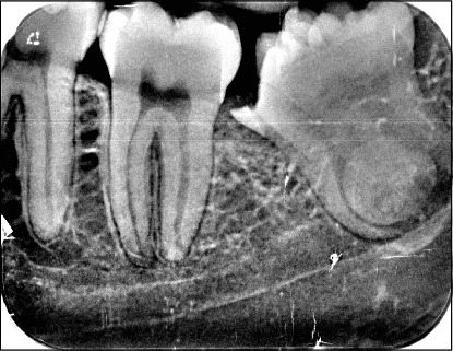
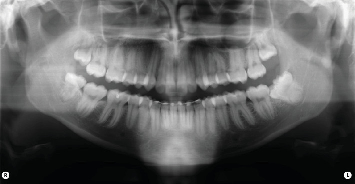
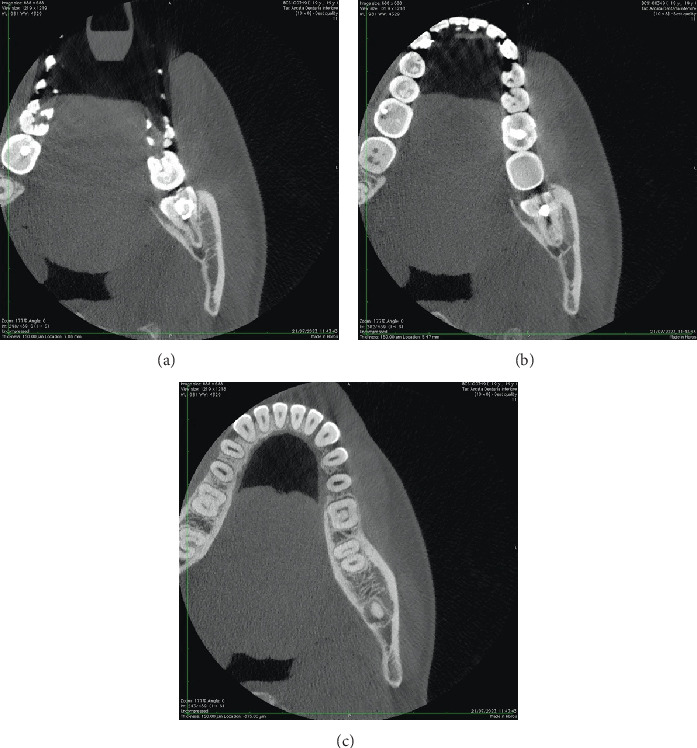
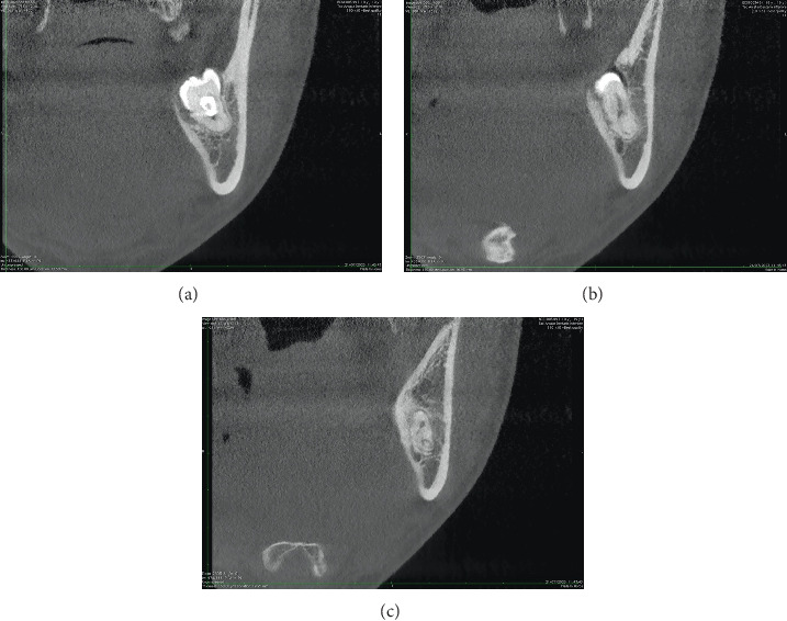
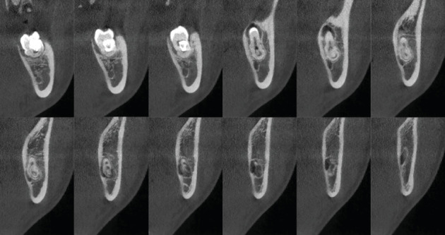
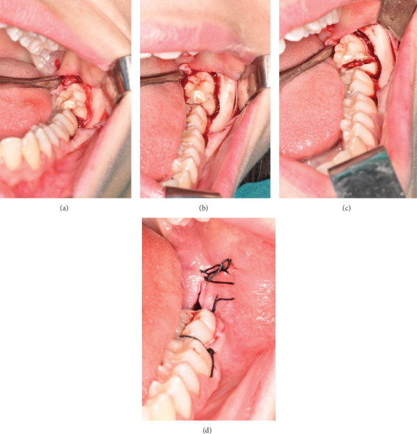
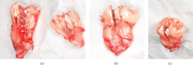
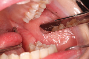
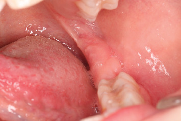
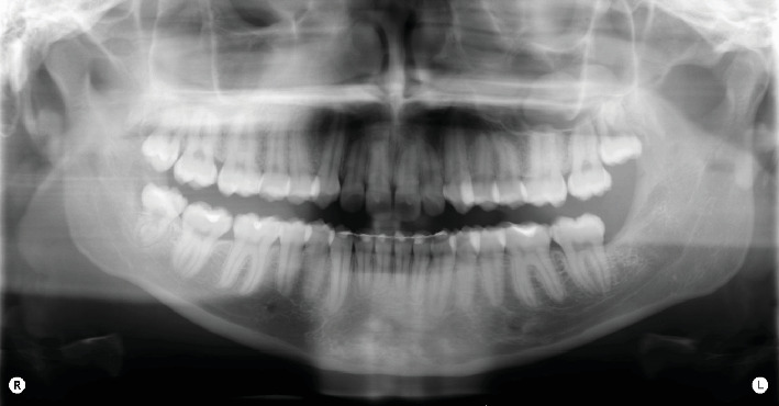
Introduction: Gemination and fusion are rare developmental anomalies that can present significant diagnostic challenges. Due to the complexity of distinguishing between these conditions, the term "double tooth" is commonly employed in clinical practice. The precise etiology of these anomalies remains uncertain, and their occurrence in permanent dentition-particularly involving molars-is exceptionally rare. This report describes an uncommon case of gemination affecting the mandibular left third molar (tooth 3.8) and provides a comprehensive discussion contextualized within existing literature. The case report was prepared following the CARE guidelines to ensure methodological rigor and completeness. Methods: After an intraoral examination and radiographic assessment-including orthopantomography, periapical radiographs, and cone beam computed tomography (CBCT)-the patient underwent surgical extraction. The procedure involved administering a truncal nerve block to anesthetize the inferior alveolar and lingual nerves, supplemented by local infiltration anesthesia of the buccal nerve. A full-thickness mucoperiosteal flap was elevated, followed by ostectomy and odontotomy to facilitate extraction. The tooth was subsequently removed using a combination of elevators and forceps. Results: Postoperative evaluations conducted at 1.5 and 3 months confirmed complete healing of the surgical site. A detailed analysis of pre- and postoperative radiographic and clinical findings validated the diagnosis of gemination, characterized by coronal continuity with a single root and root canal. Conclusions: Gemination of third molars is exceedingly rare, with only a few cases documented in the literature. To the best of our knowledge, this is the first reported instance of gemination involving the mandibular left third molar (tooth 3.8). This report contributes to the growing body of knowledge on developmental dental anomalies and highlights the importance of thorough differential diagnosis in similar clinical scenarios.

Keywords: dental anomalies; dental gemination; dentistry; double tooth; oral surgery; third molar.


Conflict of interest statement

The authors declare no conflicts of interest.

Figures

Figure 1
Figure 1
Intraoral objective examination assessment of postpulpotomy element 3.8.
Figure 2
Figure 2
Endoral x-ray shows the absence of enamel and dentin tissue, the relationship between the tooth root and the mandibular canal, and coronal-distal radiolucency.
Figure 3
Figure 3
OPG shows altered coronal and root anatomy of element 3.8, deviation of the mandibular canal at the root apex, and a coronal-distal radiolucency.
Figure 4
Figure 4
(a–c) Axial CBCT sections of the 3.8 tooth element where the continuity relationship of the root apex with the NVB can be appreciated.
Figure 5
Figure 5
(a–c) Coronal CBCT sections of the 3.8 tooth element illustrate the continuity relationship between the root apex and the NVB.
Figure 6
Figure 6
Sagittal CBCT cross-sections of the 3.8 tooth element depict the continuity relationship between the root apex and the NVB.
Figure 7
Figure 7
(a) Intraoral photos of the full-thickness mucoperiosteal cleaved surgical flap, (b) vestibulo-distal ostectomy, (c) vestibulo-lingual odontotomy, and (d) postoperative suturing with 3/0 silk.
Figure 8
Figure 8
Postoperative photograph (a) of the sectioned 3.8 tooth element and (b, c) of the coronoradicular reconstruction.
Figure 9
Figure 9
Intraoral photograph 1.5 months after the surgical extraction.
Figure 10
Figure 10
Photograph taken intraorally 3 months postsurgical extraction.
Figure 11
Figure 11
OPG 3 months after surgery shows complete bone healing in zone 3.8, confirmed by the presence of lamellar bone tissue with a high level of ossification.

Similar articles

References

    1. Brauer H. U., Bartols A. Gemination of an Erupted Mandibular Third Molar: A Short Presentation of an Exceptionally Rare Clinical Occurrence. European Journal of Dentistry . 2024;18(2):687–691. doi: 10.1055/s-0043-1772248. - DOI - PMC - PubMed
    1. Türkaslan S., Gökçe H. S., Dalkız M. Esthetic Rehabilitation of Bilateral Geminated Teeth: A Case Report. European Journal of Dentistry . 2007;1(3):188–191. doi: 10.1055/s-0039-1698337. - DOI - PMC - PubMed
    1. Nandini D. B., Deepak B. S., Selvamani M., Puneeth H. K. Diagnostic Dilemma of a Double Tooth: A Rare Case Report and Review. Journal of Clinical and Diagnostic Research . 2014;8(1):271–272. doi: 10.7860/JCDR/2014/6556.3928. - DOI - PMC - PubMed
    1. Ramezani M., Asgari S., Adel M. Endodontic Management of a Rare Case of the Geminated Maxillary Second Molar Tooth Using CBCT. Clinical Case Reports . 2021;9(8) doi: 10.1002/ccr3.4496.e04496 - DOI - PMC - PubMed
    1. Nunes E., de Moraes I. G., de Oliveira Novaes P. M., de Sousa S. M. G. Bilateral Fusion of Mandibular Second Molars With Supernumerary Teeth: Case Report. Brazilian Dental Journal . 2002;13(2):137–141. doi: 10.1590/S0103-64402002000200012. - DOI - PubMed

Publication types

LinkOut - more resources