. 2025 May 13:13:1596421.
doi: 10.3389/fcell.2025.1596421. eCollection 2025.

Increased reproductive outcomes after optimized sperm preparation

Affiliations

Increased reproductive outcomes after optimized sperm preparation

Matías D Gómez-Elías et al. Front Cell Dev Biol. .

Abstract

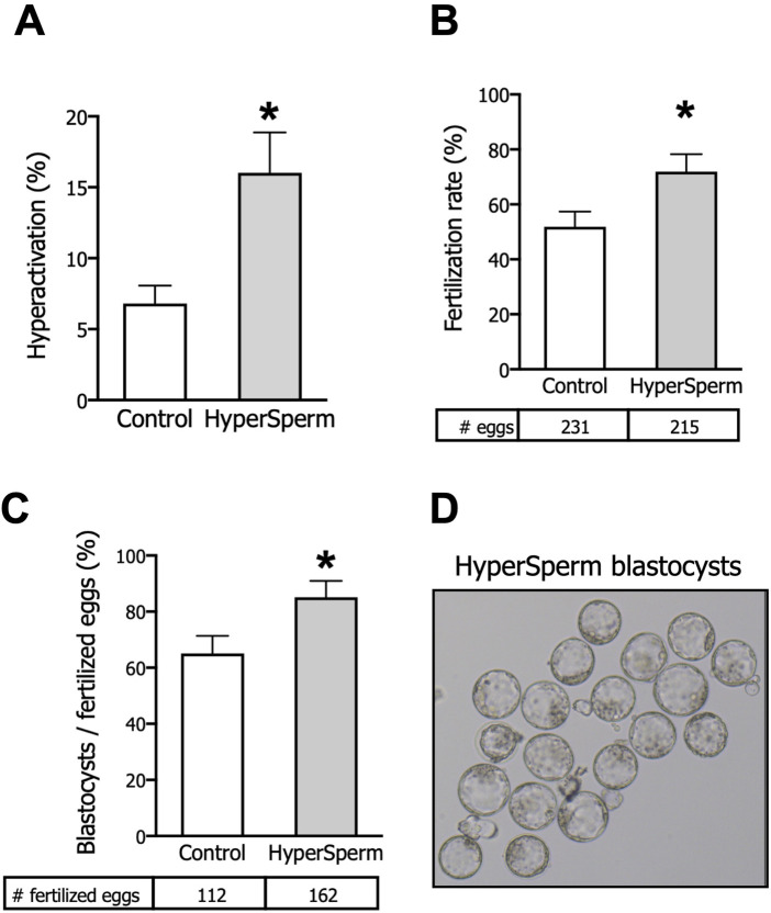
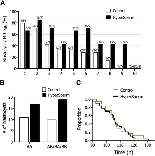
A key factor to the success of in vitro fertilization (IVF) is the preparation of human sperm, a critical step that directly impacts the efficacy of the procedure. This proof-of-concept study evaluated the effect of HyperSperm, a novel sperm preparation technique designed to enhance sperm function, on fertilization, embryo development, and pregnancy outcomes in both a mouse model and a first-in-human trial following IVF. In mice, HyperSperm treatment significantly increased hyperactivated motility (p < 0.05), leading to improved fertilization and blastocyst development (p < 0.05), as well as higher implantation rates (p < 0.05) and larger litter sizes (p < 0.05). Offspring displayed normal growth and fertility. In human sperm samples from normozoospermic men, HyperSperm exhibited a high safety profile, with motility, acrosome reaction, viability, and DNA fragmentation comparable to controls. A first-in-human, prospective, single-center, split-oocyte study in 10 couples undergoing IVF with donated oocytes demonstrated similar fertilization rates between HyperSperm and control groups (p = 0.425), but significantly higher usable blastocyst rates in the HyperSperm arm (43.8% vs. 67.9%, p = 0.0122). Morphokinetic parameters were comparable between groups. These results suggest that HyperSperm enhances sperm hyperactivation, a hallmark of capacitation, leading to improved embryo development in both mice and humans. This technique represents a promising approach to optimizing sperm preparation in assisted reproduction, warranting further clinical investigation.

Keywords: assisted reproductive technology; capacitation; implantation; in vitro fertilization; live birth.


Conflict of interest statement

Authors MG-E, GL, NO-S, OB, IK, RV, DK and MB were employed by Fecundis Lab. MB, DK, MG-E, GL and RV are shareholders of Fecundis. The remaining authors declare that the research was conducted in the absence of any commercial or financial relationships that could be construed as a potential conflict of interest. The author DK declared that they were an editorial board member of Frontiers, at the time of submission. This had no impact on the peer review process and the final decision.

Figures

FIGURE 1
FIGURE 1
HyperSperm increases fertilization and embryo development rates. (A) Hyperactivation, measured by CASA, n = 5. (B) Fertilization rates (2-cell embryos/eggs), n = 10. (C) Embryo development rates (blastocysts/fertilized eggs), n = 10. (D) Representative image of HyperSperm-derived blastocysts. Paired t-test was performed, *p < 0.05 represents statistical significance vs. control.
FIGURE 2
FIGURE 2
HyperSperm increases the efficiency of embryo implantation. (A) Implantation rates (implantation sites at day 7/blastocysts transferred), n = 6 for Control and n = 8 for HyperSperm. (B) Representative image of implantation sites (*) obtained with Control (upper panel) and HyperSperm (lower panel) treatments. (C) Litter size (% of pups born per blastocyst transferred), n = 8 for Control and n = 7 for HyperSperm. (D) Representative image sequence of HyperSperm pups on different days after birth. Data represents the mean ± SEM. Unpaired t-test was performed, *p < 0.05.
FIGURE 3
FIGURE 3
HyperSperm produces pups of normal weight. (A) Gestation period (days from embryo transfer to birth). Data represents the mean ± SEM, n = 3 litters for Control and n = 6 litters for HyperSperm. Unpaired t-test was performed. (B) Percentage of males born relative to the total offspring for each treatment. Data represents the mean. (C) Pup weight at birth (day 1) and at weaning (day 21). Data represent the mean ± SEM. (D) Body weight of females and males at 6 months. Data represent the mean ± SEM.
FIGURE 4
FIGURE 4
HyperSperm generates fertile progeny. (A) Litter size (number of pups/litter) obtained by mating Control or HyperSperm-derived females or males with hybrid F1 mice. (B) Sperm concentration of male born as a result of treatment. (C) Sperm motility (%) of male born as a result of treatment. In all cases, data represents the mean ± SEM of at least 3 matings, and Student’s t-test was performed.
FIGURE 5
FIGURE 5
Assessment of safety after treatment with standard conditions (Control) or HyperSperm. (A) Total motility, measured by CASA, n = 12. (B) Hyperactivation, measured by CASA, n = 12. (C) Percentage of viable cells, using eosin-Y staining, n = 9. (D) Survival measured as sperm vitality after overnight incubation, n = 5. (E) Acrosomal integrity, using FITC-PSA staining, n = 14. (F) DNA fragmentation, using the TUNEL assay, n = 17. Wilcoxon matched-pairs signed rank test was performed, *p < 0.05; **p < 0.01; ***p < 0.005.
FIGURE 6
FIGURE 6
HyperSperm increased blastocyst production in humans. (A) Effect of HyperSperm on individual outcomes. Bars indicate the percentage of blastocysts per mature egg for each patient, ordered from the highest (1) to the lowest (10) value in the Control arm. (B) Effect of HyperSperm on embryo quality. Bars indicate the total number of blastocysts obtained in each group according to the Gardner grading system. AA: top quality embryos; AB/BB/BA: high quality embryos. Fisher’s exact test was performed, p-value = 0.787. (C) Effect of HyperSperm on embryo morphokinetics. Median time point at which embryos reached blastocyst stage in the Control and HyperSperm groups. Median tB: 106.2 vs. 106.0 h (Control vs. HyperSperm), n = 18 Control and n = 32 HyperSperm-derived blastocysts, p > 0.05.

References

    1. Alvau A., Battistone M. A., Gervasi M. G., Navarrete F. A., Xu X., Sánchez-Cárdenas C., et al. (2016). The tyrosine kinase FER is responsible for the capacitation-associated increase in tyrosine phosphorylation in murine sperm. Dev. (Cambrigde) 143, 2325–2333. 10.1242/dev.136499 - DOI - PMC - PubMed
    1. Austin C. R. (1951). Observations on the penetration of the sperm in the mammalian egg. Aust. J. Sci. Res. 4, 581–596. 10.1071/BI9510581 - DOI - PubMed
    1. Avidor-Reiss T., Carr A., Fishman E. L. (2020). The sperm centrioles. Mol. Cell Endocrinol. 518, 110987. 10.1016/J.MCE.2020.110987 - DOI - PMC - PubMed
    1. Balli M., Cecchele A., Pisaturo V., Makieva S., Carullo G., Somigliana E., et al. (2022). Opportunities and limits of conventional IVF versus ICSI: it is time to come off the fence. J. Clin. Med. 11, 5722. 10.3390/JCM11195722 - DOI - PMC - PubMed
    1. Banker M., Dyer S., Chambers G. M., Ishihara O., Kupka M., de Mouzon J., et al. (2021). International committee for monitoring assisted reproductive technologies (ICMART): world report on assisted reproductive technologies, 2013. Fertil. Steril. 116, 741–756. 10.1016/J.FERTNSTERT.2021.03.039 - DOI - PubMed

LinkOut - more resources