

Human and Deep Learning Predictions of Peripheral Lung Cancer Using a 1.3 mm Video Endoscopic Probe
- PMID: 40433758
- PMCID: PMC12437996
- DOI: 10.1111/resp.70057
Human and Deep Learning Predictions of Peripheral Lung Cancer Using a 1.3 mm Video Endoscopic Probe
Abstract
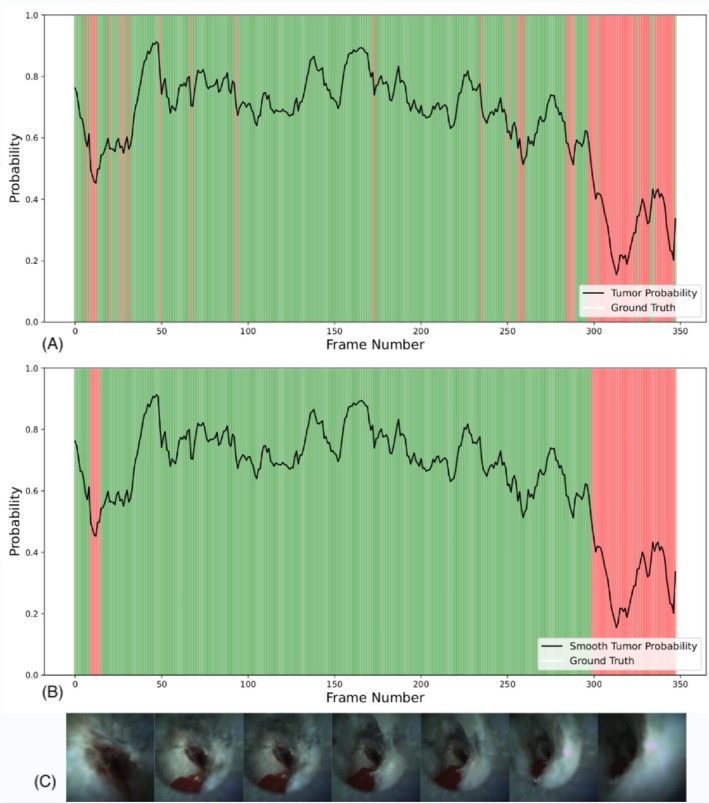
Background and objective: Iriscope, a 1.3 mm video endoscopic probe introduced through an r-EBUS catheter, allows for the direct visualisation of small peripheral pulmonary nodules (PPNs). This study assessed the ability of physicians with different levels of experience in bronchoscopy, and the ability of artificial intelligence (AI) to predict the malignant nature of small PPNs during Iriscope peripheral endoscopy.
Methods: Patients undergoing bronchoscopy with r-EBUS and Iriscope for peripheral PPNs < 20 mm with a definite diagnosis were analysed. Senior and Junior physicians independently interpreted video-recorded Iriscope sequences, classifying them as tumoral (malignant) or non-tumoral, blind to the final diagnosis. A deep learning (DL) model was also trained on Iriscope images and tested on a different set of patients for comparison with human interpretation. Diagnostic accuracy, sensitivity, specificity, and F1 score were calculated.
Results: Sixty-one patients with small PPNs (median size 15 mm, IQR: 11-20 mm) were included. The technique allowed for the direct visualisation of the lesions in all cases. The final diagnosis was cancer for 37 cases and a benign lesion in 24 cases. Senior physicians outperformed junior physicians in recognising tumoral Iriscope images, with a balanced accuracy of 85.4% versus 66.7%, respectively, when compared with the final diagnosis. The DL model outperformed junior physicians with a balanced accuracy of 71.5% but was not superior to senior physicians.
Conclusion: Iriscope could be a valuable tool in PPNs management, especially for experienced operators. Applied to Iriscope images, DL could enhance overall performance of less experienced physicians in diagnosing malignancy.
Keywords: artificial intelligence; bronchoscopy; deep learning; imaging; peripheral pulmonary nodules; radial‐EBUS.
© 2025 The Author(s). Respirology published by John Wiley & Sons Australia, Ltd on behalf of Asian Pacific Society of Respirology.
Conflict of interest statement
S.L. received consulting fees in Lys medical, Olympus and Fujifilm. The remaining authors declare no conflicts of interest.
Figures

References
-
- Callister M. E. J., Baldwin D. R., Akram A. R., et al., “British Thoracic Society Guidelines for the Investigation and Management of Pulmonary Nodules,” Thorax 70, no. Suppl 2 (2015): ii1–ii54. - PubMed
MeSH terms
LinkOut - more resources
Full Text Sources
Medical
