. 2025 Jun 17;99(6):e0008725.
doi: 10.1128/jvi.00087-25. Epub 2025 May 28.

Hydrophobic residue substitutions enhance the stability and in vivo immunogenicity of respiratory syncytial virus fusion protein

Affiliations

Hydrophobic residue substitutions enhance the stability and in vivo immunogenicity of respiratory syncytial virus fusion protein

Qiaoyun Song et al. J Virol. .

Abstract

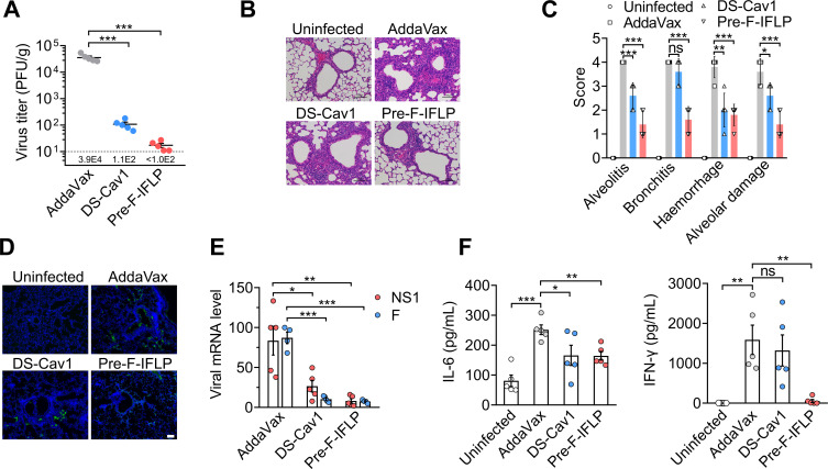
Respiratory syncytial virus (RSV) entry into host cells is facilitated by viral fusion, wherein the metastable RSV fusion (F) protein undergoes a conformational change from a prefusion state to a highly stable postfusion structure. The prefusion F elicits a more robust human antibody response than its postfusion F and is a primary target for RSV vaccine development. However, the inherent instability of the prefusion F trimer and its low protein expression level in host cells are a significant challenge for developing a high-potency RSV vaccine. Here, we report that the introduction of four hydrophobic residue substitutions in the RSV F protein resulted in a highly stable prefusion F trimer (pre-F-IFLP). This engineered variant exhibits enhanced expression and stability compared to DS-Cav1, with improved thermal stability, increased resistance to acid and base, and extended storage life. Furthermore, pre-F-IFLP induced neutralizing antibody responses 72-fold higher than those elicited by DS-Cav1 following a second booster immunization and fully protected mice against RSV infection.

Importance: In this study, we demonstrate that introducing four hydrophobic residue substitutions into the RSV F protein leads to the generation of a highly stable prefusion F trimer (pre-F-IFLP) with improved expression levels in cultured cells and superior stability compared to DS-Cav1, the first-generation prefusion F-stabilized RSV vaccine. Furthermore, pre-F-IFLP induced significantly higher neutralizing antibody responses than DS-Cav1 following both the first and second booster immunizations and conferred complete protection against RSV infection in a mouse model. These findings present an alternative approach for stabilizing the trimeric prefusion F protein, enhancing its expression, and significantly improving its protective efficacy for the prevention of RSV infection in vivo.

Keywords: prefusion F; respiratory syncytial virus; vaccine; viral fusion.


Conflict of interest statement

The authors declare no conflict of interest.

Figures

Fig 1
Fig 1
Mutations to stabilize the prefusion RSV F protein. HEK293T cells were transfected with pcDNA3.1 vectors encoding various RSV F variants. Cells were stained with D25 for prefusion F (A) and motavizumab for prefusion/postfusion F (B), followed by incubation with an Alexa 488-conjugated secondary antibody. Scale bars, 100 µm. (C, D, E) Following incubation for 10 min at 37°C or 55°C, the prefusion and postfusion F in cells were determined using flow cytometry. MFI, mean fluorescence intensity. (F) Cell-cell fusion activity in transfected cells was assessed using a dual-luciferase reporter assay. (C–F) Data are presented as mean ± SD, n = 3 biological replicates.
Fig 2
Fig 2
Characterization of the physical stability of the prefusion F variants. (A, B) Purified RSV F ectodomains were transferred to a nitrocellulose membrane and incubated for 10 min at 37°C or 55°C. (C, D) The RSV F constructs on the nitrocellulose membrane were treated with PBS-HCl (pH 3.5) or PBS-NaOH (pH 10) solution for 30 min at 25°C. (E–H) The nitrocellulose membrane with RSV proteins was stored at 25°C for 1, 5, 10, and 30 days. All RSV F constructs on the nitrocellulose membrane were incubated with D25 and motavizumab, followed by detection with horseradish peroxidase-conjugated secondary antibodies with ECL solution. (A–H) Data are presented as mean ± SD, n = 3 biological replicates.
Fig 3
Fig 3
Antigenic characterization of prefusion F variants. Binding curves for D25, AM14, and motavizumab to pre-F-IFLP and DS-Cav1 were generated using surface plasmon resonance assays. RSV F-specific antibodies were covalently immobilized onto the surface of CM5 sensor chips. Soluble recombinant proteins were diluted in PBS and flowed over the chips. Data were analyzed using Biacore S200 Evaluation Software, and the KD values were calculated to assess the binding affinity of RSV F constructs with anti-RSV F-specific antibodies. (A, C) Pre-F-IFLP was tested at concentrations of 3.125, 6.25, 12.5, 25, and 50 nM; (B) DS-Cav1 at 12.5, 25, 50, 100, and 200 nM; and (D, E, F) both pre-F-IFLP and DS-Cav1 were tested at 6.25, 12.5, 25, 50, and 100 nM.
Fig 4
Fig 4
Neutralizing efficacy of mouse serum after first boost immunization. (A) Schematic showing the schedule of animal studies. (B) The mouse serum after twice immunization was collected on day 24 and prepared for detection of neutralizing titer. Dilutions of the mouse serum to neutralize 50% of the RSV infection (ID50) were measured. (C) Following the first boost immunization, antibody titers in the mouse serum were detected. (D) Ratio of binding titers to neutralizing titers derived from the data in (B) and (C). Each dot represents the ratio of ED50/ID50 for each mouse. (B–D) Each dot represents individual mice. Data are mean ± SEM, n = 6. An unpaired two-tailed t-test was used to measure the statistical differences between the compared groups. *, P < 0.05; ***, P < 0.001.
Fig 5
Fig 5
Neutralizing efficacy of mouse serum after second boost immunization. (A) On day 34, mouse serum was collected following three rounds of immunization and prepared for analysis of neutralizing efficacy. Neutralizing titers of the mouse serum to inhibit RSV infection were determined. ID50 is the serum dilutions required to reduce RSV infection by 50%. (B) Binding titers of neutralizing antibodies in the mouse serum to respective immunogens. (C) Ratios of binding titers to neutralizing titers were calculated. (D) The ectodomain of RSV F (amino acids 1–526) was expressed, stored in a high-ionic strength buffer (20 mM Tris, 500 mM NaCl, pH 7.4, 10% glycerinum), and then pre-incubated with sera from immunized mice. The mixtures were diluted in PBS and subsequently incubated with HEp-2 cells for 1 h. Surface binding of the RSV F ectodomains to HEp-2 cells was stained using motavizumab and Alexa Fluor 488-conjugated secondary antibody and subsequently detected by flow cytometry. Mean fluorescence intensity (MFI) was analyzed using FlowJo v.10. (E) HEK293T cells were transfected with plasmids encoding full-length RSV F and then treated with diluted sera from immunized mice. Syncytia and cell nuclei were stained with RSV F-specific antibody and 4′,6-diamidino-2-phenylindole, respectively. Bar, 100 µm. (A–D) Data are presented as mean ± SEM, n = 5. An unpaired two-tailed t-test was used for comparisons between two groups. *, P < 0.05; **, P < 0.01; ***, P < 0.001.
Fig 6
Fig 6
Protective effects of pre-F-IFLP and DS-Cav1 immunizations against RSV infection in mice. (A) Mice were intratracheally infected with RSV following three immunizations with either pre-F-IFLP or DS-Cav1. On day 5 postinfection (d.p.i.), viral loads in the lung homogenates of immunized mice were determined. (B) Representative hematoxylin and eosin staining images of lung tissue sections were assessed. Scale bars, 100 µm. (C) Pathological changes in lung tissues were scored, and representative features of lung lesions were measured. (D) Representative immunostaining images of RSV F antigens in lung tissues are shown. Scale bars, 100 µm. (E) The mRNA levels of viral NS1 and F genes were detected by RT-PCR. (F) On day 5 d.p.i., secretion levels of IL-6 and IFN- γ in mouse serum were measured by ELISA. (A, C, E, F) Each dot represents an individual mouse. Data are presented as mean ± SEM, n = 5. An unpaired two-tailed t-test was used for comparisons between two groups. *, P < 0.05; **, P < 0.01; ***, P < 0.001. ns, no significant difference.

Similar articles

References

    1. Kaler J, Hussain A, Patel K, Hernandez T, Ray S. 2019. Respiratory syncytial virus (RSV): a scourge from infancy to old age. Thorax 74:986–993. doi: 10.7759/cureus.36342 - DOI - PubMed
    1. Cong B, Dighero I, Zhang T, Chung A, Nair H, Li Y. 2023. Understanding the age spectrum of respiratory syncytial virus associated hospitalisation and mortality burden based on statistical modelling methods: a systematic analysis. BMC Med 21:224. doi: 10.1186/s12916-023-02932-5 - DOI - PMC - PubMed
    1. Shi T, McAllister DA, O’Brien KL, Simoes EAF, Madhi SA, Gessner BD, Polack FP, Balsells E, Acacio S, Aguayo C, et al. 2017. Global, regional, and national disease burden estimates of acute lower respiratory infections due to respiratory syncytial virus in young children in 2015: a systematic review and modelling study. Lancet 390:946–958. doi: 10.1016/S0140-6736(17)30938-8 - DOI - PMC - PubMed
    1. Vieira SE, Gilio AE, Durigon EL, Baraldi E, Ejzenberg B. 2015. Lower respiratory tract infection caused by respiratory syncytial virus: current management and new therapeutics. Lancet Resp Med 3:888–900. doi: 10.1590/S1807-59322007000600009 - DOI - PubMed
    1. Nair H, Nokes DJ, Gessner BD, Dherani M, Madhi SA, Singleton RJ, O’Brien KL, Roca A, Wright PF, Bruce N, Chandran A, Theodoratou E, Sutanto A, Sedyaningsih ER, Ngama M, Munywoki PK, Kartasasmita C, Simões EAF, Rudan I, Weber MW, Campbell H. 2010. Global burden of acute lower respiratory infections due to respiratory syncytial virus in young children: a systematic review and meta-analysis. Lancet 375:1545–1555. doi: 10.1016/S0140-6736(10)60206-1 - DOI - PMC - PubMed

MeSH terms

Substances

LinkOut - more resources