Review
. 2025 Aug;37(33):e2417564.
doi: 10.1002/adma.202417564. Epub 2025 May 28.

From Mechanoelectric Conversion to Tissue Regeneration: Translational Progress in Piezoelectric Materials

Affiliations
Review

From Mechanoelectric Conversion to Tissue Regeneration: Translational Progress in Piezoelectric Materials

Xinyu Wang et al. Adv Mater. 2025 Aug.

Abstract

Piezoelectric materials, capable of converting mechanical stimuli into electrical signals, have emerged as promising tools in regenerative medicine due to their potential to stimulate tissue repair. Despite a surge in research on piezoelectric biomaterials, systematic insights to direct their translational optimization remain limited. This review addresses the current landscape by bridging fundamental principles with clinical potential. The biomimetic basis of piezoelectricity, key molecular pathways involved in the synergy between mechanical and electrical stimulation for enhanced tissue regeneration, and critical considerations for material optimization, structural design, and biosafety is discussed. More importantly, the current status and translational quagmire of mechanisms and applications in recent years are explored. A mechanism-driven strategy is proposed for the therapeutic application of piezoelectric biomaterials for tissue repair and identify future directions for accelerated clinical applications.

Keywords: advanced materials for translational medicine; biosensors; piezoelectricity; regenerative medicine; tissue engineering.


Conflict of interest statement

The authors declare no conflict of interest.

Figures

Figure 1
Figure 1
Possible synergistic signaling pathways for mechanical and electrical signals in tissue regeneration. There are seven possible pathways: integrin/FAK/Ras, Hippo (YAP/TAZ), wnt/β‐catenin, TGF‐β, ion channels, calcium signaling, and piezocatalytic effects. Complex cross‐talk exists between pathways, especially between the integrin pathway and the Hippo pathway. The calcium signaling pathway plays a predominantly downstream role and also has a regulatory role on upstream pathways. Created in BioRender. (2024) https://BioRender.com/a48v522.
Figure 2
Figure 2
Brief history of piezoelectric materials. Piezoelectric materials have gone through three main stages: piezoelectric ceramics, artificial piezoelectric polymers, and natural piezoelectric polymers. Piezoelectric ceramics are represented by PZT and BaTiO3, and artificial piezoelectric polymers are represented by PVDF and PLLA. Natural piezoelectric polymers are emerging, including chitosan and glycine. Created in BioRender. (2024) https://BioRender.com/y99j480.
Figure 3
Figure 3
Schematic diagram of the fabrication of piezoelectric composite hydrogel and scaffold. The first example is the enhancement of BaTiO₃ nanoparticles by domain optimization, phase boundary engineering, and defect engineering, followed by a combination with nanofibers and hydrogel matrix to form a composite piezoelectric hydrogel. The second example is the enhancement of PVDF films by stretching, polarization, annealing, and optimization of electrostatic spinning parameters, which are then combined with porous scaffolds to form composite piezoelectric scaffolds. Existing approaches emphasize the improvement of piezoelectric properties through material processing and combinatorial techniques. Created in BioRender. (2024) https://BioRender.com/g01t330.
Figure 4
Figure 4
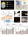
Topological materials for bone, cartilage, and nerve regeneration. a) SEM images of randomized, aligned, and latticed poly (lactate‐co‐glycolate)/fish collagen/nano‐HA fibrous membranes. b) Latticed structure has the strongest ability to repair cranial defects. Reproduced with permission.[ 129b ] Copyright 2021, Elsevier. c) Gradient‐sized diamond‐pored microstructure induces heterogeneous differentiation of BMSC. The inner is fibroblast‐like cells, and the outer is chondrocyte‐like cells. Scale bars, 50 µm. Reproduced with permission.[ 129c ] Copyright 2024, Ke Ai Publishing. d) Process of making micropatterned/aligned PCL scaffolds. e) Micropatterned/aligned PCL scaffolds induce directed growth of Schwann cells. Reproduced with permission.[ 129d ] Copyright 2021, American Association for the Advancement of Science.
Figure 5
Figure 5
The cases for piezoelectric materials for bone regeneration. a) Structure of the mineralized PLLA scaffolds. b) Mineralized PLLA scaffolds promote bone regeneration in vivo. Reproduced with permission.[ 157 ] Copyright 2024, Elsevier. c) Structure of the PCL/KNN@PDA films. d) PCL/KNN@PDA films promote bone regeneration in diabetic mice. Reproduced with permission.[ 162c ] Copyright 2023, American Chemical Society.
Figure 6
Figure 6
The cases for piezoelectric materials for bone regeneration. a) Structure of the self‐generating bone device. b) In vitro osteogenic capacity of the device. c) In vivo bone repair capacity of the device. Reproduced with permission.[ 165 ] Copyright 2024, American Association for the Advancement of Science. d) Structure of the injectable piezoelectric bone cement. b) In vivo bone repair capacity of the piezoelectric bone cement. Reproduced with permission.[ 167a ] Copyright 2023, Wiley‐VCH.
Figure 7
Figure 7
The cases for piezoelectric materials for cartilage regeneration. a) Structure of 3‐layer PLLA‐collagen scaffold. b) The scaffold promotes ADSC deposition of GAGs under different pressures. Scale bars, 200 µm. Reproduced with permission.[ 169b ] Copyright 2022, American Association for the Advancement of Science. c) Illustration of the injectable piezoelectric hydrogel. d) The pro‐chondrogenic capacity of the hydrogel in vivo. Reproduced with permission.[ 169c ] Copyright 2023, Nature Pub. Group.
Figure 8
Figure 8
The cases for piezoelectric materials for peripheral nerve regeneration. a) Production process of the piezoelectric hydrogel conduit. b) Pro‐neural differentiation capacity in vitro. c) Promotion of neural markers S100β and NF200 in vivo. Reproduced with permission.[ 171c ] Copyright 2024, Wiley‐VCH. d) Structure of the self‐generating electrical nerve stimulation system. e) Promotion of neural markers MBP and S100 in vivo. f) Promotion of nerve regeneration motor function recovery in vivo. Reproduced with permission.[ 173a ] Copyright 2021, Wiley‐VCH. g) Structure of the microneedle nerve conduit. h) Promotion of neurogenesis in vivo. PG, PCL/rGO. PZG, PCL/Zno NPs/rGO. Reproduced with permission.[ 173c ] Copyright 2024, American Chemical Society.
Figure 9
Figure 9
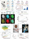
The cases for piezoelectric materials for central nervous system repair. a) Structure of the 3D piezoelectric PLA/KNN@PDA scaffold. b) Promotion of spinal cord injury recovery in vivo. Reproduced with permission.[ 176a ] Copyright 2022, American Chemical Society. c) Structure of the Au@BT nanoparticles. Scale bar, 100 nm. d) Motor function recovery properties in vivo. Reproduced with permission.[ 176b ] Copyright 2024, Wiley‐VCH. e) Structure of the photo‐sensitive artificial retina. f) Capacitance response curve under different lighting conditions. Reproduced with permission.[ 174c ] Copyright 2016, Wiley‐VCH. g) Artificial retina implantation surgery. h) Enhanced flash visual evoked potential. Reproduced with permission.[ 174a ] Copyright 2020, American Chemical Society. i) Capacitance response curves under different lighting and bending conditions. Reproduced with permission.[ 174b ] Copyright 2022, Springer Nature.
Figure 10
Figure 10
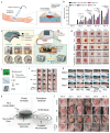
The cases for piezoelectric materials for wound healing. a) Illustration of the PLLA skin‐wound scaffold. b) Promotion of wound healing in vivo. Reproduced with permission.[ 182b ] Copyright 2023, Elsevier. c) Structure of the piezoelectric skin patch. d) Promotion of wound healing in vivo. Reproduced with permission.[ 182c ] Copyright 2023, American Chemical Society. e) Structure of the moxa‐modified zinc oxide nanosheets. f,g) Anti‐superficial and deep fungal resistance of nanosheets in vivo. CM, carbonized moxa. CMZ, carbonized moxa@ZnO. KTZ, ketoconazole. Reproduced with permission.[ 182d ] Copyright 2024, American Chemical Society. h) Structure of the γ‐glycine/PVA composite device. i) Promotion of wound healing in vivo. Scale bar, 6 mm. Reproduced with permission.[ 182e ] Copyright 2024, American Association for the Advancement of Science.
Figure 11
Figure 11
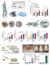
The cases for piezoelectric materials for dental tissue regeneration. a) Structure of the t‐BT/GelMA hydrogel.: b) Promotion of the repair of inflammatory periodontal defects. Reproduced with permission.[ 183b ] Copyright 2024, Ke Ai Publishing. c) Structure of the strontium‐containing P(VDF‐TrFE) film. d) Promotion of the differentiation of dental pulp stem cells. e,f) Promotion of dentin regeneration in vivo. Reproduced with permission.[ 185 ] Copyright 2024, Wiley‐VCH.
Figure 12
Figure 12
The cases for piezoelectric materials for tendon, myocardial, and corneal regeneration. a) Self‐powered piezoelectric tendon device promotes tendon recovery in vivo. Reproduced with permission.[ 186a ] Copyright 2021, Wiley‐VCH. b) Structure of the 3D printing of the cardiac serpentine scaffold. c) Improvement of myocardial infarction in vivo. Reproduced with permission.[ 187 ] Copyright 2024, Wiley‐VCH. d) Structure of the Blink‐driven piezoelectric contact lens. Reproduced with permission.[ 188 ] Copyright 2023, Nature.
Figure 13
Figure 13
The mechanism‐driven strategy and current issues of the tissue application of piezoelectric materials. The four sections in the center are the four core elements of the mechanism‐driven strategy. The peripheral sections are the issues at this stage.

Similar articles

Cited by

References

    1. F. D. I. S. C. O. B. Edition , FDA approval of Ryoncil (remestemcel‐L‐rknd) for steroid‐refractory acute graft versus host disease in pediatric patients, https://www.fda.gov/drugs/resources‐information‐approved‐drugs/fda‐disco... (accessed: February 2025).
    1. E. M. Agency , Alofisel withdrawn from the EU market, https://www.ema.europa.eu/en/news/alofisel‐withdrawn‐eu‐market (accessed: February 2025).
    1. B. Carmen Pope , Ryoncil, https://www.drugs.com/ryoncil.html (accessed: February 2025).
    1. a) Levin M., Cell 2021, 184, 1971; - PubMed
    2. b) Levin M., Semin. Cell Dev. Biol. 2009, 20, 543. - PMC - PubMed
    1. a) Mao Y. L., Wickström S. A., Nat. Rev. Mol. Cell Biol. 2024; - PubMed
    2. b) Maurer M., Lammerding J., Annu. Rev. Biomed. Eng. 2019, 21, 443; - PMC - PubMed
    3. c) Di X., Gao X., Peng L., Ai J., Jin X., Qi S., Li H., Wang K., Luo D., Signal. Transduct. Target Ther. 2023, 8, 282; - PMC - PubMed
    4. d) Du H., Bartleson J. M., Butenko S., Alonso V., Liu W. F., Winer D. A., Butte M. J., Nat. Rev. Immunol. 2023, 23, 174. - PMC - PubMed

Substances

LinkOut - more resources