. 2025 Oct;19(10):2978-2988.
doi: 10.1002/1878-0261.70062. Epub 2025 May 28.

Therapeutic applications of a novel humanized monoclonal antibody targeting chemokine receptor CCR9 in pancreatic cancer

Affiliations

Therapeutic applications of a novel humanized monoclonal antibody targeting chemokine receptor CCR9 in pancreatic cancer

Hannah G McDonald et al. Mol Oncol. 2025 Oct.

Abstract

The relative failure of immune checkpoint inhibitors in pancreatic ductal adenocarcinoma (PDAC) despite having a dense, immunosuppressive tumor microenvironment highlights the need to target alternate/escape pathways. We have previously examined C-C chemokine receptor type 9 (CCR9) as a candidate immune checkpoint and developed a targeted, humanized monoclonal antibody (SRB2). Cytotoxicity of SRB2 was evaluated in vitro and in vivo. CCR9 expression on PDAC cells/tissues, immune components of patient-derived organoids (PDOs), and antibody-dependent cell-mediated cytotoxicity were examined. In PANC-1 and MIA PaCa-2 cell lines, we demonstrated highest CCR9 expression; however, no direct cytotoxic effect was observed with SRB2 treatment. In PANC-1 cells, NK cell-mediated cytotoxicity was promoted by SRB2. Dose-dependent SRB2 cytotoxicity was observed in PDAC PDOs. In patient-derived xenograft mouse models, cytotoxicity of SRB2 monotherapy and in combination with oxaliplatin was also shown. In humanized immune-competent mouse models, SRB2 efficacy was similar to other drugs, but two mice in this cohort had complete tumor regression. Our current studies suggest that therapeutic targeting of CCR9 may improve PDAC outcomes, and additional studies are underway to evaluate SRB2 for clinical use.

Keywords: chemokine receptor CCR9; immune checkpoint; pancreatic cancer.


Conflict of interest statement

The authors disclose that DRL, EAC, PG, and LS are employees of SunRock Biopharma and have a financial interest in SRB2 (anti‐CCR9 monoclonal antibody). Other authors declare no conflict of interest.

Figures

Fig. 1
Fig. 1
SRB2 and PDAC cell proliferation. The results of this assay demonstrated that SRB2 had no effect on CCL25‐induced PDAC cell proliferation. CCL25 (400 ng/mL) induced MIAPaCa‐2 (A) and PANC‐1 (B) cell proliferation, while SRB2 did not attenuate proliferation in either cell line. MG132 (10 μM) was used as a positive cytotoxic control. There was one biological replicate per group, and data are presented as mean ± SEM.
Fig. 2
Fig. 2
ADCC assay in PANC‐1 cells. NK cell‐mediated cytotoxicity was promoted by SRB2. PANC‐1 cell lysis was measured after treatment with SRB2 (10 μg/mL) and NK cell co‐culture. The results showed significantly higher cell lysis in CCR9+ cells compared to CCR9 cells. There were 3 biological replicates included, and data is presented as mean ± SEM and compared with an unpaired Student's t‐test.
Fig. 3
Fig. 3
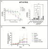
In vitro and in vivo effects of SRB2. Here, SRB2 was administered alone and in combination with cytotoxic drugs in PDAC PDOs and PDX animals. Dose–response curves of SRB2, irinotecan, oxaliplatin, and paclitaxel are shown for hPT15 PDOs (A). Data include three biological replicates and are presented as mean ± SD. The bar graph shows the effects of SRB2 and/or cytotoxic drugs (n = 4 biological replicates per treatment group) (A). Data are presented as mean ± SD and compared using two‐way ANOVA. Then, the effects of SRB2 and cytotoxic/targeted drugs were evaluated in CCR9+ PDAC PDX animals. We observed trends toward decreased tumor growth in the experimental treatment groups compared with the control groups (B). N = 8 mice were tested per treatment group. PDX data are presented as mean ± SD and compared using mixed‐effects analysis.
Fig. 4
Fig. 4
IHC measurements of proliferation (Ki‐67) and necrosis (CC3). Excised tumors from two mice per treatment group (total n = 12) were sectioned and IHC analysis performed for Ki‐67 (blue) and CC3 (orange). The results showed the highest necrosis (CC3) with SRB2 and the lowest proliferation (Ki‐67) with SRB2 + oxaliplatin. Data shown as mean ± SEM.
Fig. 5
Fig. 5
Immune cell populations in PDAC PDOs and PDXs. (A) IF staining for CK19 (green) and CD3 (red) in hPT15 PDOs showed retained immune and cancer cells in early passage PDOs. (B) IF staining for CD3 and PD1 in hPT15 PDX tissue slides showed staining for CD3 (green) and PD1 (red). Overlay (yellow) of both markers indicate immune cells. Scale bar in A and B represents 10 μm. Pseudocolor dot plot (C) and contour plot (D) show proportions of CCR9 and CD45 single positive (Q1,3) and double positive (Q2), as well as double negative (Q4) populations within the tumors. Flow cytometry was performed using BD FACSymphony A3 machine and analyzed using FlowJo v10 Software.
Fig. 6
Fig. 6
Tumor growth curves in NCG humanized mice. NCG mice with xenografted PDACs were treated with vehicle, SRB2 (25 mg/kg), pembrolizumab (8 mg/kg) or gemcitabine (50 mg/kg) + nab‐paclitaxel (5 mg/kg). Tumor volumes were lowest for SRB2 compared to vehicle (P = 0.031). Mean ± SEM for tumor volume for each treatment group were expressed in mm3 with N = 8 mice per group. Groups were compared with two‐way ANOVA.

References

    1. Fan J‐q, Wang M‐F, Chen H‐L, Shang D, Das JK, Song J. Current advances and outlooks in immunotherapy for pancreatic ductal adenocarcinoma. Mol Cancer. 2020;19(1):32. 10.1186/s12943-020-01151-3 - DOI - PMC - PubMed
    1. He X, Xu C. Immune checkpoint signaling and cancer immunotherapy. Cell Res. 2020;30(8):660–669. 10.1038/s41422-020-0343-4 - DOI - PMC - PubMed
    1. Macherla S, Laks S, Naqash AR, Bulumulle A, Zervos E, Muzaffar M. Emerging role of immune checkpoint blockade in pancreatic cancer. Int J Mol Sci. 2018;19(11):3505. 10.3390/ijms19113505 - DOI - PMC - PubMed
    1. Shen X, Mailey B, Ellenhorn JD, Chu PG, Lowy AM, Kim J. CC chemokine receptor 9 enhances proliferation in pancreatic intraepithelial neoplasia and pancreatic cancer cells. J Gastrointest Surg. 2009;13(11):1955–1962. 10.1007/s11605-009-1002-8 - DOI - PubMed
    1. Heinrich EL, Arrington AK, Ko ME, Luu C, Lee W, Lu J, et al. Paracrine activation of chemokine receptor CCR9 enhances the invasiveness of pancreatic cancer cells. Cancer Microenviron. 2013;6(3):241–245. 10.1007/s12307-013-0130-6 - DOI - PMC - PubMed

MeSH terms

Substances