

Wound state monitoring by multiplexed, electrochemical, real-time, localized, inflammation-tracking nitric oxide sensor (MERLIN)
- PMID: 40435247
- PMCID: PMC12118596
- DOI: 10.1126/sciadv.adv2385
Wound state monitoring by multiplexed, electrochemical, real-time, localized, inflammation-tracking nitric oxide sensor (MERLIN)
Abstract
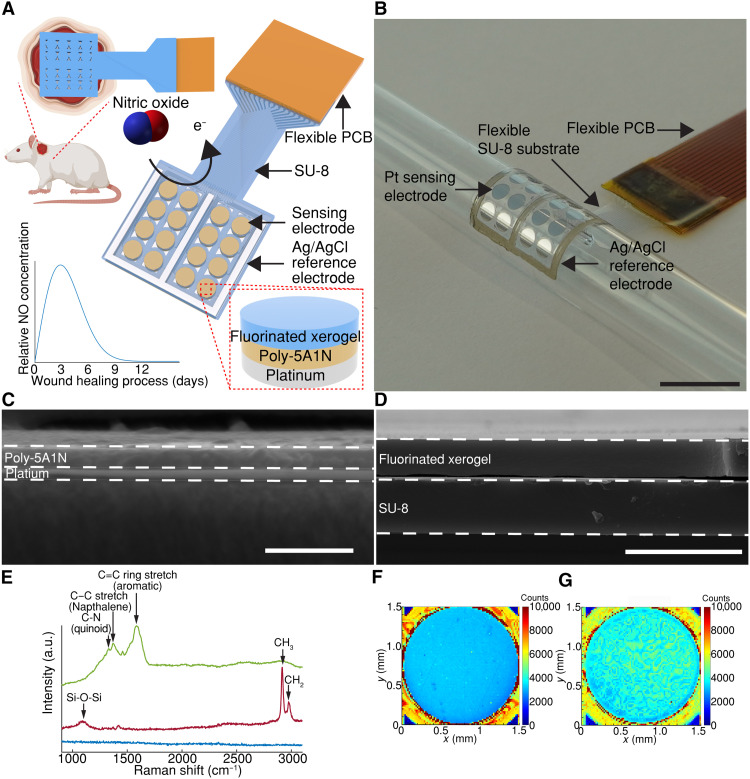
Nitric oxide (NO) released endogenously by induced NO synthase (iNOS) in macrophages is a key regulatory biomarker for wound inflammation. Detecting NO directly on the wound bed is challenging due to its short half-life time (6 to 50 seconds), low physiological concentration (nanomolar to micromolar), and interferences in the complex wound environment. Here, we present a compliant, multiplexed, electrochemical, real-time, localized, inflammation-tracking NO sensor (MERLIN) array for in vivo spatiotemporal measurement of NO, with high sensitivity (883 ± 283 nanoamperes per micromolar per square centimeter); selectivity against nitrites (~27,900-fold), ascorbic acid (~3800-fold), and uric acid (~6900-fold); and low limit of detection (~8.00 nM). MERLIN spatiotemporally tracked NO on rat skin wounds for 7 days, and results indicated that NO peaks on day 3, in line with previously reported iNOS activity. MERLIN allows spatial mapping of the NO gradient across the wound bed, which can be used to provide diagnostic information to assist wound care.
Figures


Similar articles
-
Nitric oxide in the healing wound: a time-course study.J Surg Res. 2001 Nov;101(1):104-8. doi: 10.1006/jsre.2001.6261. J Surg Res. 2001. PMID: 11676563
-
Topical application of dressing with amino acids improves cutaneous wound healing in aged rats.Acta Histochem. 2010 Sep;112(5):497-507. doi: 10.1016/j.acthis.2009.05.003. Epub 2009 Jun 28. Acta Histochem. 2010. PMID: 19560799
-
Cellular and physiological upregulation of inducible nitric oxide synthase, arginase, and inducible cyclooxygenase in wound healing.J Cell Physiol. 2019 Dec;234(12):23618-23632. doi: 10.1002/jcp.28930. Epub 2019 Jun 3. J Cell Physiol. 2019. PMID: 31161614
-
Electrochemical Nitric Oxide Sensors: Principles of Design and Characterization.Chem Rev. 2019 Nov 27;119(22):11551-11575. doi: 10.1021/acs.chemrev.8b00797. Epub 2019 Sep 25. Chem Rev. 2019. PMID: 31553169 Review.
-
Role of nitric oxide in wound repair.Am J Surg. 2002 Apr;183(4):406-12. doi: 10.1016/s0002-9610(02)00815-2. Am J Surg. 2002. PMID: 11975928 Review.
References
-
- Picón-Pagès P., Garcia-Buendia J., Munoz F. J., Functions and dysfunctions of nitric oxide in brain. Biochim. Biophys. Acta Mol. Basis Dis. 1865, 1949–1967 (2019). - PubMed
-
- Ziche M., Morbidelli L., Nitric oxide and angiogenesis. J. Neuro-Oncol. 50, 139–148 (2000). - PubMed
-
- Coleman J. W., Nitric oxide in immunity and inflammation. Int. J. Immunopharmacol. 1, 1397–1406 (2001). - PubMed
MeSH terms
Substances
LinkOut - more resources
Full Text Sources
Medical
