. 2025 May 28;20(5):e0316983.
doi: 10.1371/journal.pone.0316983. eCollection 2025.

The antiangiogenic peptide VIAN-c4551 inhibits lung melanoma metastasis in mice by reducing pulmonary vascular permeability

Affiliations

The antiangiogenic peptide VIAN-c4551 inhibits lung melanoma metastasis in mice by reducing pulmonary vascular permeability

Alma Lorena Perez et al. PLoS One. .

Abstract

Introduction: Cancer cells drive the increase in vascular permeability mediating tumor cell extravasation and metastatic seeding. VIAN-c4551, an antiangiogenic peptide analog of vasoinhibin, inhibits the growth and vascularization of melanoma tumors in mice. Because VIAN-c4551 is a potent inhibitor of vascular permeability, we evaluated whether its antitumor action extended to a reduction in metastasis generation.

Methods: Circulating levels of vascular endothelial growth factor (VEGF), lung vascular permeability, melanoma cell extravasation, and melanoma pulmonary nodules were assessed in C57BL/6J mice intravenously inoculated with murine melanoma B16-F10 cells after acute treatment with VIAN-c4551. VEGF levels, transendothelial electrical resistance, and transendothelial migration in cocultures of B16-F10 cells and endothelial cell monolayers supported the findings.

Results: B16-F10 cells increased circulating VEGF levels and elevated lung vascular permeability 2 hours after inoculation. VIAN-c4551 prevented enhanced vascular permeability and reduced melanoma cell extravasation after 2 hours and the number and size of macroscopic and microscopic melanoma tumors in lungs after 17 days. In vitro, VIAN-c4551 suppressed the B16-F10 cell-induced and VEGF mediated increase in endothelial cell monolayer permeability and the transendothelial migration of B16-F10 cells. No detrimental effect of VIAN-c4551 was observed on hematological, biochemical, and histological parameters after its intravenous administration in mice for 14 days.

Conclusions: These findings support the inhibition of distant vascular permeability for the prevention of tumor metastasis and unveil the anti-vascular permeability factor VIAN-c4551 as a potential and safe therapeutic drug able to prevent metastasis generation by lowering the extravasation of melanoma cells.


Conflict of interest statement

JPR, MZ, TB, JT, GME, and CC are inventors of a submitted patent application (WO/2021/098996), which is owned by the Universidad Nacional Autónoma de México (UNAM) and JT. JPR is the CEO and founder of VIAN Therapeutics Inc. MZ and CC are consultants for VIAN Therapeutics. Inc. This does not alter our adherence to PLOS ONE policies on sharing data and materials.

Figures

Fig 1
Fig 1. B16-F10 cells stimulate lung vascular permeability.
(a) Timeline of the evaluation of pulmonary vascular permeability after intravenous inoculation of B16-F10 cells. (b) Photographs of representative lungs showing the accumulation of Evans blue-stained albumin at different times post-B16-F10 cell delivery. (c) Quantification of Evans blue-stained albumin as index of lung vascular permeability. Data from 2 independent experiments (n = 4) is graphed using a box and a whisker plot; the box frames the interquartile range, the horizontal line indicates the median, and the whiskers the min and max values. The mean is indicated by +. *P = 0.02, **P = 0.01, ***P < 0.001 (One-way ANOVA, Tukey’s multiple comparison test).
Fig 2
Fig 2. VIAN-c4551 prevents the increase of vascular permeability stimulated by melanoma cells.
(a) Timeline of the experiment: VIAN-c4551 or vehicle (Veh) were i.v. injected 30 minutes before the i.v. delivery of B16-F10 cells or PBS. Pulmonary vascular permeability or VEGF levels were evaluated 2 hours post-tumor cells or PBS. (b) Photographs of representative lungs showing the accumulation of Evans blue-stained albumin as index of pulmonary vascular permeability. (c) Quantification of lung vascular permeability in Veh- and VIAN-c4551-treated mice after i.v. injection of PBS or B16-F10 cell inoculation. Data from 3 independent experiments (n = 9) are graphed in a box and a whisker plot; the box frames the interquartile range, the horizontal line indicates the median, and the whiskers the min and max values. The mean is indicated by +. ***P < 0.001 (Two-way ANOVA, Sidak’s multiple comparison test). (d) VEGF levels in serum from Veh- and VIAN-c4551-treated mice after injection of PBS or B16-F10 cell inoculation. Values are box and whiskers plot from 2 independent experiments (n = 6), ***P < 0.001 (Two-way ANOVA, Sidak’s multiple comparison test).
Fig 3
Fig 3. A single administration of VIAN-c4551 reduces the number and size of lung melanoma metastases in mice.
(a) Timeline of the experiment: VIAN-c4551 or vehicle (Veh) were injected i.v. 30 minutes before the i.v. inoculation of B16-F10 cells. Lung melanoma nodules were evaluated 17 days post-tumor cell delivery. (b) Representative ventral and dorsal views of the left lung and the four right lobes of a mice treated with Veh or VIAN-c4551. Quantification of the number (c) and size (d) of macroscopic melanoma nodules on the lung surface. (e) Representative lung sections stained with hematoxylin/eosin showing microscopic melanoma nodules (arrows) (scale = 200 μm). Quantification of the number (f) and size (g) of internal microscopic melanoma nodules in lungs. Numbers of nodules are graphed in a box and a whisker plot; the box represents the interquartile range, the horizontal line indicates the median, and the whiskers the min and max values. Size is graphed in a violin plot showing median and quartiles. The mean is indicated by +. Data from 3 independent experiments (n = 13). **P = 0.008, ***P < 0.001 vs Veh (unpaired t-test).
Fig 4
Fig 4. VIAN-c4551 inhibits the melanoma cell-induced permeability of endothelial cell monolayers mediated by VEGF.
(a) Effect of B16-F10 cells on the transendothelial electrical resistance (TEER) of bovine umbilical vein endothelial cell line (BUVEC-E6E7) monolayers in the absence or presence of VIAN-c4551 or anti-VEGF over a 6-hour period. *P = 0.0168, **P = 0.0081 vs Veh. (b) Effect of B16-F10 conditioned media (B16-F10-CM) on TEER of BUVEC-E6E7 monolayers. *P = 0.0182 vs Veh. (Repeated measurements one-way ANOVA, Dunnett’s multiple comparisons test). Values are means ± SD of 3 independent experiments. (c) VEGF levels in the conditioned medium of B16-F10 melanoma cells treated or not with VIAN-c4551 throughout a 24-hour incubation period. Values are means ± SD of 3 independent experiments. (d) Representative images of the actin cytoskeleton distribution in BUVEC-E6E7 monolayers treated or not with B16-F10-CM in the absence or presence of VIAN-c4551 or anti-VEGF (scale = 100 μm). (e) Effect of 3T3 conditioned media (3T3-CM) and B16-F10 conditioned media (B16-F10-CM) on TEER of BUVEC-E6E7 monolayers. *P = 0.0105 vs Veh. (Repeated measurements one-way ANOVA, Dunnett’s multiple comparisons test). (f) Effect of B16-F10 (B16-F10-CM), MDA-MB-231 (MDA-MB-231-CM), and MCF-7 (MCF-7-CM) conditioned media on TEER of human umbilical vein endothelial cell (HUVEC) monolayers. *P < 0.001 vs Veh. (Two-way ANOVA, Dunnett’s multiple comparisons test). Values are means ± SD of 3 independent experiments.
Fig 5
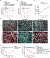
Fig 5. VIAN-c4551 reduces the in vitro transendothelial migration and the lung extravasation of B16-F10 melanoma cells.
(a) Representative images showing the B16-F10 cells expressing GFP that migrated across a confluent endothelial cell monolayer in the presence of vehicle (Veh) or VIAN-c4551. (b) Quantification of the number of migrated B16-F10-GFP cells. Values are box and whiskers plot from 4 independent experiments. The mean is indicated by +. **P = 0.0042 vs Veh (unpaired t-test) (scale = 120 μm). (c) Representative lung sections showing extravasated B16-F10-GFP cells 2 hours after their inoculation (arrows) (scale = 100 μm). (d) GFP mRNA levels in lungs from Veh- and VIAN-c4551-treated mice. Values are box and whiskers plot from 2 independent experiments (n = 6). The mean is indicated by +. **P = 0.0014 vs Veh (unpaired t-test).

Similar articles

References

    1. Anderson RL, Balasas T, Callaghan J, Coombes RC, Evans J, Hall JA, et al.. A framework for the development of effective anti-metastatic agents. Nat Rev Clin Oncol. 2019;16(3):185–204. doi: 10.1038/s41571-018-0134-8 - DOI - PMC - PubMed
    1. Tomita T, Kato M, Hiratsuka S. Regulation of vascular permeability in cancer metastasis. Cancer Sci. 2021;112(8):2966–74. doi: 10.1111/cas.14942 - DOI - PMC - PubMed
    1. Li X, Padhan N, Sjöström EO, Roche FP, Testini C, Honkura N, et al.. VEGFR2 pY949 signalling regulates adherens junction integrity and metastatic spread. Nat Commun. 2016;7:11017. doi: 10.1038/ncomms11017 - DOI - PMC - PubMed
    1. Robles JP, Zamora M, Siqueiros-Marquez L, Adan-Castro E, Ramirez-Hernandez G, Nuñez FF, et al.. The HGR motif is the antiangiogenic determinant of vasoinhibin: implications for a therapeutic orally active oligopeptide. Angiogenesis. 2022;25(1):57–70. doi: 10.1007/s10456-021-09800-x - DOI - PMC - PubMed
    1. Clapp C, Thebault S, Macotela Y, Moreno-Carranza B, Triebel J, Martínez de la Escalera G. Regulation of blood vessels by prolactin and vasoinhibins. Adv Exp Med Biol. 2015;846:83–95. doi: 10.1007/978-3-319-12114-7_4 - DOI - PubMed

MeSH terms

Substances