. 2025 Aug:66:101186.
doi: 10.1016/j.neo.2025.101186. Epub 2025 May 27.

CYP1B1 promotes angiogenesis and sunitinib resistance in clear cell renal cell carcinoma via USP5-mediated HIF2α deubiquitination

Affiliations

CYP1B1 promotes angiogenesis and sunitinib resistance in clear cell renal cell carcinoma via USP5-mediated HIF2α deubiquitination

Ke Ma et al. Neoplasia. 2025 Aug.

Abstract

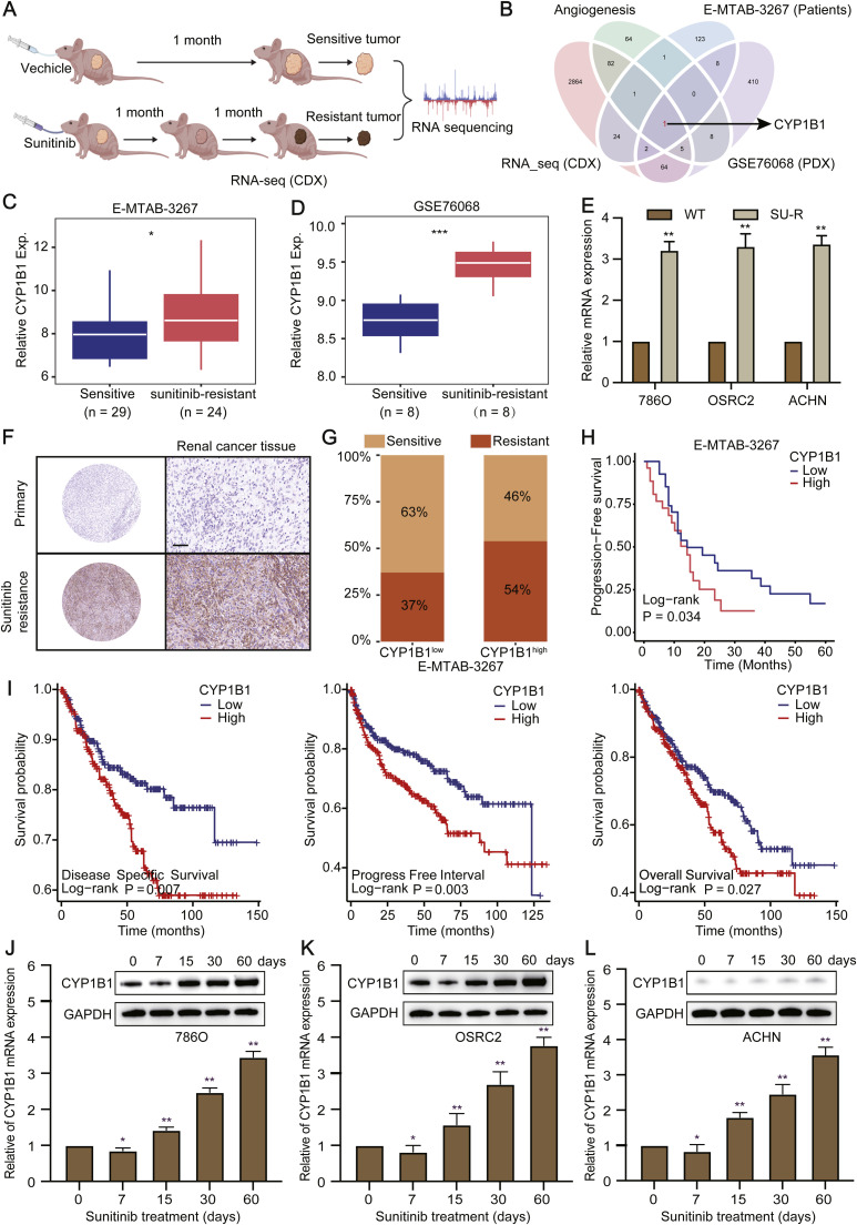
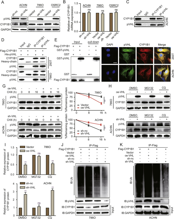
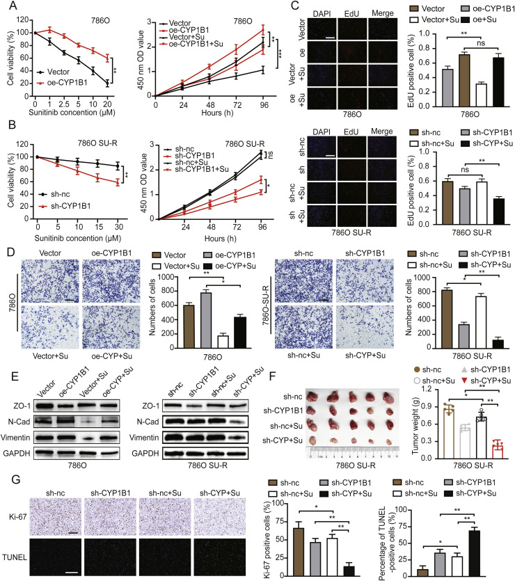
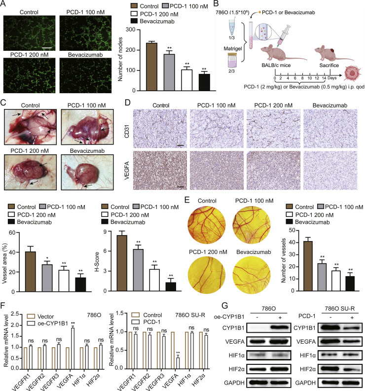
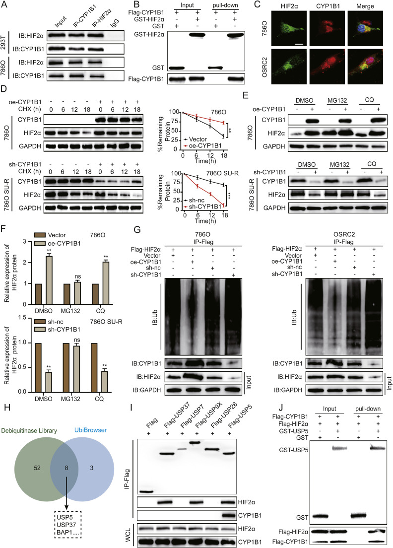
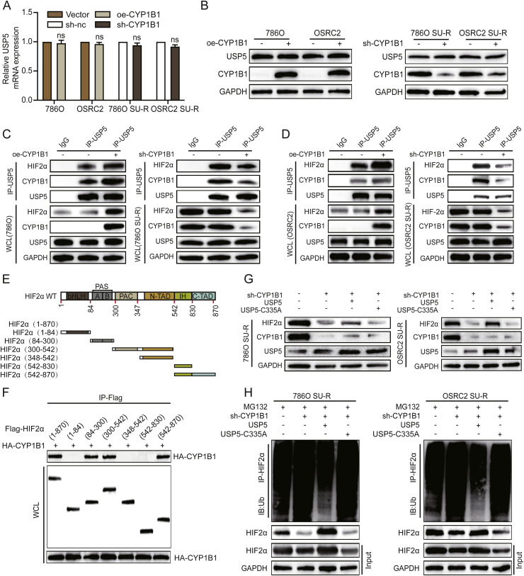
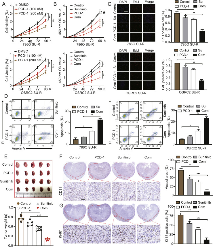
Clear cell renal cell carcinoma (ccRCC) is strongly aetiologically associated with von Hippel‒Lindau (VHL) tumour suppressor gene mutations, which result in constitutive activation of hypoxia-inducible factors and pathological angiogenesis. Although accumulating evidence indicates that antiangiogenic therapies targeting VEGF signalling can prolong the survival of ccRCC patients, the frequent development of therapeutic resistance to tyrosine kinase inhibitors such as sunitinib remains a critical clinical limitation. Through integrated multiomics analyses of sunitinib-resistant cell models, patient-derived xenografts, and clinical specimens, we systematically identified CYP1B1 as a central mediator of treatment resistance. Transcriptomic and genomic profiling revealed that CYP1B1 overexpression in resistant tumours functionally contributes to enhanced angiogenic potential and maintenance of the resistant phenotype. Mechanistic investigations demonstrated that CYP1B1 stabilizes hypoxia-inducible factor 2α (HIF2α) by facilitating USP5-mediated deubiquitination, thereby preventing proteasomal degradation. Notably, we identified VHL as a novel E3 ubiquitin ligase that regulates CYP1B1 turnover; notably, VHL deficiency in ccRCC promotes CYP1B1 protein accumulation by suppressing ubiquitination. These findings establish a feed-forward regulatory axis in which VHL loss-induced CYP1B1 stabilization promotes HIF2α signalling persistence, ultimately driving sunitinib resistance. Our study delineated the CYP1B1-USP5-HIF2α signalling cascade as a critical resistance mechanism and thus reveals a targetable vulnerability in treatment-refractory ccRCC.

Keywords: Angiogenesis; Clear cell renal cell carcinoma; HIF2α; Sunitinib resistance; Ubiquitination.


Conflict of interest statement

Declaration of competing interest The authors declare that they have no known competing financial interests or personal relationships that could have appeared to influence the work reported in this paper.

Figures

Fig 1
Fig. 1
Elevated CYP1B1 expression in SU-R RCC. A Graphical representation of sunitinib-resistant CDX models and differential gene expression analysis between sunitinib-sensitive and sunitinib-resistant tumours via RNA-seq. B Screening of angiogenesis-related genes in the CDX model and two independent SU-R RCC datasets revealed CYP1B1 as a critical gene in sunitinib resistance. C and D Comparison of CYP1B1 expression in sunitinib-sensitive versus sunitinib-resistant tumours across the E-MTAB-3267 and GSE76068 datasets. E CYP1B1 mRNA levels in three pairs of wild-type (WT) and SU-R RCC cell lines. F CYP1B1 protein expression was analysed in primary and SU-R tumour tissues from 12 patients. Scale bar: 100 μm. G and H Correlations between CYP1B1 levels and sunitinib resistance and progression-free survival in SU-R RCC patients in the E-MTAB-3267 cohort. I Differences in overall survival, progression-free survival, and disease-specific survival between CYP1B1high and CYP1B1low patients in the TCGA cohort. J-L CYP1B1 induction by sunitinib (2.5 μM) over a 0–60-day period in three RCC cell lines, with mRNA and protein levels assessed at specified time points via RT‒qPCR and western blotting (ns, not significant; *, p < 0.05; **, p < 0.01).
Fig 2
Fig. 2
pVHL mediates the ubiquitination and degradation of CYP1B1. A and B Assessment of CYP1B1 mRNA and protein levels in ACHN cells with stable VHL knockdown and 786O and OSRC2 cells with stable VHL overexpression via western blotting and RT‒qPCR. C Coimmunoprecipitation of ACHN cell whole-cell lysates with CYP1B1, pVHL or IgG antibodies, followed by western blotting with the respective antibodies. D 293T and 786O cells were transfected with Flag-CYP1B1 and His-pVHL plasmids. Coimmunoprecipitation with CYP1B1, pVHL or IgG antibodies was followed by western blotting with the indicated antibodies. E GST affinity-isolation assays with purified tagged pVHL and CYP1B1 proteins, followed by western blotting analysis. F Cotransfection of 293T and ACHN cells with VHL and CYP1B1 overexpression plasmids and subsequent immunofluorescence staining with anti-pVHL and anti-CYP1B1 antibodies. Scale bar: 10 μm. G Protein synthesis inhibition via CHX (10 μM) in 786O cells with VHL overexpression and ACHN cells with VHL knockdown; CYP1B1 protein levels were assessed by western blotting at specified time points. H and I Treatment of 786O and ACHN cells with VHL overexpression or knockdown plasmids, followed by exposure to DMSO, chloroquine (CQ, 10 μM), or MG132 (20 μM) for 8 hours; CYP1B1 protein levels were analysed by western blotting. J and K Immunoprecipitation of cells with anti-Flag CYP1B1 antibodies, followed by western blotting with the indicated antibodies (ns, not significant; **, p < 0.01).
Fig 3
Fig. 3
CYP1B1 modulates sunitinib sensitivity in ccRCC. A and B Establishment of stable CYP1B1-overexpressing WT 786O cells and CYP1B1-knockdown SU-R 786O cells. The cells were treated with DMSO or sunitinib, and cell viability was assessed via CCK-8 assays. C and D Evaluation of cell proliferation and migration in the indicated groups via EdU incorporation and Transwell assays. Scale bar: 50 μm. E Expression of epithelial‒mesenchymal transition markers in the different treatment groups. F Nude mice bearing SU-R xenografts with stable CYP1B1 knockdown were treated with vehicle control or sunitinib (20 mg/kg, p.o.) for 4 weeks. The tumour size was monitored every four days, and the tumours were weighed and photographed at the endpoint. G IHC staining of Ki-67 in tumours and a TUNEL assay for apoptosis analysis in the four treatment groups. Scale bar: 100 μm (ns, not significant; *, p < 0.05; **, p < 0.01; ***, p < 0.001).
Fig 4
Fig. 4
CYP1B1 promotes VM and enhances HIF2α/VEGFA signalling in ccRCC. A Impact of the CYP1B1 inhibitor PCD-1 on the tube formation of HUVECs. Scale bar: 50 μm. B and C Graphical representation and representative images of Matrigel plug assays following treatment with DMSO, PCD-1, or bevacizumab. D IHC staining of VEGFA and CD31 in Matrigel plugs from the indicated groups, with quantification of vessel area and VEGFA-positive cells. Scale bar: 100 μm. E Inhibition of new vessel formation by PCD-1 in the CAM assay. F and G RT‒qPCR and western blotting analysis of HIF1α, HIF2α, VEGFA, VEGFR1, VEGFR2, and VEGFR3 expression in ccRCC cells treated with DMSO and PCD-1 (100 nM) for 48 hours or transfected with vector or CYP1B1 expression plasmid (ns, not significant; *, p < 0.05; **, p < 0.01).
Fig 5
Fig. 5
CYP1B1 regulates HIF2α protein stability through the ubiquitin–proteasome pathway. A Immunoprecipitation of 293T and 786O cell lysates with CYP1B1 or HIF2α antibodies, followed by western blotting with the indicated antibodies. B The purified tagged proteins were subjected to GST pull-down assays with the indicated antibodies. C Confocal microscopy analysis of HIF2α and CYP1B1 colocalization in 786O and OSRC2 cells. Scale bar: 50 μm. D Protein synthesis inhibition in 786O cells overexpressing CYP1B1 and SU-R 786O cells with CYP1B1 knockdown via CHX (10 μM); HIF2α protein levels were determined by western blotting at specified time points. E and F Treatment of 786O cells overexpressing CYP1B1 and SU-R 786O cells with CYP1B1 knockdown with DMSO, chloroquine (10 μM), or MG132 (20 μM) for 8 hours; HIF2α protein levels were analysed by western blotting. G Immunoprecipitation of the indicated cell lysates with anti-Flag antibodies, followed by western blotting with the indicated antibodies. H Venn diagram illustrating potential deubiquitinating enzymes that regulate the degradation of ubiquitinated HIF2α. I Co-IP assays in 786O cells were performed to assess the interaction between the five deubiquitinating enzymes and HIF2α. J The purified tagged proteins were subjected to GST pull-down assays with the indicated antibodies (ns, not significant; **, p < 0.01; ***, p < 0.001).
Fig 6
Fig. 6
CYP1B1 promotes HIF2α protein stabilization by enhancing the interaction between USP5 and HIF2α. A and B The mRNA and protein levels of CYP1B1 in RCC cells were analysed when the cells were subjected to CYP1B1 knockdown or overexpression. C and D WT and SU-R RCC cells were transfected with a CYP1B1 expression plasmid or shRNA for knockdown. Co-IP with IgG or USP5 antibody was followed by western blotting with the indicated antibodies. E and F Co-IP assays were conducted on 293T cells transfected with HA-labelled CYP1B1, Flag-labelled HIF2α and structural domain plasmids (Flag-HIF2α-bHLH, Flag-HIF2α-PAS, Flag-HIF2α-NTAD, Flag-HIF2α-IH, Flag-HIF2α-PAC—NTAD, and Flag-HIF2α-IH—CTAD). G SU-R RCC cells were transfected with the CYP1B1 knockdown plasmid alone or in combination with the USP5 wild-type or mutant USP5-C335A plasmid. Western blotting was performed with the indicated antibodies. H These cell lysates were immunoprecipitated with IgG or HIF2α antibodies and then subjected to western blotting with the indicated antibodies (ns, no significance).
Fig 7
Fig. 7
Inhibition of CYP1B1 impedes the progression of SU-R ccRCC. A Assessment of the viability of SU-R ccRCC cells treated with DMSO or PCD-1 (100 nM or 200 nM) at various time points via CCK-8 assays. B-D SU-R ccRCC cells were treated with DMSO, sunitinib (10 μM), PCD-1 (100 nM), or their combination for 0–96 hours. Cell viability was evaluated via CCK-8 assays, and cell proliferation and apoptosis were assessed via EdU incorporation and flow cytometry assays. Scale bar: 50 μm. E Establishment and treatment of an orthotopic xenograft model using SU-R 786O cells (1 × 10⁶). The mice were treated with vehicle, sunitinib (20 mg/kg), PCD-1 (2 mg/kg), or their combination for 28 days, after which they were sacrificed, and the tumour weights were measured. F and G IHC staining of Ki-67 and CD31 expression levels in tumours from the four treatment groups. Scale bar: 50 μm (ns, not significant; *, p < 0.05; **, p < 0.01; ***, p < 0.001).
Fig 8
Fig. 8
Schematic representation of the mechanistic role of CYP1B1 in ccRCC: upregulation of CYP1B1 in VHL-mutated ccRCC promotes ccRCC progression and angiogenesis. CYP1B1 interacts directly with the HIF2α protein, impeding its degradation in a USP5-dependent process, thereby sustaining the HIF2α/VEGFA signalling axis and facilitating the emergence of sunitinib resistance.

References

    1. Bray F., Laversanne M., Sung H., Ferlay J., Siegel R.L., Soerjomataram I., Jemal A. Global cancer statistics 2022: GLOBOCAN estimates of incidence and mortality worldwide for 36 cancers in 185 countries. CA Cancer J. Clin. 2024;74:229–263. doi: 10.3322/caac.21834. - DOI - PubMed
    1. Yao X., Tan J., Lim K.J., Koh J., Ooi W.F., Li Z., Huang D., Xing M., Chan Y.S., Qu J.Z., Tay S.T., Wijaya G., Lam Y.N., Hong J.H., Lee-Lim A.P., Guan P., Ng M.S.W., He C.Z., Lin J.S., Nandi T., Qamra A., Xu C., Myint S.S., Davies J.O.J., Goh J.Y., Loh G., Tan B.C., Rozen S.G., Yu Q., Tan I.B.H., Cheng C.W.S., Li S., Chang K.T.E., Tan P.H., Silver D.L., Lezhava A., Steger G., Hughes J.R., Teh B.T., Tan P. VHL deficiency drives enhancer activation of oncogenes in clear cell renal cell carcinoma. Cancer Discov. 2017;7:1284–1305. doi: 10.1158/2159-8290.CD-17-0375. - DOI - PubMed
    1. Kaur G., Roy B. Decoding tumor angiogenesis for therapeutic advancements: mechanistic insigh. Biomedicines. 2024;12:827. doi: 10.3390/biomedicines12040827. - DOI - PMC - PubMed
    1. Hirsch L., Flippot R., Escudier B., Albiges L. Immunomodulatory roles of VEGF pathway inhibitors in renal cell carcinoma. Drugs. 2020;80:1169–1181. doi: 10.1007/s40265-020-01327-7. - DOI - PubMed
    1. Liu Y., Li Y., Wang Y., Lin C., Zhang D., Chen J., Ouyang L., Wu F., Zhang J., Chen L. Recent progress on vascular endothelial growth factor receptor inhibitors with dual targeting capabilities for tumor therapy. J. Hematol. Oncol. 2022;15:89. doi: 10.1186/s13045-022-01310-7. - DOI - PMC - PubMed

MeSH terms

Substances