. 2025 Jul;643(8074):1352-1362.
doi: 10.1038/s41586-025-09179-5. Epub 2025 May 28.

Protein-primed homopolymer synthesis by an antiviral reverse transcriptase

Affiliations

Protein-primed homopolymer synthesis by an antiviral reverse transcriptase

Stephen Tang et al. Nature. 2025 Jul.

Abstract

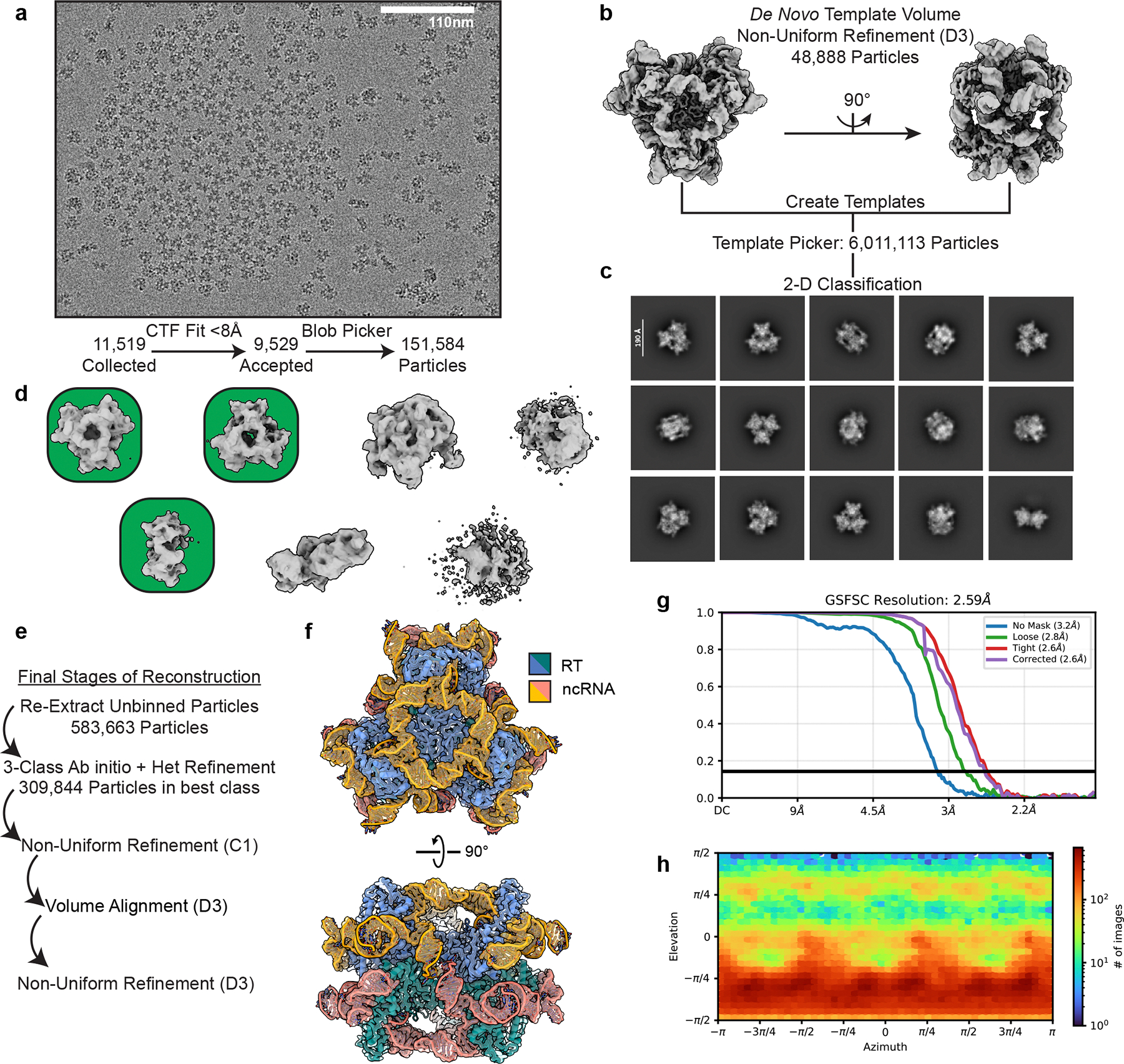
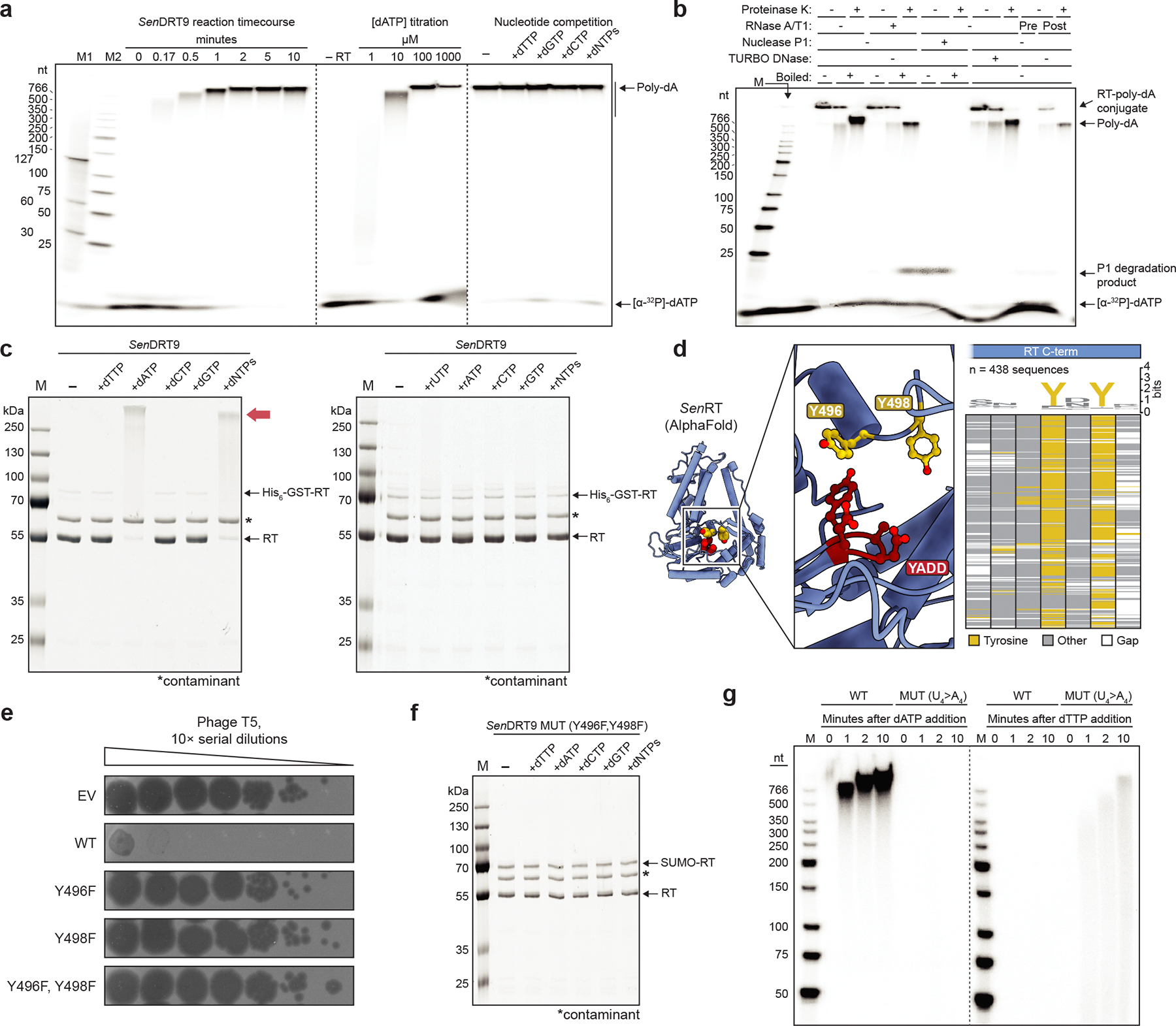
Bacteria defend themselves from viral predation using diverse immune systems, many of which target foreign DNA for degradation1. Defence-associated reverse transcriptase (DRT) systems provide an intriguing counterpoint to this strategy by using DNA synthesis instead2,3. We and others recently showed that DRT2 systems use an RNA template to assemble a de novo gene that encodes the antiviral effector protein Neo4,5. It remains unclear whether similar mechanisms of defence are used by other related DRT families. Here, we show that DRT9 systems defend against phage using DNA homopolymer synthesis. Viral infection triggers polydeoxyadenylate (poly-dA) accumulation in the cell, driving abortive infection and population-level immunity. Cryo-electron microscopy structures reveal how a non-coding RNA serves as both a structural scaffold and reverse transcription template to direct hexameric complex assembly and poly-dA synthesis. Notably, biochemical and functional experiments identify tyrosine residues within the reverse transcriptase itself that probably prime DNA synthesis, leading to the formation of protein-DNA covalent adducts. Synthesis of poly-dA by DRT9 in vivo is regulated by the competing activities of phage-encoded triggers and host-encoded silencers. Collectively, our study identifies a nucleic-acid-driven defence system that expands the paradigm of bacterial immunity and broadens the known functions of reverse transcriptases.


Conflict of interest statement

Competing interests: S.H.S. is a co-founder and scientific advisor to Dahlia Biosciences, a scientific advisor to CrisprBits and Prime Medicine, and an equity holder in Dahlia Biosciences and CrisprBits. B.W. is the founder of SurGene and is listed as an inventor on patent applications related to CRISPR–Cas systems and applications thereof. The other authors declare no competing interests.

Figures

Extended Data Figure 1 |
Extended Data Figure 1 |. Screening and identification of active DRT9 immune systems.
a, Phylogenetic tree of DRT9-encoded RT homologs. Outer rings show SLATT protein and ncRNA association within nearby genomic neighborhoods, and the inner ring shows previously identified DRT9 (UG28) homologs; systems selected for experimental testing are indicated with red circles. b, Heatmap of pairwise amino acid sequence identity percentages among DRT9-encoded RT homologs tested in this study (left), and heatmap of phage defense activity for the same DRT9 systems tested against 10 diverse E. coli phages (right); RT proteins encoded N-terminal FLAG tags. c, Representative plaque assays demonstrating that SenDRT9 and PsaDRT9 exhibit broad defense against phages from the Tequatrovirus and Tequintavirus genera, as compared to an empty vector (EV) control. d, Plaque assays demonstrating that defense activity against T2 phage is completely abolished for both SenDRT9 and PsaDRT9 systems encoding catalytically inactive RT mutants (MUT). e, Growth curves of cells expressing WT or MUT SenDRT9 +/− T5 phage at the indicated multiplicity of infection (MOI). Data are shown as mean ± s.d. for n = 3 independent biological replicates. f, Plaque forming unit (PFU; top) and colony forming unit (CFU; bottom) measurements from an infection time course experiment in which cells expressing WT or MUT SenDRT9 were infected with T5 phage at a high MOI. Cells expressing the WT defense system attenuated phage replication but were unable to recover from infection, indicating that defense activation leads to abortive infection and cell death. Data are shown as mean ± s.d. for n = 3 independent biological replicates. g, Plaque assays demonstrating that SenDRT9 encoding an RT with an N-terminal FLAG, but not C-terminal FLAG, retains WT defense against T5 phage. h, AlphaFold 3 structure prediction of the RT monomer from SenDRT9, highlighting the predicted positions of the N- and C-termini (orange spheres) and YADD active site (red spheres).
Extended Data Figure 2 |
Extended Data Figure 2 |. Discovery and characterization of homopolymeric DNA products elicited by DRT9 immune systems.
a, Unmapped reads from a cDIP-seq dataset of SenDRT9-expressing cells infected with T5 phage, showing uninterrupted strings of poly-dT and poly-dA. b, RIP-seq and cDIP-seq coverage tracks (top to bottom) for either WT or RT-inactive (MUT) PsaDRT9, in the presence of T5 phage infection. A schematic of the genomic locus is shown below; data are normalized for sequencing depth and plotted as counts per million reads (CPM). c, MEME analysis results revealing a poly-dT motif enriched in unmapped reads from the WT + T5 cDIP-seq dataset in b. E, E-value significance; n, number of contributing sites. d, Methylene blue-stained membrane used for the Southern blot shown in Fig. 1h. Representative data are shown for experiments repeated at least two times with similar results. e, Schematic of oligo spike-in experiment to address potential bias in next-generation sequencing-based detection of poly-dA and poly-dT on the AVITI platform; P, phosphorylated 5′ end. f, Bar graph of dA25 and dT25 counts from total DNA sequencing of oligo spike-in experiments schematized in e; the apparent bias against poly-dA capture and sequencing leads to artificially elevated levels of dT25-containing reads relative to dA25-containing reads. g, Bar graph of normalized homopolymer counts from total RNA-seq datasets of WT and MUT SenDRT9-expressing cells +/− T5 phage infection. These data demonstrate that poly-dA and poly-dT cDNAs are not transcribed, in contrast to the cDNA products of DRT2. Data in f,g are shown as mean ± s.d. for n = 3 independent biological replicates.
Extended Data Figure 3 |
Extended Data Figure 3 |. Additional characterization of ncRNA sequence perturbations on SenDRT9 defense activity.
a, Covariance model of the DRT9 ncRNA from an analysis of 201 homologous systems (left), and WebLogo from a multiple sequence alignment centered around the putative U-rich template region (top right). b, Comparison of SenDRT9 (left) and KpnDRT2 (right) ncRNAs, highlighting the scaffold (grey) and template (orange) regions. Both ncRNAs template cDNA synthesis from a similar location relative to SL2 (reverse transcription start site, in red), and program repetitive cDNA synthesis across the template region. c, Representative plaque assays for the data shown in Fig. 2b. d, Heat map quantifying SenDRT9 defense activity against T5 phage for the indicated ncRNA mutations and deletions within the 3′-proximal SL6 and U-rich region, quantified as the fold reduction in EOP relative to an empty vector (EV) control; data are shown as the mean of n = 2 technical replicates. e, RIP-seq coverage tracks for SenDRT9 with WT ncRNA or the indicated ncRNA mutations in uninfected cells; the bottom three variants are mutated in the putative template region (residues 123–126). A schematic of the genomic locus is shown below; data are normalized for sequencing depth and plotted as counts per million reads (CPM). f, Heat map quantifying SenDRT9 defense activity against T2 phage for the same ncRNA mutations tested against T5 phage in Fig. 2b–d and panel d; data are shown as in d. g, Methylene blue-stained membrane used for the Southern blot shown in Fig. 2f. Representative data are shown for experiments repeated at least two times with similar results.
Extended Data Figure 4 |
Extended Data Figure 4 |. Purification and characterization of the SenDRT9-encoded RT-ncRNA complex.
a, E. coli expression vector design for the SenDRT9-encoded ncRNA and His6-GST-tagged RT. b, Size-exclusion chromatogram of the His6-GST-tagged RT-ncRNA complex separated on a Superdex 200 10/300 column (left), and SDS-PAGE analysis of the void volume and labeled peaks (right); the high A260:A280 ratio is consistent with a protein-nucleic acid complex. c, Denaturing, SYBR Gold-stained 10% urea-PAGE analysis of the ~150-nt ncRNA species co-purifying with SenRT after RNase or DNase treatment (top), and RNA-seq analysis of the ncRNA (middle). The mature ncRNA carries an extraneous 5′-G resulting from the T7 promoter and lacks SL6 at the 3′ end (bottom), suggesting that SL6 may be involved in transcriptional termination in vivo while being dispensable for RT-mediated poly-dA synthesis. d, Plaque assay showing loss of SenDRT9 defense activity for a His6-GST-tagged RT variant against T5 phage (left), but not T2 phage (right); EV, empty vector. e, Overlaid chromatograms from gel filtration experiments with RT-ncRNA complexes before and after TEV protease treatment of the His6-GST-RT fusion protein, revealing a shift to earlier retention volume and thus increased oligomeric state; the persistent high A260:A280 ratio suggests that the RT-ncRNA interaction remains intact. f, Denaturing 8% urea-PAGE analysis of DNA polymerization assays that contained 150 nM RT-ncRNA complex and increasing amounts of dATP per RT monomer, as indicated. Reactions were incubated at 37 °C for 60 min, followed by proteinase K treatment and phenol-chloroform extraction prior to electrophoretic separation; the gel was stained with SYBR Gold. For b,c,f, representative data are shown for experiments repeated at least two times with similar results.
Extended Data Figure 5 |
Extended Data Figure 5 |. Roles of C-terminal tyrosine residues in protein-primed reverse transcription and DRT9 phage defense.
a, Southern blot analysis of total DNA isolated from T5-infected cells expressing WT SenDRT9 +/− proteinase K treatment. The blot was probed with oligo-dT40 (left) to detect poly-dA species; the methylene blue-stained membrane after transfer is shown at right; M denotes a ladder marker. b, Overlaid chromatograms from gel filtration analysis of RT-ncRNA complexes +/− dATP incubation, revealing an increased A260:A280 ratio and a dramatic shift in retention volume in the presence of dATP. c, Volcano plot of differential peptide abundance from mass spectrometry analysis of WT versus catalytically inactive (MUT) RT proteins immunoprecipitated from T5-infected cells expressing SenDRT9. Each dot indicates a SenRT peptide, and blue dots indicate significantly depleted peptides with log2(fold change) < −1.5 and adjusted p-value < 0.05, as determined by unpaired two-tailed t-test with correction for multiple comparisons using the Benjamini-Hochberg method; the most depleted peptide, IMNPQSLNYDYE, contains two tyrosine residues likely involved in priming of poly-dA synthesis. d, Plaque assay showing loss of SenDRT9 defense activity against T2 phage for a double Y496F,Y498F mutant, but not single Y496F or Y498F mutants; EV, empty vector. e, RIP-seq coverage tracks for SenDRT9 in T5-infected cells with WT and single or double Y496F/Y498F RT mutants, as indicated. A schematic of the genomic locus is shown below; data are normalized for sequencing depth and plotted as counts per million reads (CPM). f, Denaturing 5% urea-PAGE analysis of DNA polymerization assays with WT (left) and Y496F,Y498F mutant (right) RT. All reactions contained 20 nM RT-ncRNA and 100 μM [α-32P]-dATP; M denotes a DNA ladder marker. g, Southern blot analysis of total DNA isolated from T5-infected cells expressing SenDRT9 with WT and single or double Y496F/Y498F RT mutants, as indicated. The blot was probed with oligo-dT40 (left) to detect poly-dA species; the methylene blue-stained membrane after transfer is shown at right; M denotes a DNA ladder marker. h, SDS-PAGE analysis of DNA polymerization assays with either WT SenDRT9 or a U4>A4 mutant ncRNA. RT-ncRNA complexes (0.15 μM) were incubated with the indicated dNTPs (0.9 mM). The red arrow denotes high-molecular weight (MW) protein-DNA conjugates; the SUMO-RT band arises from incomplete tag removal; *, purification contaminant; M, protein ladder marker. i, SDS-PAGE analysis of DNA polymerization assays as in h, but with a U4>G4 mutant ncRNA; no high-MW species are observed. j, Denaturing, SYBR Gold-stained 10% urea-PAGE analysis (left) of WT, U4>A4, and U4>G4 ncRNAs co-purifying with SenRT. The absence of a band for the U4>C4 ncRNA, despite a clear band corresponding to the RT by SDS-PAGE (right), indicates that the U4>C4 mutation disrupts RT-ncRNA complex formation. For a,f-j, representative data are shown for experiments repeated at least two times with similar results.
Extended Data Figure 6 |
Extended Data Figure 6 |. Cryo-EM analysis and image processing workflow for trimeric SenDRT9 RT-ncRNA complex.
a, Representative micrograph from SenDRT9 RT-ncRNA complex data collection with the His6-GST fusion protein, revealing a densely-packed field of particles in vitreous ice. b, Initial consensus volume after processing blob-picked particles, which refined to 3.7 Å resolution (C3-reconstruction) for de novo template generation and subsequent particle picking. c, Representative selected 2D classes from 5,235,086 initial particles picked using the template in panel b. d, Four-class ab initio reconstruction from 2,530,970 particles in selected classes to remove junk particles. e, Results of a five-class 3D classification without a mask and filtered at 8 Å resolution, with a class similarity of 0.1 to remove remaining poorly aligned particles. The best volume, highlighted by a green background, contained 367,640 particles that were selected for further processing. f, View of the final reconstruction in g shown at high contour (top), revealing noisy density at the N-terminus of each RT monomer corresponding to the His6-GST fusion used for expression and purification. We hypothesized that the GST fusion would prevent the formation of higher-order oligomers (bottom). g, Final (C3) reconstruction that refined to 3 Å resolution and was used for building the trimeric RT-ncRNA complex model. The map is shown with partial transparency and colored according to the model, with RT subunits in blue and ncRNA subunits in orange. h, Plot of Cryosparc’s gold standard Fourier shell correlation for the map in g. i, Plot of particle view distribution reveals mild anisotropy but no missing views. j, SDS-PAGE analysis of DNA polymerization assays with His6-GST-RT sample, in which RT-ncRNA complexes were incubated with the indicated dNTP(s) before reactions were quenched and resolved electrophoretically. Red arrows indicate preliminary evidence of protein-DNA conjugates in reactions that contained dATP. Representative data are shown for experiments repeated at least two times with similar results.
Extended Data Figure 7 |
Extended Data Figure 7 |. Cryo-EM analysis and image processing workflow for hexameric SenDRT9 RT-ncRNA complex.
a, Representative micrograph from the SenDRT9 RT-ncRNA hexameric complex, revealing a monolayer of evenly distributed particles. b, Initial reconstruction from on-the-fly image analysis that was used to generate de novo templates used for particle picking. c, Representative 2D Classes from 6,011,113 template-picked particles. d, Initial volumes from a 7-class ab initio reconstruction of 1,411,369 particles. Selected volumes for downstream processing are shown with a green background. e, Final steps used to sort selected particles in d to yield the final reconstruction. f, Final (D3) reconstruction of the SenDRT9 RT-ncRNA hexamer that refined to 2.6 Å and was used for model building. The map is shown as a partially transparent surface, with RT subunits shown in blue/cyan and ncRNA subunits shown in orange/salmon. g, Plot of Cryosparc’s gold standard Fourier shell correlation for the map in f. h, Plot of particle view distribution reveals mild anisotropy but no missing views.
Extended Data Figure 8 |
Extended Data Figure 8 |. Additional features of the SenDRT9 hexamer visualized by cryo-EM.
a, Local resolution map of the SenDRT9 RT-ncRNA hexamer (FSC cutoff 0.143), showing that resolution falls off near the periphery of the complex in flexible regions such as SL3 and SL4. b, Semi-transparent density highlighting a network of protein-RNA interactions that stabilize SL5. c, Semi-transparent density highlighting protein-protein interactions that stabilize the back-to-back assembly of trimers. d, Difference map generated by subtracting a calculated map of the model from the experimental density. Unmodelled density (grey) is visible in the map directly adjacent to the YADD active site (red), which may correspond to a small stretch of poly-dA product; repeated efforts to sort these particles failed to separate distinct conformational states. e, A close-up view of the SenRT active site (YADD residues in red). Density for the RT is shown as a semi-transparent surface, and the density map for the ncRNA is omitted here for simplicity. f, Semi-transparent density map highlighting the ncRNA poly-G track (residues 80–86) that was used to establish register for ModelAngelo.
Extended Data Figure 9 |
Extended Data Figure 9 |. Activation of DRT9 immunity by phage-encoded factors.
a, Representative plaque assays with WT or escaper T5 phage variants tested in strains expressing either the WT or RT-inactive (MUT) SenDRT9 system. b, Bar graphs of relative nucleotide levels for the indicated species quantified by LC-MS/MS, in lysates from cells expressing SenDRT9 or an empty vector (EV) control +/− T5 phage infection. Data are shown as the mean for n = 2 independent biological replicates. c, Bar graphs of plaque forming units per mL (PFU/mL) of T5 phage lysates after liquid culture infection of cells expressing SenDRT9 or an EV control, in the presence of the indicated supplemented nucleosides; data are shown as the mean for n = 2 independent biological replicates. d, Methylene blue-stained membrane of total DNA from cells expressing WT or MUT SenDRT9 +/− gp58 induction, for the Southern blot shown in Fig. 5f. e, Western blot analysis of SenRT co-immunoprecipitation with gp58. Immunoprecipitation was performed with a FLAG antibody on lysates from cells co-expressing SenRT (WT or MUT, FLAG-tagged or untagged) with V5-tagged gp58, followed by Western blot analysis using a V5 antibody. The unique presence of a band corresponding to gp58 in the IP eluate from cells expressing WT SenDRT9 indicates a DNA-dependent interaction between the RT and gp58. f, Denaturing 5% urea-PAGE analysis of DNA polymerization assays, after reactions were treated with ExoI (left) or Nuclease P1 (right) in the presence of increasing concentrations of T5 gp58. All reactions contained 20 nM RT-ncRNA (U4>A4) and 100 μM [α-32P]-dTTP and were incubated at 37 °C for 60 min prior to addition of gp58 and ExoI or Nuclease P1. For d-f, representative data are shown for experiments repeated at least two times with similar results.
Figure 1 |
Figure 1 |. Systematic discovery of DRT9 reverse transcription substrates and products in vivo.
a, Phylogenetic tree of bacterial reverse transcriptase (RT) homologs within the UG family, based on Mestre et al.. DRT9 (UG28) and DRT2 (UG2) are highlighted, as are other experimentally studied systems; genetic architectures of archetypal systems are shown below. b, Schematic of RNA immunoprecipitation (RIP) and cDNA immunoprecipitation (cDIP) sequencing approaches to identify nucleic acid templates and products of SenDRT9. c, MA plots showing enriched RNA (top) and DNA (bottom) loci from RIP-seq and cDIP-seq experiments, respectively, relative to input controls; red dots indicate transcripts with log2(enrichment) > 5 and false discovery rate (FDR) < 0.05. d, RIP-seq and cDIP-seq coverage tracks for WT or catalytically inactive (MUT) RTs in T5 phage-infected cells expressing SenDRT9 (left) or KpnDRT2 (right). Genomic locus schematics are shown below; data are normalized for sequencing depth and plotted as counts per million reads (CPM). e, Bar graph analyzing the percentage of unmapped cDIP-seq reads from WT or MUT SenDRT9 cDIP-seq datasets +/− T5 phage infection. f, Schematic of unmapped read analytical pipeline (top), and MEME results that revealed poly-dT and poly-dA motifs enriched in unmapped reads from the WT + T5 cDIP-seq dataset in e (bottom). E, E-value significance; n, number of contributing sites. g, Bar graph of normalized homopolymer counts from WT and MUT cDIP-seq datasets +/− T5 phage infection. h, Southern blot analysis of total DNA isolated from cells expressing WT or MUT SenDRT9 +/− T5 phage infection. Duplicate blots were probed with either oligo-dT40 (left) or oligo-dA40 (right); sizes from a double-stranded DNA (dsDNA) ladder are marked. i, Bar graph of chimeric dA10dT10 and dT10dA10 counts from WT and MUT cDIP-seq datasets in the presence of T5 phage infection. For e-g,i, data are mean ± s.d. for n = 3 independent biological replicates.
Figure 2 |
Figure 2 |. ncRNA sequence determinants of SenDRT9-mediated phage defense and poly-dA synthesis.
a, Predicted secondary structure of the SenDRT9 ncRNA. Stem-loop (SL) regions and uridine nucleotides implicated in RNA-templated DNA synthesis are labeled; coordinates are numbered based on the mature ncRNA species identified by RIP-seq. b, Bar graph quantifying SenDRT9 defense activity against T5 phage for scrambled ncRNA SL mutants (MUT), quantified as the fold reduction in efficiency of plating (EOP) relative to an empty vector (EV) control. Data are from n = 2 technical replicates. c, Heat map quantifying SenDRT9 defense activity for the indicated ncRNA point mutations, quantified as in b. Uridine nucleotides implicated in RNA-templated DNA synthesis are highlighted in bold; data are shown as the mean of n = 2 technical replicates. d, Heat map quantifying SenDRT9 defense activity for the indicated ncRNA mutations, shown as in c. e, Plaque assay showing loss of SenDRT9 defense activity against T5 phage for substitutions within the putative ncRNA template region; residues U123–U126 (U4) were mutated to each of the indicated nucleotides. f, Southern blot analysis of total DNA isolated from cells expressing the WT or U4>A4 MUT ncRNA from e, +/− T5 phage infection. Duplicate blots were probed with either oligo-dT40 (left) or oligo-dA40 (right); sizes from a dsDNA ladder are marked. Representative data are shown for experiments repeated at least two times with similar results.
Figure 3 |
Figure 3 |. The SenDRT9 RT-ncRNA complex performs protein-primed, RNA-templated DNA homopolymer synthesis.
a, Denaturing 5% urea-PAGE analysis of DNA polymerization assays testing the effects of time (left), dATP concentration (middle), and nucleotide competition (right) on poly-dA synthesis. All reactions contained 20 nM RT-ncRNA complex; Left, reactions contained 100 μM dATP; Middle, reactions were incubated for 10 min; Right, reactions were incubated for 10 min with 100 μM radiolabeled dATP and unlabeled nucleotides at 100 μM. M1 and M2 denote DNA ladder markers. b, Denaturing 5% urea-PAGE analysis of DNA polymerization assays, after reactions were treated with the indicated proteinase or nuclease reagents +/− boiling. All reactions contained 20 nM RT-ncRNA and 100 μM [α-32P]-dATP, and were incubated at 37 °C for 10 min prior to proteinase/nuclease addition, with the exception of lane 15, which was pre-treated with RNase A/T1 prior to dATP addition. c, SDS-PAGE analysis of DNA polymerization assays, in which RT-ncRNA complexes (0.15 μM) were incubated with the indicated dNTPs or rNTPs (0.9 mM) for 60 min at 37 °C. The red arrow denotes high-molecular weight (MW) protein-DNA conjugates in reactions that contained dATP; the His6-GST-RT band arises from incomplete tag removal; * purification contaminant; M, protein marker. d, SenRT AlphaFold 3 structure prediction (left), with magnified view (middle) showing the close proximity of C-terminal residues Y496 and Y498 (yellow) to the YADD active site (red); the multiple sequence alignment (right) highlights their strong conservation. e, Plaque assay showing loss of SenDRT9 defense activity against T5 phage for the indicated mutants; EV, empty vector. f, SDS-PAGE analysis of DNA polymerization assays as in c, but with a Y496F,Y498F RT mutant. g, Denaturing 5% urea-PAGE analysis of DNA polymerization assays with either WT or U4>A4 mutant ncRNA in the presence of [α-32P]-dATP (left) or [α-32P]-dTTP (right). All reactions contained 20 nM RT-ncRNA complexes and 100 μM [α-32P]-dATP or [α-32P]-dTTP. For a-c,f,g, representative data are shown for experiments repeated at least two times with similar results.
Figure 4 |
Figure 4 |. Cryo-EM structure of the hexameric SenDRT9 RT-ncRNA complex.
a, Domain architecture (left) of SenDRT9 RT (PDB: 9NLX, this study) compared to AbiK (PDB: 7R06); the SenRT has an N-terminal extension (red) that reaches toward the thumb domain (right), creating a triangular architecture. b, Surface representation of a single RT monomer colored by relative electrostatic potential; the 3′ end of the ncRNA (red ribbon) threads through the center of the RT triangle. c, Cartoon representation of the ncRNA secondary structure observed in the cryo-EM density, with features involved in inter-subunit interactions highlighted with asterisks. The ncRNA interacts with each domain of the RT, represented as rectangles colored as in a. d, A 2.6 Å semi-transparent map and resulting model of the hexameric SenDRT9 RT-ncRNA complex (PDB: 9NLV, this study). The top view looks down the three-fold symmetric axis of one trimer, and the side view reveals the back-to-back arrangement of two trimers. RTs and ncRNAs in the two trimers are colored in blue/green and yellow/salmon, respectively. e, Semi-transparent density map and model of the kissing loop interactions between flipped out bases in SL5 (C93) and SL5.1 (G113) of adjacent ncRNAs. f, Semi-transparent density map and model showing tetraloops in SL4. g, A close-up view of the templating nucleotides U123-U126 and the polymerase active site (red, YADD); density for the RT is hidden for simplicity.
Figure 5 |
Figure 5 |. Identification of a viral trigger that activates SenDRT9-mediated abortive infection.
a, Schematic of workflow to isolate and sequence phage variants that escape detection and/or elimination by a SenDRT9 immune response. b, Representative coverage tracks from whole-genome sequencing of T5 ‘escaper’ phage variants, shown above the corresponding genomic locus; all mutation classes shown perturb the putative P11 promoter controlling the expression of four protein-coding genes (green). c, Bar graph quantifying P11 operon transcript levels during infection of SenDRT9-expressing cells with WT or escaper T5 phage variants, measured via RNA-seq and quantified as transcripts per million reads (TPM). d, Scatterplot comparing transcriptome-wide expression levels between WT T5 and T5.e17 escaper phage during infection of SenDRT9-expressing cells, quantified as in c; P11 operon transcripts are colored in green. e, Colony formation assays to evaluate cell viability upon co-expression of WT or RT-inactive (MUT) SenDRT9 with candidate phage genes regulated by the P11 promoter. f, Southern blot analysis of total DNA isolated from cells expressing WT or MUT SenDRT9 +/− gp58 induction. DNA was probed with oligo-dT40 to detect poly-dA species; sizes from a dsDNA ladder are marked. Representative data are shown for experiments repeated at least two times with similar results. g, Predicted AlphaFold 3 structure of T5 gp58 (green) superimposed onto the structure of E. coli PriA (grey) bound to a replication fork DNA substrate (orange; PDB ID: 8FAK). The magnified inset highlights gp58 residues predicted to bind DNA 3′ ends; RMSD = 0.95 Å over 47 Cα atoms. h, Bar graphs quantifying cell viability in colony forming units (CFU) upon co-expression of the indicated gp58 alanine substitution variants with WT (left) or MUT (right) SenDRT9; mutations predicted to affect DNA 3′ end recognition are indicated with red text. Data are shown as mean ± s.d. for n = 3 technical replicates.
Figure 6 |
Figure 6 |. Host regulation of and response to poly-dA synthesis by DRT9 immune systems.
a, Bar graph quantifying cell viability in colony forming units (CFU) upon transformation of WT or ΔsbcB E. coli with WT or RT-inactive (MUT) SenDRT9, demonstrating that the sbcB gene product (ExoI) neutralizes the otherwise toxic properties of DRT9 synthesis products. Data are shown as mean ± s.d. for n = 3 technical replicates. b, Denaturing 5% urea-PAGE analysis of DNA polymerization assays, after reactions were treated with ExoI (left) or Nuclease P1 (right) in the presence of increasing concentrations of T5 gp58. Reactions were incubated at 37 °C for 10 min prior to addition of gp58 and ExoI or Nuclease P1. Representative data are shown for experiments repeated at least two times with similar results. c, Schematic of co-IP MS experiments to identify protein interactors of SenDRT9 and its DNA synthesis products. d, UpSet plot showing the number of overlapping and unique protein interactors identified from co-IP MS experiments with WT or MUT SenDRT9 +/− T5 phage infection; significantly enriched proteins were identified as those exhibiting >20-fold enrichment with FDR < 0.05. Phage proteins (orange) and host proteins (blue) enriched uniquely in the WT + T5 condition are labeled. e, Bar graph showing normalized protein intensity values for T5 SSB and E. coli H-NS and StpA across the indicated co-IP MS experiments from d, including an untagged control. Data are shown as mean ± s.d. for n = 3 independent biological replicates. f, Model for the antiphage defense mechanism of DRT9 systems. The RT-ncRNA hexamer constitutively synthesizes poly-dA using a U-rich template within the ncRNA. Host ExoI degrades poly-dA in uninfected cells, but phage infection leads to expression of a trigger protein (gp58 in T5) that competes for 3′ end binding, leading to poly-dA accumulation along with partial second-strand synthesis of poly-dT. Additional viral and host factors (SSB, H-NS, and StpA) bind poly-dA and/or poly-dT products, leading to programmed cell death.

Update of

References

MAIN TEXT REFERENCES

    1. Georjon H & Bernheim A The highly diverse antiphage defence systems of bacteria. Nat. Rev. Microbiol. 21, 686–700 (2023). - PubMed
    1. Gao L et al. Diverse enzymatic activities mediate antiviral immunity in prokaryotes. Science 369, 1077–1084 (2020). - PMC - PubMed
    1. Mestre MR et al. UG/Abi: a highly diverse family of prokaryotic reverse transcriptases associated with defense functions. Nucleic Acids Res. 50, 6084–6101 (2022). - PMC - PubMed
    1. Tang S et al. De novo gene synthesis by an antiviral reverse transcriptase. Science 386, eadq0876 (2024). - PMC - PubMed
    1. Wilkinson ME, Li D, Gao A, Macrae RK & Zhang F Phage-triggered reverse transcription assembles a toxic repetitive gene from a noncoding RNA. Science 386, eadq3977 (2024). - PMC - PubMed

ADDITIONAL REFERENCES

    1. Baba T et al. Construction of Escherichia coli K-12 in-frame, single-gene knockout mutants: the Keio collection. Mol. Syst. Biol. 2, 2006.0008 (2006). - PMC - PubMed
    1. Katoh K, Misawa K, Kuma K & Miyata T MAFFT: a novel method for rapid multiple sequence alignment based on fast Fourier transform. Nucleic Acids Res. 30, 3059–3066 (2002). - PMC - PubMed
    1. Price MN, Dehal PS & Arkin AP FastTree 2 – Approximately Maximum-Likelihood Trees for Large Alignments. PLOS ONE 5, e9490 (2010). - PMC - PubMed
    1. Cantalapiedra CP, Hernández-Plaza A, Letunic I, Bork P & Huerta-Cepas J eggNOG-mapper v2: Functional Annotation, Orthology Assignments, and Domain Prediction at the Metagenomic Scale. Mol. Biol. Evol. 38, 5825–5829 (2021). - PMC - PubMed
    1. Eddy SR Accelerated Profile HMM Searches. PLoS Comput. Biol. 7, e1002195 (2011). - PMC - PubMed

MeSH terms

LinkOut - more resources