. 2025 Sep:233:106748.
doi: 10.1016/j.pep.2025.106748. Epub 2025 May 26.

Optimization of CYP27A1 recombinant protein expression

Affiliations

Optimization of CYP27A1 recombinant protein expression

Johanna E Papa et al. Protein Expr Purif. 2025 Sep.

Abstract

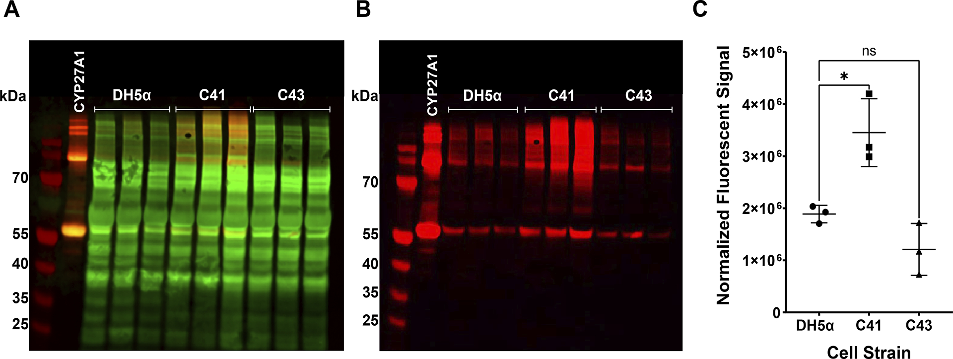
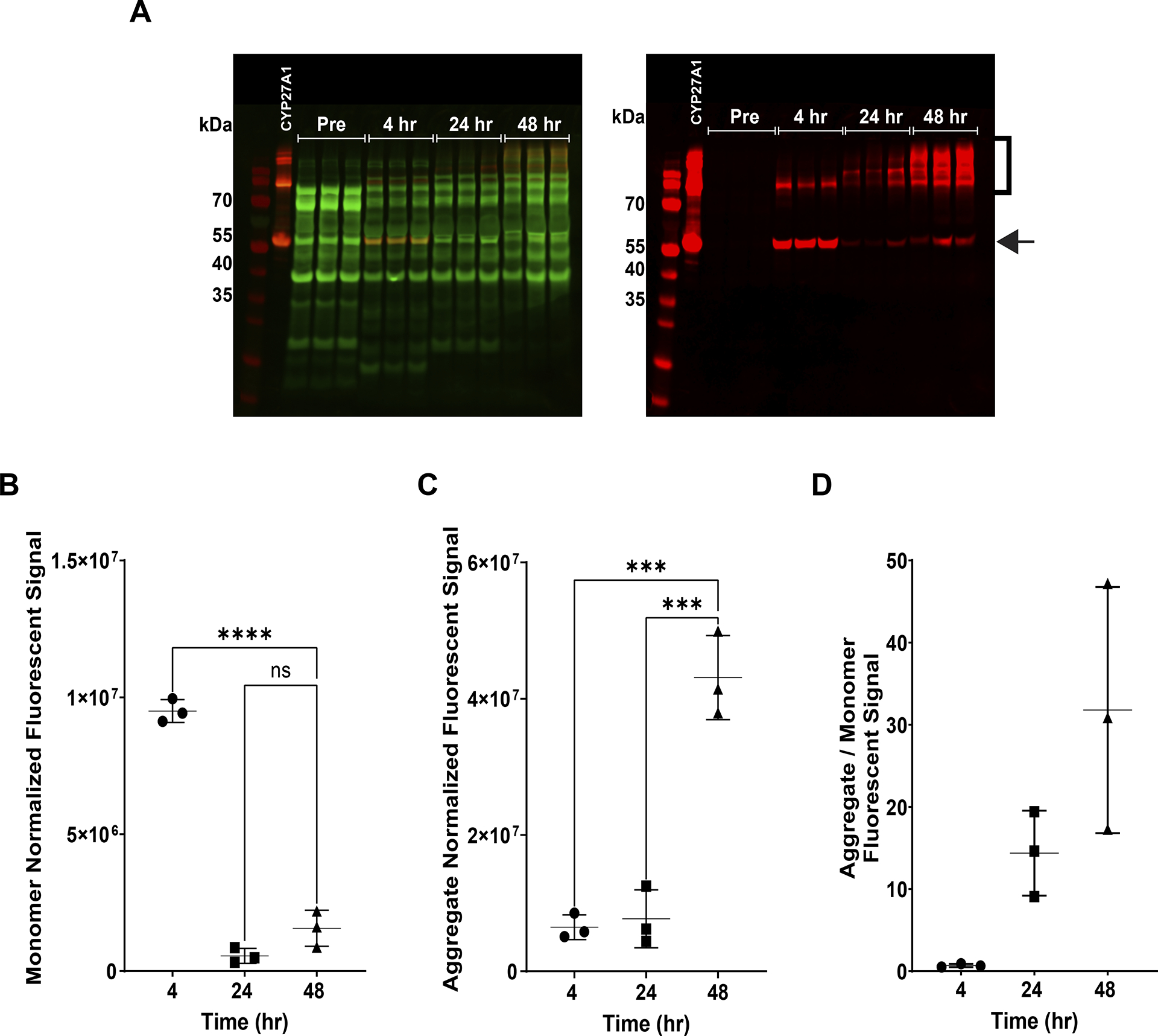
Human mitochondrial cytochrome P450 27A1 is a monooxygenase enzyme that oxidizes bile acids and other sterol derivatives. The enzyme plays an important role in sterol metabolism and is a potential target for clinical therapies related to metabolic conditions and certain cancers. To support the development of such therapies, detailed structural and functional studies of the enzyme should be pursued. Producing large quantities of purified, recombinant enzyme would enable these studies. Recombinant production of human cytochrome P450 27A1 in E. coli is challenging due to the enzyme being membrane associated. This work explores the optimization of human cytochrome P450 27A1 expression in E. coli by systematically testing the effects of cell strain, expression temperature, concentrations of induction reagents, and expression times. Western blot analysis is used to investigate the effects of variable changes prior to purification. E. coli cell strain (switching to C41(DE3)) appears to have the largest positive effect on overall yield. Increasing δ-aminolevulinic acid concentration (induces heme synthesis) also leads to significantly increased yields. Decreasing expression time decreases the amount of higher order cytochrome P450 aggregates that are formed. The combination of these changes is a more robust expression protocol with three major advantages: decreased expression time, lower aggregate to monomer ratios, and increased overall yield.


Conflict of interest statement

Declaration of competing interest The authors declare the following financial interests/personal relationships which may be considered as potential competing interests: Michael Reddish reports financial support was provided by National Institutes of Health. If there are other authors, they declare that they have no known competing financial interests or personal relationships that could have appeared to influence the work reported in this paper.

Figures

Fig. 1.
Fig. 1.
CYP27A1 expression in DH5α (control strain) and BL21 derived strains C41(DE3) and C43(DE3). Western blot of cell lysates (N = 3 biological replicates) A) overlay of total protein stain signal (green) with conjugated donkey anti-mouse IgG secondary antibody signal (red), B) secondary antibody only. C) Replicates plotted with average ± SD. Quantification of CYP27A1 band signal. Significance determined by one-way ANOVA, *P ≤ 0.05, ns = P > 0.05. Control conditions: 1 mM IPTG, 1 mM δALA, 1 mg/mL arabinose, expression at 28 °C for 48 h.
Fig. 2.
Fig. 2.
Effect of additives on C41-CYP27A1 expression. A) Representative western blot of cell lysates (top), left: overlay of total protein stain signal (green) with conjugated donkey anti-mouse IgG secondary antibody signal (red), right: secondary antibody only. Western blot shown is CYP27A1 expression in the presence of varying [δALA]. N = 3 biological replicates plotted with average ± SD. B) Quantification of CYP27A1 band signal with 1 mM IPTG as [control]. C) Quantification of CYP27A1 band signal with 1 mg/mL arabinose as [control]. D) Quantification of CYP27A1 band signal with 1 mM δALA as [control]. Significance determined by one-way ANOVA, *P ≤ 0.05. *P ≤ 0.05, **P ≤ 0.01, ns = P > 0.05.
Fig. 3.
Fig. 3.
C41-CYP27A1 expression at 4 h at tested temperatures. Western blot of cell lysates (N = 3 biological replicates) A) overlay of total protein stain signal (green) with conjugated donkey anti-mouse IgG secondary antibody signal (red), B) secondary antibody only. C) Replicates plotted with average ± SD. Quantification of CYP27A1 band signal. Significance determined by one-way ANOVA, *P ≤ 0.05, **P ≤ 0.01, ns = P > 0.05. Control: C41-CYP27A1 expressed at 28 °C for 48hr.
Fig. 4.
Fig. 4.
CYP27A1 expression over time at 28 °C. A) Western blot of cell lysates (N = 3 biological replicates), left: overlay of total protein stain signal (green) with conjugated donkey anti-mouse IgG secondary antibody signal (red), right: secondary antibody only. B) Quantification of CYP27A1 monomeric band signal (arrow). C) Replicates plotted with average ± SD. Quantification of CYP27A1 aggregate band signal (bracket). Significance determined by one-way ANOVA, ***P ≤ 0.001 ****P ≤ 0.0001, ns = P > 0.05. D) Time correlation between aggregate to monomer band ratio.
Fig. 5.
Fig. 5.
CYP27A1 expression in DH5α compared to C41(DE3) with new expression parameters. Western blot of cell lysates (N = 3 biological replicates), A) overlay of total protein stain signal (green) with conjugated donkey anti-mouse IgG secondary antibody signal (red), B) secondary antibody only. C) Replicates plotted with average ± SD. Quantification of CYP27A1 band signal. Significance determined by Student’s t-test, ****P ≤ 0.0001.
Fig. 6.
Fig. 6.
Vitamin D3 kinetics curve. Activity of 0.2 μM human CYP27A1 in the presence of increasing Vitamin D3 concentrations with an equal concentration of bovine adrenodoxin reductase and a 10-fold greater concentration of human adrenodoxin. Data represents two experiments with three technical replicates each. The generated curve was fit to the Michaelis-Menten equation using GraphPad Prism version 10.4.1 (Boston, Massachusetts USA).
Fig. 7.
Fig. 7.
Carbon monoxide binding spectrum of CYP27A1 lysate from A) DH5α, protein expression 48 h, B) C41 (DE3), protein expression 4 h.

References

    1. Pikuleva IA, Björkhem I, Waterman MR, Expression, purification, and enzymatic properties of recombinant human cytochrome P450c27 (CYP27), Arch. Biochem. Biophys 343 (1) (Jul. 1997) 123–130, 10.1006/abbi.1997.0142. - DOI - PubMed
    1. Axén E, Postlind H, Sjöberg H, Wikvall K, Liver mitochondrial cytochrome P450 CYP27 and recombinant-expressed human CYP27 catalyze 1 alpha-hydroxylation of 25-hydroxyvitamin D3, Proc. Natl. Acad. Sci. U. S. A 91 (21) (Oct. 1994) 10014–10018, 10.1073/pnas.91.21.10014. - DOI - PMC - PubMed
    1. Andersson S, Davis DL, Dahlbäck H, Jörnvall H, Russell DW, Cloning, structure, and expression of the mitochondrial cytochrome P-450 sterol 26-hydroxylase, a bile acid biosynthetic enzyme, J. Biol. Chem 264 (14) (May 1989) 8222–8229. - PubMed
    1. Ma L, Cho W, Nelson ER, Our evolving understanding of how 27-hydroxycholesterol influences cancer, Biochem. Pharmacol 196 (Feb. 2022) 114621, 10.1016/j.bcp.2021.114621. - DOI - PMC - PubMed
    1. Mast N, Lin JB, Pikuleva IA, Marketed drugs can inhibit cytochrome P450 27A1, a potential new target for breast cancer adjuvant therapy, Mol. Pharmacol 88 (3) (Sep. 2015) 428–436, 10.1124/mol.115.099598. - DOI - PMC - PubMed

MeSH terms

LinkOut - more resources