Comparative Study
. 2025 Jul;301(7):110288.
doi: 10.1016/j.jbc.2025.110288. Epub 2025 May 26.

Mechanisms of USP18 specificity toward ISG15 revealed by paralog sequence analysis comparison

Affiliations
Comparative Study

Mechanisms of USP18 specificity toward ISG15 revealed by paralog sequence analysis comparison

Thomas Bonacci et al. J Biol Chem. 2025 Jul.

Abstract

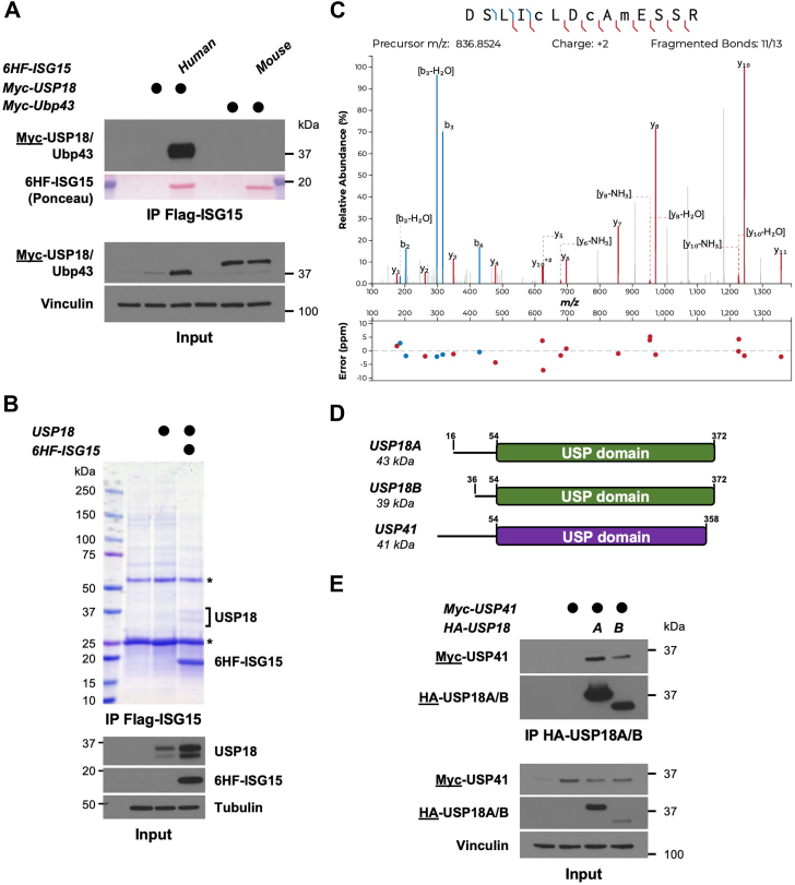
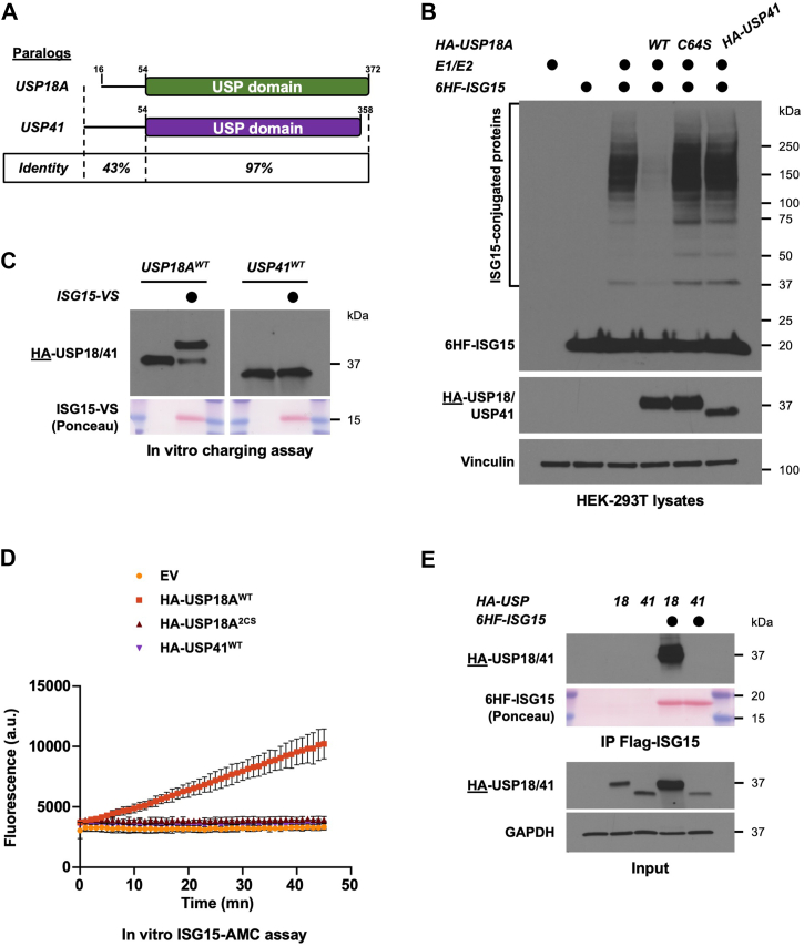
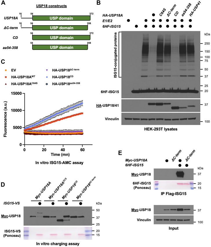
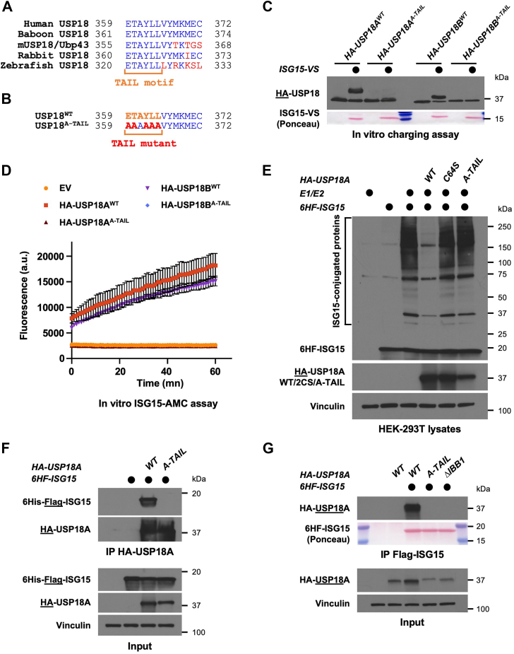
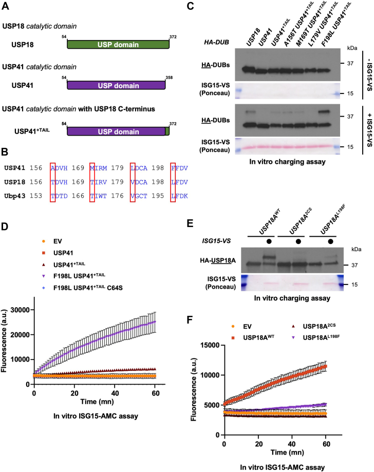
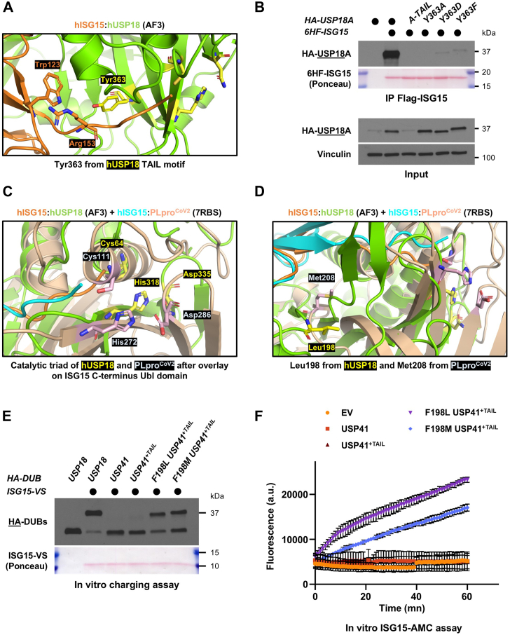
The ubiquitin-like protein ISG15 is activated in response to type 1 interferons, and its conjugation to proteins regulates the response to bacterial and viral infection. Its subsequent deconjugation, which is broadly achieved by the human enzyme USP18, critically controls interferon signaling and the defense against pathogens. However, the molecular determinants underlying USP18 specificity for ISG15 remain elusive. To identify such features, we took advantage of USP18's paralog USP41, which has a strikingly similar catalytic domain and yet lacks deISGylating activity. By performing a comparative sequence analysis coupled with biochemical and enzymatic assays, we identified hallmarks specific to USP18 that are critical for its enzymatic function and ISG15 recognition. Accordingly, AlphaFold-guided analysis suggests that these features mediate USP18-ISG15 interactions, underlining their importance for deISGylating activities. Thus, our results reveal important mechanistic insights into USP18-mediated ISG15 hydrolysis and could inform the development of deISGylase inhibitors relevant to infection and other interferon-related diseases.

Keywords: ISG15; USP18; USP41; alphafold; deISGylating enzymes; deubiquitinating enzymes (DUBs).


Conflict of interest statement

Conflict of interest The authors declare that they have no conflicts of interest with the contents of this article.

Figures

Figure 1
Figure 1
Identification of the uncharacterized DUB USP41 as a USP18 interacting protein. A, Myc-USP18, or its mouse ortholog Myc-Ubp43, were ectopically expressed in HEK-293T cells alone or with human or mouse ISG15, respectively. After 24 h, precleared lysates of transfected cells were subjected to immunoprecipitation on anti-FLAG beads. IP and input samples were separated by SDS-PAGE and analyzed by western blotting. Immunoblotted antigen is underlined to the left of blots. Representative of >3 independent, biological replicates. B, untagged USP18 was ectopically expressed in HEK-293T cells, either alone or with a 6His-FLAG-tagged ISG15 (6HF-ISG15). After 48 h, precleared lysates were used to immunoprecipitate ISG15 on anti-FLAG beads. Immunoprecipitates were separated by SDS-PAGE and the gel was stained using Colloidal Coomassie Blue staining, while inputs were analyzed by western blotting. The two USP18 isoforms that co-precipitated with ISG15 were excised, digested with trypsin, and sent for mass-spectrometry analysis. Asterisks indicate heavy and light chains of the FLAG antibody used for IP. Representative of >3 independent, biological replicates. C, MS/MS spectrum of the doubly charged ion (m/z 836.8524) corresponding to USP41 tryptic peptide DSLICLDCAMESSR. Carbamidomethylation is present on C5 and C8 corresponding to C178 and C181 in USP41. Oxidation is present on M10 corresponding to M183 in UPS41. D, cartoon showing the organization of USP18A, USP18B and USP41. E, Myc-USP41 and HA-USP18 (both isoforms) were ectopically expressed in HEK-293T cells. After 24 h, precleared lysates of transfected cells were used to immunoprecipitate USP18A and B on anti-HA beads. IP and input samples were separated by SDS-PAGE and analyzed by western blotting. Immunoblotted antigen is underlined to the left of blots. Representative of >3 independent, biological replicates.
Figure 2
Figure 2
USP41 is a paralog of US18 which does not react with ISG15.A, cartoon showing the sequence identity of USP18 and USP41. B, protein ISGylation in HEK-293T cells was reconstituted by ectopically expressing V5-UBA7 (E1), V5-UbcH8 (E2) and 6His-FLAG-ISG15 (6HF-ISG15). Where indicated, HA-USP18 WT, a catalytically inactive version (C64S), or HA-USP41, were co-transfected. After 24 h, lysates of transfected cells were prepared then analyzed by SDS-PAGE and Western blot. Immunoblotted antigen is underlined to the left of blots. Representative of >3 independent, biological replicates. C, HA-USP18 and HA-USP41 were ectopically expressed in HEK-293T cells and after 24 h, precleared lysates of transfected cells were used to immunoprecipitate USP18 or USP41 on anti-HA beads. Immunoprecipitates were mixed with reaction buffer containing an ISG15 activity-based probe (ISG15-VS) or not, and reaction products were analyzed by SDS-PAGE and Western blot. Representative of >3 independent, biological replicates. D, the indicated constructs were ectopically expressed in HEK-293T cells and after 24 h, precleared lysates of transfected cells were used to immunoprecipitate USP18 WT, USP18 2CS, or USP41 WT on anti-HA beads. Immunoprecipitates were mixed with reaction buffer containing the fluorogenic substrate ISG15-AMC, and fluorescence increase was monitored using a plate reader at the appropriate excitation and emission wavelengths. Representative of >3 independent, biological replicates. E, HA-USP18 and HA-USP41 were ectopically expressed in HEK-293T cells, either alone or with 6His-FLAG-ISG15. After 24 h, precleared lysates of transfected cells were used to immunoprecipitate ISG15 on anti-FLAG beads. Immunoblotted antigen is underlined to the left of blots. Representative of >3 biological replicates.
Figure 3
Figure 3
The C-terminus of USP18 is necessary for its function. A, cartoon showing the different truncation constructs of USP18 that were generated. The ΔC-term lacks the last 14 amino acids of the USP domain, and the CD leaves only the catalytic domain. The construct aa54 to 358 represents USP18 catalytic domain without the last 14 amino acids. B, protein ISGylation in HEK-293T cells was reconstituted by transfection of the ISG15 machinery (E1/E2/ISG15) and where indicated, HA-tagged USP18 constructs were co-transfected. After 24 h, lysates of transfected cells were prepared then analyzed by SDS-PAGE and Western blot. Immunoblotted antigen is underlined to the left of blots. Representative of >3 independent, biological replicates. C, the indicated constructs were ectopically expressed in HEK-293T cells and after 48 h, precleared lysates of transfected cells were used to immunoprecipitate HA-tagged USP18 variants on anti-HA beads. Immunoprecipitates were mixed with reaction buffer containing the fluorogenic substrate ISG15-AMC, and fluorescence increase was monitored as previously described. Representative of >3 independent, biological replicates. D, the indicated Myc-tagged USP18 constructs were ectopically expressed in HEK-293T cells and after 48 h, precleared lysates of transfected cells were used to immunoprecipitate USP18 on anti-Myc beads. Immunoprecipitates were mixed with reaction buffer containing ISG15-VS or not, and reaction products were analyzed by SDS-PAGE and Western blot. Representative of >3 independent, biological replicates. E, HA-USP18 FL or HA-USP18 ΔC-term were ectopically expressed in HEK-293T cells, either alone or with 6HF-ISG15. After 24 h, precleared lysates of transfected cells were used to immunoprecipitate ISG15 on anti-FLAG beads. Immunoblotted antigen is underlined to the left of blots. Representative of >3 independent, biological replicates.
Figure 4
Figure 4
Identification of the TAIL motif as a molecular determinant of ISG15 binding. A, Sequence alignment of the C-termini of human USP18, baboon USP18, mUSP18/Ubp43, rabbit USP18 and zebrafish USP18. Red indicates residues that are divergent, while the TAIL motif is indicated in orange. B, sequence of USP18 A-TAIL mutant that was generated compared to its WT counterpart. C, HA-tagged USP18 WT or A-TAIL were ectopically expressed in HEK-293T cells then immunoprecipitated on anti-HA beads. Immunoprecipitates were mixed with reaction buffer containing ISG15-VS or not, and reaction products were analyzed by SDS-PAGE and Western blot. Representative of >3 independent, biological replicates. D, the indicated constructs were ectopically expressed and purified from HEK-293T cells as in (C). Immunoprecipitates were mixed with reaction buffer containing the fluorogenic substrate ISG15-AMC, and fluorescence increase was monitored as previously described. Representative of >3 independent, biological replicates. E, protein ISGylation in HEK-293T cells was reconstituted by transfection of the ISG15 machinery (E1/E2/ISG15) and where indicated, HA-tagged USP18 constructs were co-transfected. After 24 h, lysates of transfected cells were prepared then analyzed by SDS-PAGE and Western blot. Immunoblotted antigen is underlined to the left of blots. Representative of >3 independent, biological replicates. F, 6HF-ISG15 was ectopically expressed in HEK-293T cells, either alone, with WT or A-TAIL versions of HA-USP18A. At 24 h post-transfection, precleared lysates were used to immunoprecipitate HA-USP18 on anti-HA beads. Immunoblotted antigen is underlined to the left of blots. Representative of >3 independent, biological replicates. G, WT, A-TAIL or ΔIBB1 versions of HA-USP18A were ectopically expressed in HEK-293T cells in combination with 6HF-ISG15. After 24 h, precleared lysates of transfected cells were used to immunoprecipitate ISG15 on anti-FLAG magnetic beads. Immunoblotted antigen is underlined to the left of the blots. Representative of two independent, biological replicates.
Figure 5
Figure 5
Engineering USP41 into a deISGylase identifies Leu198 of USP18 as crucial for activity.A, cartoon showing the different truncation constructs of USP18 and USP41 that were generated. USP41+TAIL is a chimeric construct where the last 14 amino acids of USP18 were added to the USP domain of USP41. B, sequence alignment of Ubp43, USP18 and USP41. Red boxes highlight residues that are different between USP18 and USP41 but conserved between USP18 and mUSP18/Ubp43. C, the indicated HA-tagged constructs were ectopically expressed in HEK-293T cells and after 24 h, precleared lysates of transfected cells were used to immunoprecipitate these HA-DUBs on anti-HA beads. Immunoprecipitates were mixed with reaction buffer containing ISG15-VS or not, and reaction products were analyzed by SDS-PAGE and Western blot. Representative of >3 independent, biological replicates. D, same as in (C), except that after immunoprecipitation, the indicated HA-tagged constructs were mixed with reaction buffer containing the fluorogenic substrate ISG15-AMC, and fluorescence increase was monitored as previously described. Representative of >3 independent, biological replicates. E, the indicated HA-tagged USP18A constructs were purified from HEK-293T cells on anti-HA beads 48 h after transfection. HA-USP18A immunoprecipitates were mixed with reaction buffer containing ISG15-VS or not, and reaction products were analyzed by SDS-PAGE and Western blot. Representative of >3 independent, biological replicates. F, the indicated Myc-tagged constructs were ectopically expressed in HEK-293T cells and after 24 h, precleared lysates of transfected cells were used to immunoprecipitate Myc-tagged USP18 variants on anti-Myc beads. Immunoprecipitates were mixed with reaction buffer containing the fluorogenic substrate ISG15-AMC, and fluorescence increase was monitored as previously described. Representative of >3 independent, biological replicates.
Figure 6
Figure 6
AlphaFold-guided analysis identifies Tyr363 and Leu198 as crucial for ISG15 binding and USP18 activity. A, close view of Tyr363 from USP18 TAIL motif using AlphaFold 3, and its position respective to Trp123 and Arg153 of hISG15. Residues from the catalytic triad (Cys64, His318, Asn335) are colored in yellow. B, WT, A-TAIL, or Tyr363 mutant versions of HA-USP18A were ectopically expressed in HEK-293T cells along with 6HF-ISG15. After 24 h, anti-FLAG magnetic beads were used to immunoprecipitate ISG15 from precleared lysates of transfected cells. Immunoblotted antigen is underlined to the left of the blots. Representative of >3 independent, biological replicates. C, close view of the catalytic triads of hUSP18 and PLproCoV2, by overlaying the AlphaFold structure of hISG15: hUSP18 with the crystal structure of hISG15:PLproCoV2 (PDB identifier: 7RBS) on the C-terminal Ubl domain of ISG15. The catalytic triad of USP18 (Cys64, His318, Asp335) is in yellow, and the catalytic triad of PLproCoV2 (Cys111, His272, Asp286) is in light pink. D, close view of Leu198 from hUSP18 in yellow and Met208 from PLproCoV2 in light pink, found in a similar space and orientation by overlaying the two structures on ISG15 C-terminal Ubl domain like in (C). E, the indicated HA-tagged constructs were expressed in HEK-293T cells and immunoprecipitated on anti-HA beads. Immunoprecipitates were mixed with reaction buffer containing ISG15-VS or not, and reaction products were analyzed by SDS-PAGE and Western blot. Representative of >3 independent, biological replicates. F, same as in (E), except that the purified HA-tagged DUBs were mixed with reaction buffer containing the fluorogenic substrate ISG15-AMC, and fluorescence increase was monitored as previously described. Representative of >3 independent, biological replicates.

Similar articles

References

    1. Oh E., Akopian D., Rape M. Principles of ubiquitin-dependent signaling. Annu. Rev. Cell Dev. Biol. 2018;34:137–162. - PubMed
    1. Hershko A., Ciechanover A. The ubiquitin system. Annu. Rev. Biochem. 1998;67:425–479. - PubMed
    1. McClellan A.J., Laugesen S.H., Ellgaard L. Cellular functions and molecular mechanisms of non-lysine ubiquitination. Open Biol. 2019;9:190147. - PMC - PubMed
    1. Dikic I., Schulman B.A. An expanded lexicon for the ubiquitin code. Nat. Rev. Mol. Cell Biol. 2023;24:273–287. - PMC - PubMed
    1. Akizuki Y., Kaypee S., Ohtake F., Ikeda F. The emerging roles of non-canonical ubiquitination in proteostasis and beyond. J. Cell Biol. 2024;223 - PMC - PubMed

Publication types

LinkOut - more resources