. 2025 Jun 12;129(23):5653-5663.
doi: 10.1021/acs.jpcb.5c00189. Epub 2025 May 28.

Adaptation of DNA to Protein Binding Revealed by Spectroscopy and Molecular Simulation

Affiliations

Adaptation of DNA to Protein Binding Revealed by Spectroscopy and Molecular Simulation

Thor van Heesch et al. J Phys Chem B. .

Abstract

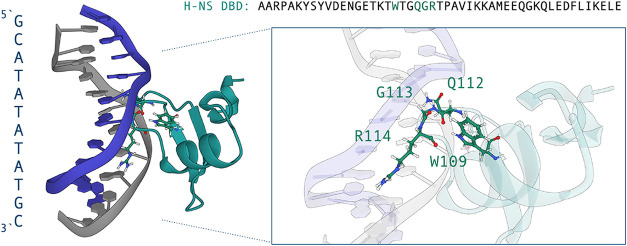
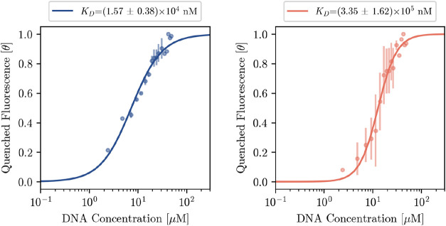
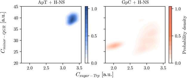
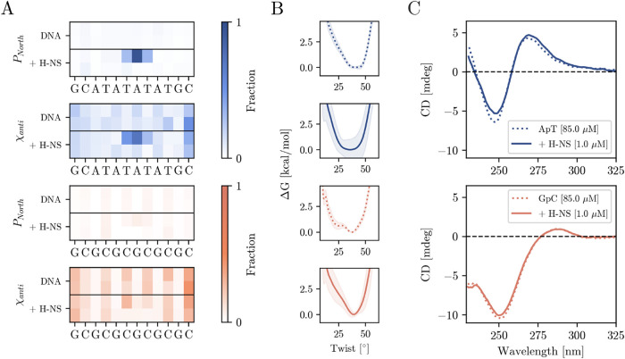
DNA demonstrates remarkable structural diversity, transitioning between conformations such as B-DNA and A-DNA under specific environmental or protein-binding conditions. These transitions are relevant for mediating cellular processes such as gene regulation, DNA organization, and stress response. In bacteria, the histone-like nucleoid structuring protein (H-NS) exemplifies the interaction between sequence-dependent DNA conformational adaptability and protein-mediated regulatory mechanisms. Despite evidence for the strong affinity of H-NS for AT-rich DNA, the specific molecular and structural interactions driving this recognition remain largely unclear. Combining fluorescence spectroscopy, circular dichroism (CD), molecular dynamics (MD) simulations, and enhanced sampling techniques, we show that H-NS exhibits a 10-fold higher affinity for ApT repeats compared to that of GpC repeats. Interestingly, selective binding of H-NS to AT-rich DNA causes a structural adaptation in the DNA, including increased bending flexibility, minor groove widening, and localized A-like DNA features, while GC-rich DNA remains closer to the canonical B-form. Our approach yielded detailed insights into how H-NS exploits the intrinsic conformational plasticity of DNA to achieve sequence-dependent binding. More broadly, this work illustrates how DNA-binding proteins can harness the structural adaptability of the DNA double helix, which may modulate regulatory outcomes, and provides insight into how the intrinsic properties of DNA shape protein-DNA interactions in diverse biological systems.


Figures

1
1
Molecular visualization of H-NS bound to the ApT DNA sequence, based on a snapshot of the molecular dynamics trajectories. The left panel shows the overall structure of the DNA duplex (blue and gray) interacting with the DNA-binding domain (DBD) of H-NS (teal). The right panel provides a zoomed-in view of the key residues involved in the interaction: W109, Q112, G113, and R114. These residues mediate contacts with the DNA bases and backbone, highlighting the specificity of H-NS for ApT-rich sequences. The sequence of the H-NS DBD is shown above for reference, with the region corresponding to the DNA-binding motif highlighted in green. Visualization has been done with Mol*Viewer. Note that snapshots of H-NS in complex with ApT and GpC are shown in Figure S1A in the Supporting Information.
2
2
Hill plots for the ApT (left) and GpC (right) of the binding interaction between H-NS and ApT and GpC are shown with the x-axis the log concentration of added DNA and y-axis the fluorescence intensity fraction θ. Hill coefficients are fitted to n ApT = 1.38 ± 0.10 and n GpC = 2.27 ± 0.17. The concentration range included in the fits is listed in the Supporting Information.
3
3
2D probability distributions showing the Trp109 and DNA backbone deoxy ribose contacts with respect to the QGR binding motif contact of H-NS and DNA minor groove acceptors. Left for the ApT sequence (blue) and right for the GpC sequence (red). Probability distributions were computed for 3 × 1 μs of MD simulations for both complexes.
4
4
(A) Fractions of the sugar phase in North configuration and χ torsion in anti configuraton for each base pair of the ApT and GpT system with H-NS bound (+ H-NS) and unbound (DNA) based on the analysis of the MD simulations. The Supporting Information contains a detailed explanation of the differences in structure of B- and A-DNA. (B) Free energy profiles of the central base pair’s twist obtained using metadynamics simulations of the ApT (blue) and GpT (orange) system with H-NS bound (solid lines) and unbound (dotted lines). (C) Circular Dichroism spectra of ApT (top) GpC sequence (bottom) in the presence of 1 μM of DBD-H-NS in 20 mM Tris-HCl (pH = 8.0) + 50 mM KCl at 298 K (solid lines) and without (dotted lines). Concentration of all sequences are maintained at 85 μM per base.

References

    1. Whelan D. R., Bambery K. R., Heraud P., Tobin M. J., Diem M., McNaughton D., Wood B. R.. Monitoring the reversible B to A-like transition of DNA in eukaryotic cells using Fourier transform infrared spectroscopy. Nucleic Acids Res. 2011;39:5439–5448. doi: 10.1093/nar/gkr175. - DOI - PMC - PubMed
    1. Tolstorukov M. Y., Jernigan R. L., Zhurkin V. B.. Protein-DNA hydrophobic recognition in the minor groove is facilitated by sugar switching. J. Mol. Biol. 2004;337:65–76. doi: 10.1016/j.jmb.2004.01.011. - DOI - PubMed
    1. Svozil D., Kalina J., Omelka M., Schneider B.. DNA conformations and their sequence preferences. Nucleic Acids Res. 2008;36:3690–3706. doi: 10.1093/nar/gkn260. - DOI - PMC - PubMed
    1. Schneider B., Černỳ J., Svozil D., Čech P., Gelly J.-C., de Brevern A. G.. Bioinformatic analysis of the protein/DNA interface. Nucleic Acids Res. 2014;42:3381–3394. doi: 10.1093/nar/gkt1273. - DOI - PMC - PubMed
    1. Ng H.-L., Kopka M. L., Dickerson R. E.. The structure of a stable intermediate in the A-B DNA helix transition. Proc. Natl. Acad. Sci. U.S.A. 2000;97:2035–2039. doi: 10.1073/pnas.040571197. - DOI - PMC - PubMed

MeSH terms

LinkOut - more resources