. 2025 May 28;16(1):422.
doi: 10.1038/s41419-025-07746-2.

VCPIP1 facilitates pancreatic adenocarcinoma progression via Hippo/YAP signaling

Affiliations

VCPIP1 facilitates pancreatic adenocarcinoma progression via Hippo/YAP signaling

Zhihao Liu et al. Cell Death Dis. .

Abstract

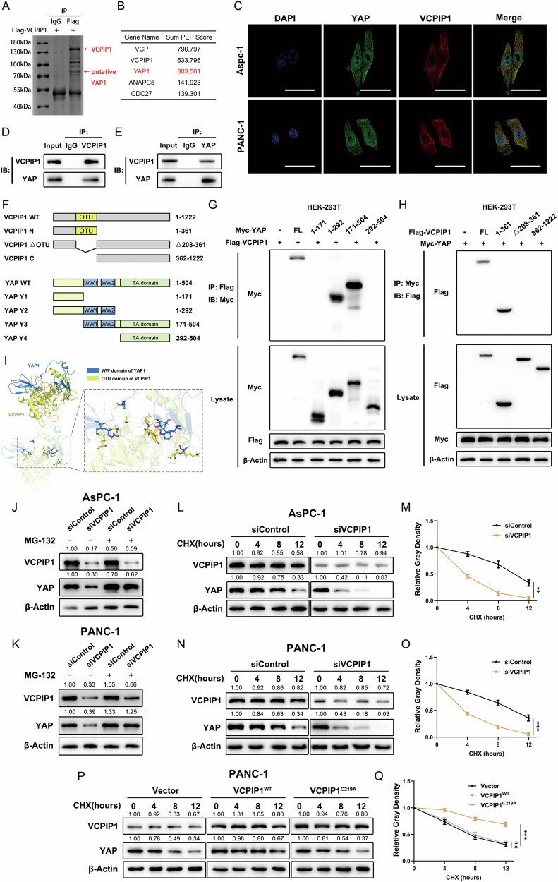
Dysregulation of Hippo signaling is observed in pancreatic adenocarcinoma (PAAD). Moreover, overactivation of YAP is crucial for tumor progression. Although the inhibitory phospho-cascade is functional, the reason for YAP hyperactivation in PAAD remains unclear. Recent studies have revealed that the ubiquitin modification of YAP also plays an important role in the Hippo/YAP axis and cancer progression. To gain a better understanding of the potential mechanisms underlying the ubiquitination and deubiquitination of YAP, we carried out siRNA screening for critical deubiquitinases in PAAD. By using a deubiquitinase (DUB) library, we identified valosin-containing protein-interacting protein 1 (VCPIP1) as an important effector of YAP function and PAAD progression. Inhibition of VCPIP1 hampered PAAD progression via Hippo signaling. Clinical data revealed that VCPIP1 was elevated in PAAD and correlated with poor survival in PAAD patients. Biochemical assays demonstrated that VCPIP1 interacted with YAP, inhibiting K48-linked polyubiquitination and thereby increasing YAP stability. YAP directly binds to the VCPIP1 promoter region, enhancing its transcription in PAAD. Our study revealed a forward feedback loop between VCPIP1 and Hippo signaling in PAAD, indicating that VCPIP1 is a potential therapeutic drug target in PAAD.


Conflict of interest statement

Competing interests: The authors declare no competing interests. Ethics approval and consent to participate: All procedures were performed in compliance with relevant laws and institutional guidelines and have been approved by the Ethics Committee of Shengjing Hospital, China Medical University. The ethical code number is 2024PS1466K for the use of clinical specimens and 2024PS1486K for animal experiments.

Figures

Fig. 1
Fig. 1. VCPIP1 is a critical regulator of the Hippo pathway in PAAD.
A Illustration of deubiquitinase screening. Data from the TCGA-PAAD cohort were subjected to GSEA. Deubiquitinases were sorted by their normalized enrichment score (NES), and VCPIP1 ranked at the top of the list. The top 16 deubiquitinases with NESs >1.5 were further screened by transfecting their siRNAs (at a concentration of 50 nM) into PANC-1 cells. The expression of the YAP target gene CTGF was used as a readout of Hippo signaling activity. B GSEA of TCGA-PAAD data. High expression of VCPIP1 is positively correlated with the YAP gene set. C Data from GTEx and TCGA-PAAD were combined to analyze the expression of VCPIP1 in normal and PAAD tissues. Data were downloaded from UCSC Xena and normalized to enable comparability. D Kaplan‒Meier plot showing the relationship between VCPIP1 expression and disease-free survival in TCGA-PAAD patients. E Heatmap showing the correlation between VCPIP1 expression and genes in the YAP signature. TCGA-PAAD data were utilized. F Heatmap showing that the expression of YAP signature genes was downregulated in the siVCPIP1 group. G Genes that were differentially expressed between the siVCPIP1 and siControl groups were visualized via a volcano plot. H GSEA was performed on our RNA-Seq data (siVCPIP1 vs siControl). The gene expression of the siVCPIP1 group was negatively correlated with the YAP gene set. I Enrichment analysis was performed on the genes with differential expression in our RNA-Seq data. The dot plot shows the top enriched KEGG pathways. J, K Immunohistochemistry (IHC) showing the expression of VCPIP1 and YAP in normal and pancreatic cancer tissues. VCPIP1 is significantly more highly expressed in pancreatic cancer samples than in normal pancreas samples. The scale bars are 400 μm for 10X and 100 μm for 40X. The P value was calculated via the chi-square test. L Kaplan‒Meier plot showing the relationship between VCPIP1 expression and overall survival in 57 pancreatic cancer patients in our hospital. M, N The correlation between VCPIP1 and YAP protein expression in pancreatic cancer patients was analyzed by immunohistochemistry. The scale bars are 400 μm for 10X and 100 μm for 40X. The P value was calculated via the chi-square test.
Fig. 2
Fig. 2. VCPIP1 is required for PAAD progression.
A PAAD cells were transfected with either siControl or siVCPIP1#1/#2 at a concentration of 50 nM. The knockdown efficiency of VCPIP1 was subsequently measured via qRT‒PCR. B The mRNA expression levels of CTGF and CYR61 in PAAD cells after transfection with siControl or siVCPIP1 were measured via qRT‒PCR. C, D A luciferase reporter gene assay was used to detect TEAD transcriptional activity in AsPC-1 and PANC-1 cells after transfection with siControl or siVCPIP1. E The protein levels of VCPIP1 and YAP in PAAD cells after transfection with siControl or siVCPIP1 were measured via Western blotting. Proliferation rates of PAAD cells were evaluated by CCK-8 (F) and EdU (G, H) assays after transfection with siControl or siVCPIP1. Nuclei in the EdU assay were stained with Hoechst 33342. The scale bar is 200 μm. Transwell assays were performed to assess the migration (I) and invasion (J) abilities of PAAD cells after transfection with siControl or siVCPIP1. K, L A wound healing assay was performed to assess the migration ability of PAAD cells after transfection with siControl or siVCPIP1. M, N PAAD cell apoptosis was evaluated via flow cytometry after the cells were transfected with siControl or siVCPIP1 and then stained with PI and FITC. OQ PANC-1 cells stably transfected with shControl or shVCPIP1 were inoculated subcutaneously into nude mice. Tumor weight (O) and volume (P) were evaluated. An IHC assay was used to evaluate Ki-67, YAP and VCPIP1 expression (Q). The percentage of Ki-67-positive cells was calculated. n = 6. The scale bar is 200 μm. The experiments were performed in triplicate. All the data are presented as the means ± SDs. Statistical methods: Student’s t test for (AD, GO, Q); two-way ANOVA for (F, P). *P < 0.05; **P < 0.01; ***P < 0.001.
Fig. 3
Fig. 3. Hydrolysis activity is dispensable for the ability of VCPIP1 to regulate YAP function.
A Schematic diagram of the C219A mutation of VCPIP1. B After the vectors or VCPIP1 WT or VCPIP1 C219A were transfected into PAAD cells, the mRNA expression levels of CTGF and CYR61 were measured via qRT‒PCR. A luciferase reporter gene assay was used to detect TEAD transcriptional activity in AsPC-1 (C) and PANC-1 (D) cells after transfection with the vector or with VCPIP1 WT or VCPIP1 C219A. E The protein levels of VCPIP1 and YAP in PAAD cells after transfection with vector, WT VCPIP1 or C219A VCPIP1 were measured via Western blotting. The proliferation rates of PAAD cells were evaluated by CCK-8 (F) and EdU (G, H) assays after the cells were transfected with the vector or with VCPIP1 WT or VCPIP1 C219A. For the EdU assay, the nuclei were stained with Hoechst 33342. The scale bar is 200 μm. Cell migration (I) and invasion (J) abilities were evaluated via Transwell assays in AsPC-1 and PANC-1 cells after transfection with vector or with VCPIP1 WT or VCPIP1 C219A. Cell migration ability was evaluated by a wound healing assay in AsPC-1 (K) and PANC-1 (L) cells after transfection with the vector or with VCPIP1 WT or VCPIP1 C219A. AsPC-1 (M) and PANC-1 (N) cells were transfected with vector, VCPIP1 WT or VCPIP1 C219A and stained with PI and FITC. Cell apoptosis was evaluated by flow cytometry. The experiments were performed in triplicate. All the data are presented as the means ± SDs. Statistical methods: Student’s t test for (BD) and (GN); two-way ANOVA for (F). *P < 0.05; **P < 0.01; ***P < 0.001.
Fig. 4
Fig. 4. YAP rescued the inhibitory effect of VCPIP1 knockdown on PAAD.
A PANC-1 cells were transfected with siControl + vector, siVCPIP1 + vector, siControl + YAP or siVCPIP1 + YAP. The protein levels of VCPIP1 and YAP were subsequently examined by immunoblotting. B The mRNA expression levels of CTGF and CYR61 in PANC-1 cells after transfection with the indicated siRNAs or plasmids were measured via qRT‒PCR. C TEAD transcriptional activity in PANC-1 cells after transfection with the indicated siRNA or plasmid was measured via a luciferase reporter gene assay. The proliferation rates of PANC-1 cells were evaluated by CCK-8 (D) and EdU (E) assays after transfection with the indicated siRNAs and plasmids. For the EdU assay, the nuclei were stained with Hoechst 33342. The scale bar is 200 μm. F, G Migration ability was evaluated by a wound healing assay in PANC-1 cells following transfection with the indicated siRNA or plasmid. H, I Cell migration and invasion abilities were evaluated via Transwell assays in PANC-1 cells following transfection with the indicated siRNAs and plasmids. J Apoptosis was evaluated by flow cytometry in PANC-1 cells after transfection with the indicated siRNA or plasmid. KN PANC-1 cells stably transfected with the indicated lentivirus were inoculated subcutaneously into nude mice. Then, the tumor weight (L) and volume (M) were measured. Subsequently, an IHC assay was used to evaluate Ki-67, YAP and VCPIP1 expression (N). The percentage of Ki-67-positive cells was calculated. n = 6. The scale bar is 200 μm. The experiments were performed in triplicate. All the data are presented as the means ± SDs. Statistical methods: Student’s t test for (B, C, EL, N); two‒way ANOVA for (D, M). *P < 0.05; **P < 0.01; ***P < 0.001.
Fig. 5
Fig. 5. VCPIP1 interacts with YAP and regulates YAP protein stability.
A PANC-1 cells transfected with Flag-VCPIP1 were lysed and immunoprecipitated with a Flag tag antibody or isotype IgG. Immunoprecipitated samples were denatured, subjected to electrophoresis and subsequently stained with Coomassie blue. The red arrow indicates the putative band of VCPIP1 and YAP. B Top 5 proteins enriched in the samples obtained via immunoprecipitation and then analyzed via protein mass spectrometry, as indicated in (A). C Immunofluorescence assays demonstrated the localization of the VCPIP1 and YAP proteins in AsPC-1 and PANC-1 cells. Green and red represent YAP and VCPIP1, respectively. Nuclei were stained with DAPI. The scale bar is 50 μm. D, E The interaction between endogenous VCPIP1 and YAP was detected through a co-IP assay. The lysates of PANC-1 cells were separately subjected to immunoprecipitation with anti-VCPIP1 (D) or anti-YAP1 (E) antibodies. Subsequently, western blotting was employed to analyze and detect the corresponding results. F The schematic illustrates wild-type and truncated VCPIP1 mutants (residues 1--361, Δ208--361, and 362--1222) as well as YAP and its truncated mutants (residues 1--171, 1--292, 171--504, and 292--504), which were used in the following Co-IP assay. Co-IP assays demonstrated that the WW domain of YAP (G) and the OTU domain of VCPIP1 (H) are required for their interaction. I The docking diagram, which depicts the interaction between the OTU domain of VCPIP1 and the WW domain of YAP, was visualized via PyMOL. MG132 abrogates the ability of siVCPIP1 to downregulate the protein level of YAP in AsPC-1 (J) and PANC-1 (K) cells. The cells were transfected with siControl or siVCPIP1 and then treated with 10 μM MG132 as indicated for 12 h. VCPIP1 and YAP were examined by immunoblotting at the protein level. Knockdown of VCPIP1 decreases YAP protein stability in AsPC-1 (L, M) and PANC-1 (N, O) cells. The cells were transfected with siControl or siVCPIP1 and then treated with CHX (100 μM) at different time points. P, Q VCPIP1 overexpression promotes YAP protein stability, whereas overexpression of the VCPIP1 C219A mutant has no significant effect on YAP protein stability. The CHX concentration was 100 μM. **P < 0.01; ***P < 0.001. Two-way ANOVA was employed for statistical analysis.
Fig. 6
Fig. 6. VCPIP1 stabilizes YAP by reducing YAP K48-linked polyubiquitination.
A, B VCPIP1 knockdown increased K48-linked polyubiquitination of the YAP protein but did not affect K63-linked polyubiquitination of the YAP protein. PANC-1 cells were transfected with HA-tagged ubiquitin or K48 ubiquitin or K63 ubiquitin plasmids (A) or K48R mutant ubiquitin or K63 R mutant ubiquitin plasmids (B) and siControl or siVCPIP1 and then treated with 20 µM MG132 for 6 h. The cells were harvested and immunoprecipitated with an anti-YAP antibody, and the level of ubiquitinated YAP protein was examined by immunoblotting with an HA tag. C, D VCPIP1 overexpression decreased total ubiquitination and K48-linked polyubiquitination of the YAP protein but did not affect K63-linked polyubiquitination of the YAP protein, whereas overexpression of the VCPIP1 C219A mutant affected neither K48 nor K63-linked polyubiquitination of the YAP protein. HEK-293T cells were transfected with HA-tagged ubiquitin or K48 ubiquitin or K63 ubiquitin plasmids (C) or K48R mutant ubiquitin or K63R mutant ubiquitin plasmids (D), along with Flag-VCPIP1 WT or Flag-VCPIP1 C219A and Myc-YAP plasmids, and then treated with MG132 (20 µM) for 6 h. The cells were harvested and immunoprecipitated with an anti-Myc antibody, and the level of ubiquitinated YAP protein was examined by immunoblotting with an HA tag. E VCPIP1 removes the polyubiquitination of the YAP protein at the K252, K280 and K315 sites. HEK293T cells were transfected with HA-tagged ubiquitin and Flag-VCPIP1 and Myc-YAP WT or Myc-tagged point mutant forms of YAP (as shown in the panel). The cells were subsequently treated with MG132 (20 µM) for 6 h, harvested and immunoprecipitated with an anti-Myc antibody. The level of ubiquitinated YAP protein was examined by immunoblotting with an HA tag antibody.
Fig. 7
Fig. 7. YAP transcriptionally regulates VCPIP1 expression in PAAD.
ChIP-seq data analysis of TEAD (A) and YAP (B) revealed prominent peaks accumulating in the VCPIP1 promoter region. The data used in the analysis were TEAD1 (GSM2534071), TEAD2 (GSE91936), TEAD3 (GSM2534286), TEAD4 (GSE170161), YAP and input (GSE61852). The numbers on the upper left of each track indicate the fold change for TEAD1-4 and the peak reads for YAP and the input. C The binding of YAP to the VCPIP1 promoter region was verified via a ChIP assay. Fixed PANC-1 cells were subjected to chromatin immunoprecipitation with an anti-YAP antibody or with isotype IgG as a negative control. The enriched DNA was analyzed by gel electrophoresis. ChIP‒qPCR assay indicating decreased enrichment of the VCPIP1 promoter in AsPC-1 (D) and PANC-1 (E) cells after YAP knockdown. The cells were fixed, lysed and then immunoprecipitated with an anti-YAP antibody or isotype IgG. The enriched DNA was subjected to qPCR analysis. F PAAD cells were transfected with siControl or siYAP1, and the protein levels of VCPIP1 and YAP1 were examined by immunoblotting. G, H The mRNA expression levels of CTGF and VCPIP1 were measured via qRT‒PCR in AsPC-1 (H) and PANC-1 (I) cells after transfection with siControl or siYAP1. I AsPC-1 and PANC-1 cells were treated with the indicated concentrations of verteporfin for 48 h, and the protein levels of VCPIP1 were examined by immunoblotting. The mRNA expression levels of VCPIP1 and CTGF were measured via qRT‒PCR in AsPC-1 (J) and PANC-1 (K) cells after treatment with the indicated concentrations of verteporfin for 48 h. L AsPC-1 and PANC-1 cells were treated with the indicated concentrations of XMU-MP-1 for 48 h, and the protein levels of VCPIP1 were subsequently examined by immunoblotting. The mRNA expression levels of CTGF and VCPIP1 were measured via qRT‒PCR in AsPC-1 (M) and PANC-1 (N) cells after treatment with the indicated concentrations of XMU-MP-1 for 48 h. The experiments were performed in triplicate. All the data are presented as the means ± SDs. Statistical methods: Student’s t test for (DN). **P < 0.01; ***P < 0.001.
Fig. 8
Fig. 8. The VCPIP1 inhibitor CAS-12290-201 restrains PAAD progression.
A Schematic diagram of the inhibitory effect of CAS-12290-201 on VCPIP1. CAS-12290-201 covalently labels the catalytic cysteine of VCPIP1 and inhibits its hydrolysis activity. B PANC-1 cells were treated with various concentrations of the VCPIP1 inhibitor CAS-12290-201 for 48 h, and the protein levels of VCPIP1 and YAP1 were examined by immunoblotting. C The mRNA expression levels of CTGF and CYR61 were measured via qRT‒PCR in PANC-1 cells after treatment with the indicated concentrations of CAS-12290-201 for 48 h. D A luciferase reporter gene assay was used to detect TEAD transcriptional activity in PANC-1 cells after treatment with the indicated concentrations of CAS-12290-201 for 48 h. The proliferation rate was assessed by CCK-8 (E) and EdU (F) assays in PANC-1 cells after treatment with the indicated concentrations of CAS-12290-201 for 48 h. The scale bar is 200 μm. G, H Migration and invasion abilities were assessed by Transwell assays in PANC-1 cells after treatment with the indicated concentrations of CAS-12290-201 for 48 h. I, J Cell migration ability was evaluated by a wound healing assay in PANC-1 cells after treatment with the indicated concentrations of CAS-12290-201 for 48 h. K PANC-1 cells were treated with 2 μM or 4 μM CAS-12290-201 for 48 h and stained with PI and FITC. Cell apoptosis was evaluated by flow cytometry. L The VCPIP1 inhibitor CAS-12290-201 increased K48-linked polyubiquitination of the YAP protein. PANC-1 cells transfected with HA-tagged ubiquitin, K48 ubiquitin or K48R mutant ubiquitin plasmids were treated with CAS-12290-201 (4 µM) for 48 h and MG132 (20 µM) for 6 h and then subjected to immunoprecipitation and immunoblotting. M PANC-1 cells were treated with 4 μM CAS-12290-201 for 48 h and 10 μM MG132 as indicated for 12 h. The protein levels of VCPIP1 and YAP were examined by immunoblotting. N, O The VCPIP1 inhibitor CAS-12290-201 decreases YAP protein stability. PANC-1 cells were treated with 4 μM CAS-12290-201 for 48 h and then treated with CHX (100 μM) at different time points. The protein levels of YAP were subsequently examined by immunoblotting at corresponding time points to assess changes in protein stability. The experiments were performed in triplicate. All the data are presented as the means ± SDs. Statistical methods: Student’s t test for (C, D, FK); two-way ANOVA for (E, O). *P < 0.05; **P < 0.01; ***P < 0.001.
Fig. 9
Fig. 9. The VCPIP1 inhibitor CAS-12290-201 sensitizes PAAD cells to gemcitabine treatment.
A AsPC-1 cells were pretreated with DMOS or 4 µM CAS-12290-201 for 48 h and then treated with the indicated concentrations of gemcitabine for 24 h. Cell viability was evaluated by a CCK-8 assay. The variable Hill slope model of nonlinear regression was employed to estimate the IC50. For the GEM group, the estimated IC50 was 143.8 nM. For the GEM + CAS-12290-201 group, the estimated IC50 was 61.42 nM. B The synergy score of the combined treatment of GEM and CAS-12290-201 in AsPC-1 cells was calculated via SynergyFinder 3.0 (synergyfinder.org). The cells were pretreated with the indicated concentrations of CAS-12290-201 for 48 h and then treated with the indicated concentrations of GEM for 24 h. Cell viability was evaluated via the CCK-8 assay, and the data were uploaded to the SynergyFinder website to calculate the synergy score. C PANC-1 cells were treated in the same way as described in (A). The estimated IC50 for the GEM group was 90.41 nM, and that for the GEM + CAS-12290-201 group was 45.34 nM. D Synergy score of the combination treatment of GEM and CAS-12290-201 in PANC-1 cells. Details are described in (B). E PANC-1 cells were treated with DMSO, 20 nM GEM or 20 nM GEM + 2 µM CAS-12290-201. Cell proliferation was evaluated by a CCK-8 assay. F PANC-1 cells were pretreated with 4 µM CAS-12290-201 for 48 h and then treated with 50 nM or 100 nM GEM for 24 h. Cell apoptosis was evaluated by using a caspase 3/7 green fluorescence reagent. The scale bar is 200 μm. G PANC-1 cells were treated with DMSO, 20 nM GEM or 20 nM GEM + 2 µM CAS-12290-201. Cell proliferation was evaluated by an EdU assay. The scale bar is 200 μm. HL PANC-1 cells were inoculated subcutaneously into nude mice, and treatment started 7 days after inoculation. The mice were randomly assigned to three groups and subjected to different treatments. For the vehicle group, the mice were treated with vehicle solution (5% NMP, 5% Solutol, or 20% DMSO). For the GEM group, the mice were treated with gemcitabine (20 mg/kg, i.p., weekly). For the GEM + CAS-12290-201 group, the mice were treated with gemcitabine (10 mg/kg, i.p., weekly) or CAS-12290-201 (40 mg/kg, i.p., every day). Tumor weight (J) and volume (K) were measured. An IHC assay was used to evaluate Ki-67 and YAP expression (L). The percentage of Ki-67-positive cells was calculated. n = 6. The scale bar is 200 μm. The experiments were performed in triplicate. All the data are presented as the means ± SDs. Statistical methods: Student’s t test for (F, G, J, L); two-way ANOVA for (E, K). *P < 0.05; **P < 0.01; ***P < 0.001.
Fig. 10
Fig. 10. VCPIP1 forms a positive feedback loop and regulates the Hippo/YAP axis.
VCPIP1 interacts with YAP and reduces the K48-linked ubiquitination of the YAP protein. As a result, it stabilizes the YAP protein and enhances the transcription of downstream target genes. VCPIP1 was identified as one of the target genes of the YAP/TEAD complex. Transcriptionally activated VCPIP1, in turn, stabilizes YAP, forming a positive feedback loop that ultimately contributes to pancreatic cancer progression.

References

    1. Siegel RL, Giaquinto AN, Jemal A. Cancer statistics, 2024. CA Cancer J Clin. 2024;74:12–49. - PubMed
    1. Wood LD, Canto MI, Jaffee EM, Simeone DM. Pancreatic cancer: pathogenesis, screening, diagnosis, and treatment. Gastroenterology. 2022;163:386. - PMC - PubMed
    1. Mao W, Mai J, Peng H, Wan J, Sun T. YAP in pancreatic cancer: oncogenic role and therapeutic strategy. Theranostics. 2021;11:1753–62. - PMC - PubMed
    1. Justice RW, Zilian O, Woods DF, Noll M, Bryant PJ. The Drosophila tumor suppressor gene warts encodes a homolog of human myotonic dystrophy kinase and is required for the control of cell shape and proliferation. Genes Dev. 1995;9:534–46. - PubMed
    1. Xu T, Wang W, Zhang S, Stewart RA, Yu W. Identifying tumor suppressors in genetic mosaics: the Drosophila lats gene encodes a putative protein kinase. Development. 1995;121:1053–63. - PubMed

MeSH terms