. 2025 May 28;41(1):91.
doi: 10.1007/s10565-025-10038-2.

The key regulator circPDE3B promotes arsenic-induced bladder carcinogenesis by affecting STAT3 and NF-κB stability

Affiliations

The key regulator circPDE3B promotes arsenic-induced bladder carcinogenesis by affecting STAT3 and NF-κB stability

Yihong Gao et al. Cell Biol Toxicol. .

Abstract

Long-term exposure to arsenic (As), which is a ubiquitous environmental contaminant, significantly enhances the risk of multiple cancers, including bladder and lung cancers. In recent years, the important roles of circular RNAs (circRNAs) in tumorigenesis and development have attracted widespread attention. However, the specific molecular mechanisms by which circRNAs promote bladder cancer development following exposure to arsenic remain incompletely understood. This study is the first to demonstrate that circPDE3B is significantly upregulated in a cell model of transformation triggered by arsenic and that it promotes this transformation process. Our study elucidated the biological function of circPDE3B in vitro, in SV-HUC-1 cells, showing that it accelerates the malignant transformation from arsenic via increasing cell proliferation and inhibiting apoptosis. Furthermore, we delineated a novel molecular mechanism whereby circPDE3B directly binds to NF-κB and STAT3, inhibiting their ubiquitination and increasing their stability. This, in turn, affects downstream HIF-1α expression, promoting the malignant transformation of SV-HUC-1 cells and eventually resulting in bladder carcinogenesis. Our research reveals the critical regulatory role of circPDE3B in the arsenic-triggered malignant transformation within SV-HUC-1 cells. This study offers broader perspectives on the molecular mechanisms driving bladder cancer progression, while also identifying potential targets for early diagnosis and treatment of bladder tumour.

Keywords: Arsenic; Bladder cancer; NF-κB; STAT3; circPDE3B.


Conflict of interest statement

Declarations. Ethics approval and consent to participate: This study did not involve the use of human tissue or the performance of animal experiments. Consent for publication: The contents of this manuscript have not been previously published or considered for publication elsewhere. Competing interests: The authors declare no competing interests.

Figures

Fig. 1
Fig. 1
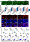
Establishment of a model of SV-HUC-1 cell malignant transformation induced by NaAsO2 and screening of circPDE3B. (A) The CCK-8 assay to identify changes in cellular viability after exposure to arsenic for different time points and at different concentrations. (B) CCK-8 assay to determine cell viability after 48 h of arsenic exposure. (C) Soft agar colony formation assay to assess the colony-forming ability of SV-HUC-1 cells cultured for 80 generations with continuous NaAsO2 exposure. (D) Statistical analysis of soft agar colony formation experiments. (E) Volcano plots showing the differential expression profiles of circRNAs in SV-HUC-1 cells and UM-UC-3 cells, with red denoting significantly upregulated circRNAs, blue denoting significantly downregulated circRNAs, and grey denoting circRNAs without differential expression. (F) qPCR measurement of circPDE3B expression in different generations. (G) Schematic diagram of the circPDE3B gene structure. (H) circPDE3B and linear PDE3B gene expression before and after RNase R treatment was measured by qPCR. (I) Agarose gel electropherograms of polymerization dispersion experiments. (J) SV-HUC-1 cells were treated with Act D for 0 h, 4 h, 8 h or 16 h. RNA was extracted for qPCR to measure circPDE3B and linear PDE3B expression. The values are presented as means ± SDs, and each experiment was performed at least three times. *, p < 0.05; ns, p ≥ 0.05
Fig. 2
Fig. 2
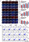
circPDE3B significantly promotes the proliferation of SV-HUC-1 cells. (A) GFP fluorescence of successfully established SV-HUC-1 cell lines in which circPDE3B was stably silenced or overexpressed. (B) Validation of the stable circPDE3B silencing or overexpression efficiency circPDE3B in cell lines. (C) CCK-8 assay to measure cell viability after circPDE3B was silenced or overexpressed in SV-HUC-1 cells. (D) EdU assay to assess cell proliferation after circPDE3B was silenced or overexpressed in SV-HUC-1 cells. (E) Statistical analysis of cell proliferation as determined by the EdU assay. (F) Flow cytometry analysis of the cell cycle after circPDE3B was silenced or overexpressed in SV-HUC-1 cells. (G) Statistical analysis of cell proliferation after the cell cycle was analysed by flow cytometry. (H) Flow cytometry analysis of cell apoptotic after the silencing and overexpression of circPDE3B in SV-HUC-1 cells. (I) Statistical analysis of apoptosis as analysed by flow cytometry. The values are presented as means ± SDs, and each experiment was performed at least three times. *, p < 0.05
Fig. 3
Fig. 3
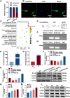
circPDE3B significantly promotes the NaAsO2-induced malignant transformation of SV-HUC-1 cells. (A) Stable circPDE3B-silenced and circPDE3B-overexpressing cell lines were exposed to NaAsO2 for the P5, P10 and P20 generations, and an EdU assay was performed to measure cell proliferation. (B) Statistical analysis of cell proliferation as determined by EdU. (C) Stable circPDE3B-silenced and circPDE3B-overexpressing cell lines were exposed to NaAsO2 for the P5, P10, and P20 generations, and the CCK-8 assay was used to measure cell viability. (D) Stable circPDE3B-silenced and circPDE3B-overexpressing cell lines were exposed to NaAsO2 for the P5, P10, and P20 generations, and flow cytometry was performed to analyse of cell apoptosis. (E) Statistical analysis of apoptosis as determined by flow cytometry. The values are presented as means ± SDs, and each experiment was performed at least three times. *, p < 0.05
Fig. 4
Fig. 4
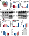
circPDE3B targets and promotes the expression of NF-κB and STAT3. (A) qPCR detection of the circPDE3B nucleoplasmic distribution in SV-HUC-1 cells. (B) Fluorescence in situ hybridization assay to determine circPDE3B subcellular localization (green fluorescence: circPDE3B; blue fluorescence: nucleus). (C) KEGG pathway enrichment analysis. (D) Schematic diagram of the RIP experiment. (E) RIP-qPCR analysis of circPDE3B directly binding to the NF-κB protein. (F) Agarose gel electrophoresis of the RIP-qPCR products of circPDE3B directly bound to the NF-κB protein. (G) RIP-qPCR analysis of circPDE3B directly bound to the STAT3 protein. (H) Agarose gel electrophoresis results of the RIP-qPCR products of circPDE3B directly bound to the STAT3 protein. (I) Western blotting analysis of NF-κB and STAT3 protein expression after the silencing and overexpression of circPDE3B. (JK) Statistical analysis of western blotting of NF-κB protein and STAT3 protein expression. (L) Western blotting analysis of NF-κB and STAT3 protein expression in the P0, P20, P40 and P80 generations during the NaAsO2-induced malignant conversion of SV-HUC-1 cells. (MN) Statistical analysis of NF-κB and STAT3 protein expression during the NaAsO2-induced malignant transformation of SV-HUC-1 cells as determined by western blotting. The values are presented as means ± SDs, and each experiment was performed at least three times. *, p < 0.05
Fig. 5
Fig. 5
circPDE3B inhibits protein degradation by reducing the levels of NF-κB and STAT3 ubiquitination. (A) Western blotting was used to measure NF-κB and STAT3 protein expression in stable circPDE3B-overexpressing cells after 0 h, 3 h, 6 h and 9 h of CHX treatment. (BC) Statistical analysis of NF-κB and STAT3 protein expression after CHX treatment of cells as determined by western blotting. (D) Western blotting was used to measure NF-κB and STAT3 protein expression in stable circPDE3B-overexpressing cells after 10 μM MG132 treatment for 0 h and 24 h. (EF) Statistical analysis of NF-κB and STAT3 protein expression after MG132 treatment as determined by western blotting. (G) Co-IP assay was conducted to detect the binding of the NF-κB protein and ubiquitin protein. (H) Co-IP assay was conducted to detect the binding of the STAT3 protein and ubiquitin protein. The values are presented as means ± SDs, and each experiment was performed at least three times. *, p < 0.05
Fig. 6
Fig. 6
circPDE3B regulates HIF-1α expression through NF-κB and STAT3. (A) Venn diagram for prediction of downstream transcriptional regulatory target genes of STAT3 and NF-κB proteins. (B) HIF-1α mRNA expression was measured by qPCR after stable silencing and overexpressing circPDE3B. (C) HIF-1α protein, P21 protein and Bcl2 protein expression was measured by western blotting after stable silencing and overexpressing circPDE3B. (DF) Statistical analysis of western blotting analysis of HIF-1α protein, P21 protein and Bcl2 protein expression. (G) Western blotting analysis of HIF-1α, Bcl2 and P21 protein expression in the P0, P20, P40 and P80 generations during the NaAsO2-induced malignant transformation of SV-HUC-1 cells. (HJ) Statistical analysis plots of HIF-1α, P21 and Bcl2 protein expression during the NaAsO2-induced malignant transformation of SV-HUC-1 cells by western blotting. (K) Soft agar colony formation assay was conducted to detect the colony formation ability of SV-HUC-1 cells after stable circPDE3B overexpression and continuously exposuring to arsenic for up to 40 generations. The values are presented as means ± SDs, and each experiment was performed at least three times. *, p < 0.05
Fig. 7
Fig. 7
circPDE3B promotes the NaAsO2-induced malignant transformation of SV-HUC-1 cells through the HIF-1α signalling pathway. (A) CCK-8 assay to determine the viability of circPDE3B-silenced and HIF-1α-overexpressed cells. (B) Fluorescence images of circPDE3B-silenced and HIF-1α-overexpressed cells as detected by the EdU assay. (C) Flow cytometry was used to analyse cell apoptosis after circPDE3B was silenced and the HIF-1α was overexpressed. (D) Western blotting analysis of P21 and Bcl2 protein expression after circPDE3B was silenced and HIF-1α was overexpressed. The values are presented as means ± SDs, and each experiment was performed at least three times. *, p < 0.05; #, p < 0.05
Fig. 8
Fig. 8
Machanism diagram. circPDE3B directly binds to STAT3 and NF-κB to enhance their stability, thereby affecting HIF-1α expression and ultimately promoting arsenic-induced bladder carcinogenesis

Similar articles

References

    1. Amara CS, Kami Reddy KR, Yuntao Y, Chan YS, Piyarathna DWB, Dobrolecki LE, Shih DJH, Shi Z, Xu J, Huang S, Ellis MJ, Apolo AB, Ballester LY, Gao J, Hansel DE, Lotan Y, Hodges HC, Lerner SP, Creighton CJ, Sreekumar A, Zheng WJ, Msaouel P, Kavuri SM, Putluri N. The IL6/JAK/STAT3 signaling axis is a therapeutic vulnerability in SMARCB1-deficient bladder cancer. Nat Commun. 2024;15:1373. 10.1038/s41467-024-45132-2. - PMC - PubMed
    1. Byeon E, Kang HM, Yoon C, Lee JS. Toxicity mechanisms of arsenic compounds in aquatic organisms. Aquat Toxicol. 2021;237:105901. 10.1016/j.aquatox.2021.105901. - PubMed
    1. Chang S, Yim S, Park H. The cancer driver genes IDH1/2, JARID1C/ KDM5C, and UTX/ KDM6A: crosstalk between histone demethylation and hypoxic reprogramming in cancer metabolism. Exp Mol Med. 2019;51:1–17. 10.1038/s12276-019-0230-6. - PMC - PubMed
    1. Chen L, Shan G. CircRNA in cancer: Fundamental mechanism and clinical potential. Cancer Lett. 2021;505:49–57. 10.1016/j.canlet.2021.02.004. - PubMed
    1. Dong J, Cheng XD, Zhang WD, Qin JJ. Recent Update on Development of Small-Molecule STAT3 Inhibitors for Cancer Therapy: From Phosphorylation Inhibition to Protein Degradation. J Med Chem. 2021;64:8884–915. 10.1021/acs.jmedchem.1c00629. - PubMed

MeSH terms