

Deep learning radiomics fusion model to predict visceral pleural invasion of clinical stage IA lung adenocarcinoma: a multicenter study
- PMID: 40437608
- PMCID: PMC12121141
- DOI: 10.1186/s13019-025-03488-6
Deep learning radiomics fusion model to predict visceral pleural invasion of clinical stage IA lung adenocarcinoma: a multicenter study
Abstract
Aim: To assess the predictive performance, risk stratification capabilities, and auxiliary diagnostic utility of radiomics, deep learning, and fusion models in identifying visceral pleural invasion (VPI) in lung adenocarcinoma.
Materials and methods: A total of 449 patients (female:male, 263:186; 59.8 ± 10.5 years) diagnosed with clinical IA stage lung adenocarcinoma (LAC) from two distinct hospitals were enrolled in the study and divided into a training cohort (n = 289) and an external test cohort (n = 160). The fusion models were constructed from the feature level and the decision level respectively. A comprehensive analysis was conducted to assess the prediction ability and prognostic value of radiomics, deep learning, and fusion models. The diagnostic performance of radiologists of varying seniority with and without the assistance of the optimal model was compared.
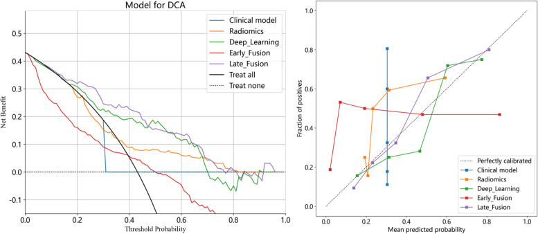
Results: The late fusion model demonstrated superior diagnostic performance (AUC = 0.812) compared to clinical (AUC = 0.650), radiomics (AUC = 0.710), deep learning (AUC = 0.770), and the early fusion models (AUC = 0.586) in the external test cohort. The multivariate Cox regression analysis showed that the VPI status predicted by the late fusion model were independently associated with patient disease-free survival (DFS) (p = 0.044). Furthermore, model assistance significantly improved radiologist performance, particularly for junior radiologists; the AUC increased by 0.133 (p < 0.001) reaching levels comparable to the senior radiologist without model assistance (AUC: 0.745 vs. 0.730, p = 0.790).
Conclusions: The proposed decision-level (late fusion) model significantly reducing the risk of overfitting and demonstrating excellent robustness in multicenter external validation, which can predict VPI status in LAC, aid in prognostic stratification, and assist radiologists in achieving higher diagnostic performance.
Keywords: Adenocarcinoma of lung; CT; Deep learning; Radiomics.
© 2025. The Author(s).
Conflict of interest statement
Declarations. Ethics approval and consent to participate: This retrospective study was approved by the Ethics Committee of Zhongshan Hospital and Shanghai Pulmonary Hospital, and the requirement to obtain informed consent from patients was waived. Consent for publication: All authors have read the manuscript and have agreed to its publication in Journal of Cardiothoracic Surgery. Competing interests: The authors declare no competing interests.
Figures


References
-
- Jiang L, Liang W, Shen J, Chen X, Shi X, He J, et al. The impact of visceral pleural invasion in node-negative non-small cell lung cancer: a systematic review and meta-analysis. Chest. 2015;148:903–11. - PubMed
-
- Yang X, Sun F, Chen L, Shi M, Shi Y, Lin Z, et al. Prognostic value of visceral pleural invasion in non-small cell lung cancer: a propensity score matching study based on the SEER registry. J Surg Oncol. 2017;116:398–406. - PubMed
-
- Inoue M, Minami M, Shiono H, Sawabata N, Ideguchi K, Okumura M. Clinicopathologic study of resected, peripheral, small-sized, non-small cell lung cancer tumors of 2 cm or less in diameter: pleural invasion and increase of serum carcinoembryonic antigen level as predictors of nodal involvement. J Thorac Cardiovasc Surg. 2006;131:988–93. - PubMed
-
- Gorai A, Sakao Y, Kuroda H, Uehara H, Mun M, Ishikawa Y, et al. The clinicopathological features associated with skip N2 metastases in patients with clinical stage IA non-small-cell lung cancer. Eur J Cardiothorac Surg. 2015;47:653–8. - PubMed
-
- Rami-Porta R, Bolejack V, Crowley J, Ball D, Kim J, Lyons G, et al. The IASLC lung cancer staging project: proposals for the revisions of the T descriptors in the forthcoming eighth edition of the TNM classification for lung cancer. J Thorac Oncol. 2015;10:990–1003. - PubMed
Publication types
MeSH terms
Grants and funding
LinkOut - more resources
Full Text Sources
Medical
