

Retinal microvasculature alteration in patients with acute pancreatitis: an observational OCTA study
- PMID: 40437618
- PMCID: PMC12121281
- DOI: 10.1186/s40001-025-02566-8
Retinal microvasculature alteration in patients with acute pancreatitis: an observational OCTA study
Abstract
Objective: To evaluate changes in retinal layer thickness and microvascular density in pancreatitis patients using optical coherence tomography angiography (OCTA).
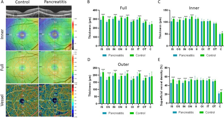
Methods: The study involved 16 pancreatitis patients and 16 healthy controls. Each participant underwent a superficial OCTA scan, with images divided into nine subregions to compare macular retinal thickness (RT) and superficial vascular density (SVD) between groups.
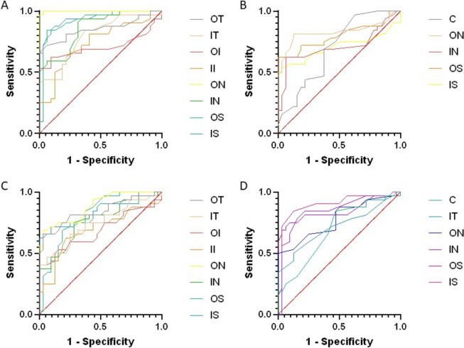
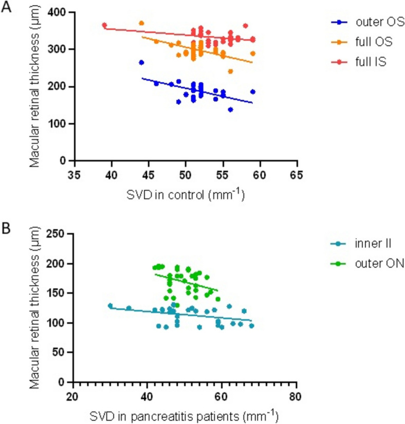
Results: Pancreatitis patients exhibited reduced retinal thickness in specific macular areas, including inner, full, and outer layers (p < 0.05). Additionally, decreased superficial vascular density was noted in inner superior (IS), outer superior (OS), inner nasal (IN), and outer nasal (ON) regions (p < 0.05). ROC curve analysis showed high diagnostic accuracy for full-layer inner superior, outer superior, and outer inferior thickness with areas under the curve of 0.9429, 0.9233, and 0.9990, respectively.
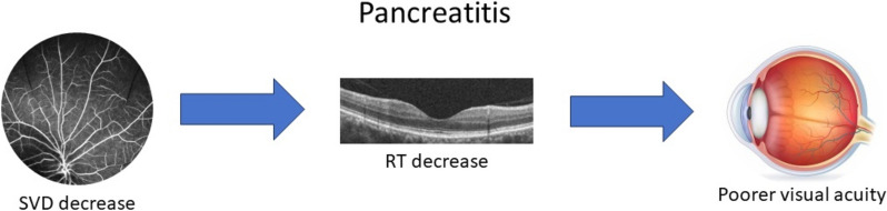
Conclusions: Pancreatitis is associated with macular retinal thinning and decreased superficial vascular density, offering potential for improved diagnostic imaging.
Keywords: Macular region; Optical coherence tomography angiography (OCTA); Pancreatitis; Retinal thickness; Superficial vascular density.
© 2025. The Author(s).
Conflict of interest statement
Declarations. Ethics approval and consent to participate: The authors are accountable for all aspects of the work in ensuring that questions related to the accuracy or integrity of any part of the work are appropriately investigated and resolved. All procedures performed in this study were in accordance with the ethical standards of the Ethics Committee of the First Affiliated Hospital of Nanchang University and with the Helsinki Declaration (as revised in 2013). Written informed consent was obtained from the patient for publication of this case report and accompanying images. A copy of the written consent is available for review by the editorial office of this journal. Competing interests: The authors declare no competing interests.
Figures
References
-
- Xiao AY, Tan ML, Wu LM, Asrani VM, Windsor JA, Yadav D, et al. Global incidence and mortality of pancreatic diseases: a systematic review, meta-analysis, and meta-regression of population-based cohort studies. Lancet Gastroenterol Hepatol. 2016;1(1):45–55. 10.1016/s2468-1253(16)30004-8. - PubMed
-
- Pendharkar SA, Mathew J, Petrov MS. Age- and sex-specific prevalence of diabetes associated with diseases of the exocrine pancreas: A population-based study. Dig Liver Dis. 2017;49(5):540–4. 10.1016/j.dld.2016.12.010. - PubMed
-
- Yadav D, Lowenfels AB. Trends in the epidemiology of the first attack of acute pancreatitis: a systematic review. Pancreas. 2006;33(4):323–30. 10.1097/01.mpa.0000236733.31617.52. - PubMed
-
- Frey C, Zhou H, Harvey D, White RH. Co-morbidity is a strong predictor of early death and multi-organ system failure among patients with acute pancreatitis. J Gastrointest Surg. 2007;11(6):733–42. 10.1007/s11605-007-0164-5. - PubMed
Publication types
MeSH terms
LinkOut - more resources
Full Text Sources
Medical
Miscellaneous
