. 2025 Jan;12(Suppl 1):S14613.
doi: 10.1117/1.NPh.12.S1.S14613. Epub 2025 May 28.

Repetitive transcranial magnetic stimulation increases synaptic plasticity of cortical axons in the APP/PS1 amyloidosis mouse model

Affiliations

Repetitive transcranial magnetic stimulation increases synaptic plasticity of cortical axons in the APP/PS1 amyloidosis mouse model

Barbora Fulopova et al. Neurophotonics. 2025 Jan.

Abstract

Significance: Growing evidence highlights the therapeutic potential of repetitive transcranial magnetic stimulation (rTMS) in diseases causing dementias such as Alzheimer's disease (AD). However, individual responses to rTMS are variable, and its underlying neural mechanisms are not fully understood.

Aim: As synaptic dysfunction is one of the key mechanisms associated with cognitive deficits in dementia, we investigated the effect of rTMS on cortical synapses using an APP/PS1 amyloidosis mouse model of AD crossed with fluorescent reporters linked to the Thy-1 promoter.

Approach: Using in vivo two-photon imaging, we characterized the plasticity of excitatory terminaux (TB) and en passant (EPB) axonal boutons at 48-h intervals for 8 days on either side of a single session of rTMS.

Results: We found both types of axonal boutons preserved the overall number of their synaptic outputs in wild type (WT) and APP/PS1 groups, pre- and post-stimulation. Both synapse types also showed a significantly reduced dynamic fraction in APP/PS1 compared with WT axons pre-stimulation. Following stimulation, the TB, but not EPB, dynamic fraction increased in both WT and APP/PS1 groups.

Conclusions: This suggests possible mechanisms of rTMS action that are cell type-specific and, together with previous findings of improved functional performance, present a potential clinical avenue for rTMS in the management of AD.

Keywords: axon; bouton; dementia; live imaging; synaptic plasticity; two-photon microscopy.


Figures

Fig. 1
Fig. 1
Experimental methodology. (a) Experimental timeline: craniotomy performed in 10- to 13-month-old mice, imaging commenced after a 3-week recovery period. In vivo imaging was conducted at 2-day intervals, with one round of stimulation shortly after day 0 imaging. Examples of repeated imaging of axonal segments rich in EPBs (c) and TBs (b) shown on maximum projection images (WT-GFP and APP-GFP). Arrowheads indicate sites of gains/losses of boutons between consecutive sessions. (WT–GFP—wild type crossed onto Thy1-GFP background; APP-GFP—APP/PS1 crossed on Thy1-GFP background; EPB—en passant bouton, TB—terminaux bouton).
Fig. 2
Fig. 2
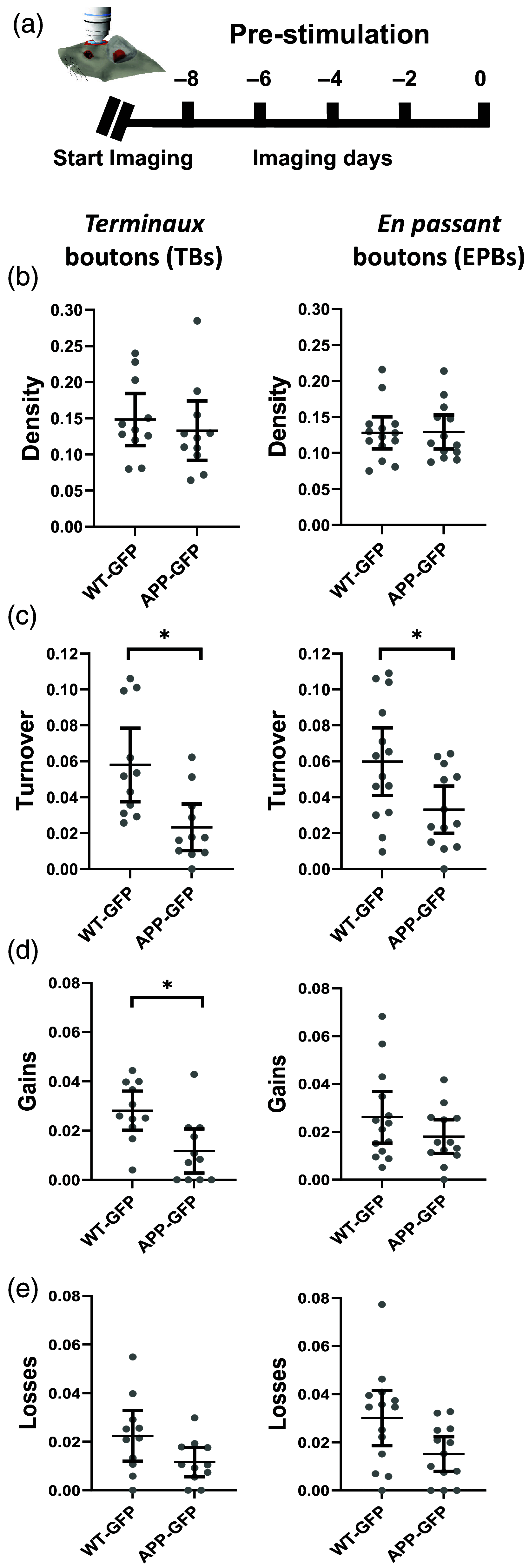
Baseline pre-stimulation properties of axonal en passant and terminaux boutons. (a) Experimental timeline. There were no significant differences between individual imaging days within each group (see also Fig. 3). Pre-stimulation data for each axon were averaged as a single pre-stimulation mean value. (b) No significant differences in the average density of TBs and EPBs between WT-GFP and APP-GFP. (c) Turnover (expressed as the combined proportion of gains and losses) was significantly lower in the APP-GFP group compared to the WT-GFP for both TBs and EPBs. (d) and (e) The proportion of gains and losses was decreased in APP-GFP TBs and EPBs, but only reached significance for TB gains. (WT–GFP—wild type animal crossed onto Thy1-GFP background; APP-GFP—APP/PS1 animal crossed on Thy1-GFP background, bars are 95% confidence intervals; *statistically significant difference).
Fig. 3
Fig. 3
Synaptic dynamics of en passant and terminaux boutons after LI-iTBS. (a) Experimental timeline. LI-iTBS did not alter the density of TBs or EPBs in neither WT-GFP (b) and (d) or APP-GFP (c) and (e) groups. LI-iTBS significantly increased TB turnover in WT-GFP (f) and APP-GFP (g) groups. In the APP-PS1 group (g), the post-stimulation increase in turnover reached the WT-GFP pre-stimulation baseline (faded line, lower panel). LI-iTBS did not significantly change turnover in the EPBs in either the WT-GFP (h) or APP-GFP (i) groups. (Line graphs show data for individual axons, and point-spread graphs immediately below show corresponding means with 95% confidence intervals; *statistically significant difference; ns—non significant after Bonferroni adjustment; WT–GFP—wild type animal crossed onto Thy1-GFP background; APP-GFP—APP/PS1 animal crossed on Thy1-GFP background.)
Fig. 4
Fig. 4
Gains and losses of terminaux and en passant boutons post-stimulation. (a) and (c) A trend towards increased gains and losses of TBs post-simulation in both WT-GFP and APP-GFP groups. Statistical significance was only observed in WT-GFP gains at day +6, and WT-GFP losses at day +4. (b) and (d) No changes in EPBs gains or losses post-stimulation. (WT–GFP—wild type animal crossed onto Thy1-GFP background; APP-GFP—APP/PS1 animal crossed on Thy1-GFP background, error bars are 95% confidence intervals; *statistically significant difference).

References

    1. Chou Y.-H., Ton That V., Sundman M., “A systematic review and meta-analysis of rTMS effects on cognitive enhancement in mild cognitive impairment and Alzheimer’s disease,” Neurobiol. Aging 86, 1–10 (2020).NEAGDO10.1016/j.neurobiolaging.2019.08.020 - DOI - PMC - PubMed
    1. Hinder M. R., et al. , “Inter- and intra-individual variability following intermittent theta burst stimulation: implications for rehabilitation and recovery,” Brain Stimul. 7(3), 365–371 (2014).10.1016/j.brs.2014.01.004 - DOI - PubMed
    1. Wischnewski M., Schutter D. J., “Efficacy and time course of theta burst stimulation in healthy humans,” Brain Stimul. 8(4), 685–692 (2015).10.1016/j.brs.2015.03.004 - DOI - PubMed
    1. Jannati A., et al. , “Assessing the mechanisms of brain plasticity by transcranial magnetic stimulation,” Neuropsychopharmacology 48(1), 191–208 (2023).NEROEW10.1038/s41386-022-01453-8 - DOI - PMC - PubMed
    1. Teo J. T., et al. , “Human theta burst stimulation enhances subsequent motor learning and increases performance variability,” Cereb. Cortex 21(7), 1627–1638 (2011).53OPAV10.1093/cercor/bhq231 - DOI - PubMed