Review
. 2025 May 28:8:0710.
doi: 10.34133/research.0710. eCollection 2025.

Nitrite Reductases in Biomedicine: From Natural Enzymes to Artificial Mimics

Affiliations
Review

Nitrite Reductases in Biomedicine: From Natural Enzymes to Artificial Mimics

Sai Zhu et al. Research (Wash D C). .

Abstract

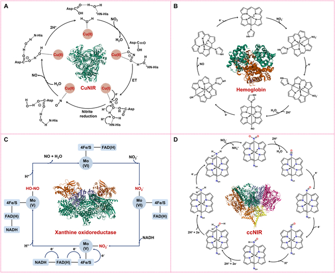
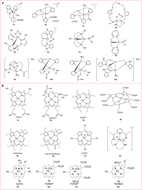
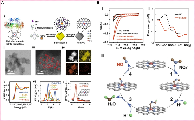
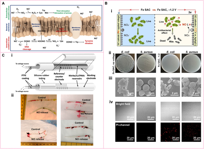
Nitrite reductases (NiRs) are natural enzymes that facilitate the reduction of nitrite. They are essential for the microbial nitrogen cycle and play a vital role in regulating numerous physiological and pathological processes associated with nitric oxide (NO) in living organisms. By the merits of protein engineering, a variety of artificial NiR mimics have been developed. These include traditional artificial proteins, metal-azacycle complexes, and nanozymes such as metal, metal oxide/sulfide nanoparticles, metal-organic frameworks, bioinorganic nanohybrids, and advanced single-atom nanozymes. This development marks an important milestone in broadening the application of enzyme-like catalytic nitrite reduction across various fields, such as biomedicine, biosensing, food science, and environmental science. In this review, we first outline the different types of NiRs, along with their active center structures and catalytic mechanisms, drawing from recent research and discoveries. We then classify the reported NiR mimic materials, discussing their active center structures and enzyme-like catalytic mechanisms. Additionally, we explore the potential future applications and challenges facing NiR mimics in the field of biomedicine.


Conflict of interest statement

Competing interests: The authors declare that they have no competing interests.

Figures

Fig. 1.
Fig. 1.
Schematic illustration of the role of nitrite reduction (route b and route e) catalyzed by NiRs in the nitrogen cycle.
Fig. 2.
Fig. 2.
Structures and active metal centers of NiRs. (A) CuNiR; (B) cd1NiR; (C) fdNiR; (D) ccNiR; (E) hemoglobin, myoglobin (Mb), neuroglobin (Ngb), cytoglobin (Cyb), cytochrome c, cytochrome bc1 complex, cytochrome c oxidoreductase, and endothelial nitric oxide synthase (eNOS); (F) YtfE; and (G) xanthine oxidoreductase, aldehyde oxidase, and sulfite oxidase.
Fig. 3.
Fig. 3.
Mechanisms of nitrite reduction catalyzed by NiRs with different active sites. (A) Nitrite reduction to NO over CuNiR, (B) nitrite reduction to NO over heme-based NiRs, (C) nitrite reduction to NO over Mo-based NiRs, and (D) nitrite reduction to NH3 over ccNiR.
Fig. 4.
Fig. 4.
De novo protein design reverse protein engineering-based mimics. (A) T2 Cu center of CuNiR. (B) Model of metallopeptide Cu(I/II)(TRIL23H)3+/2+ (i), view of the Zn(II)(H2O)(His)3 site (ii), side view of the 2 metal sites in ii (iii). Reprinted with permission from [59]. Copyright (2012) Proceedings of the National Academy of Sciences of the United States of America. (C) Model of Cu(I)(TRIW-H)3 (i); chemical structures of (top) δmHis(N(pros)-methyl-l-histidine) and (bottom) εmHis (N(tau)-methyl-l-histidine) (ii); models of the metal binding sites of (top) Cu(I)(TRIW-δmH)3 and (bottom) Cu(I)(TRIW-εmH)3 (iii). Reprinted with permission from [61]. Copyright © 2019 American Chemical Society. (D) Strategy for studying the tethering of redox partners in CuNiRs. (i) Structure (top) and mechanism (bottom) of the 2-domain AxNiR. (ii) Structure (top) and mechanism (bottom) of the 3-domain CuNiR RpNiR. In (i) and (ii), the 3 monomers of the trimeric CuNiRs are shown in green, magenta, and cyan, and cytochrome c551 is shown in yellow. (iii) Strategy for dissecting the 3-domain cytochrome c-tethered Ralstonia pickettii CuNiR into its component domains, with colors as indicated above. Reprinted with permission from [64]. Copyright © 2019 American Chemical Society.
Fig. 5.
Fig. 5.
Metal complex-based NiR mimics. (A) Structures of several typical Cu-N complexes coordinated with nitrite (1a to 12a). (B) Structures of several typical iron porphyrins (1b to 12b).
Fig. 6.
Fig. 6.
Preparation methods and morphologies of MOF-based NiRs. (A) Protein-engineering-inspired MOF nanozyme modulation. Reprinted with permission [76]. Copyright © 2020 Wiley-VCH GmbH. (B) illustration of ccNiR and its heme group with secondary sphere amino acid residues. (C) Schematic illustration of the synthesis of Zr-BTB MOF with the post-synthetic modification by heme and hydroxybenzoic acids. (D) SEM images of Zr-BTB (i), Zr-BTB@Hemin (ii), Zr-BTB@Hemin-MHBA (iii), Zr-BTB@Hemin-DHBA (iv), Zr-BTB@Hemin-THBA (v), and XRD patterns of different BTB@Hemin hybrids (vi). Reprinted with permission [78]. Copyright © 2024 The authors. Angewandte Chemie International Edition published by Wiley-VCH GmbH.
Fig. 7.
Fig. 7.
Morphology and structural characterizations of Cu NWs with corresponding catalytic mechanism. (A) SEM image and XRD pattern of copper nanowires (Cu NWs) (i); linear sweep voltammetry of nitrite electroreduction on Cu NWs (ii); chronoamperometry of Cu NWs under intermittent CO2 (iii); possible reaction pathway of NO2 reduction to NH3 in the presence of CO2 (iv); energy barriers and structures of NO2deoxygenation (v and vii); energy barriers and structures of NO deoxygenation (vi and viii). Reprinted with permission from [87]. Copyright © 2022 Wiley-VCH GmbH. (B) SEM images at 2 scales (i), HRTEM and TEM (inset) images (ii), SAED pattern (iii), and STEM and elemental mapping (iv) of Cu-RD-KOH; reaction free energy of different intermediates for NO3 reduction reaction on Cu (111) surface of Cu-RD-KOH (v); the comparison of the free energy diagram for the reduction of NO3 into NO2 and *NOOH over Cu (111) and Cu2O (111) at an applied potential of U = 0 V (vi). Simulated atomistic structure scheme shows a reaction pathway for the reduction of NO3 to NH3 and the corresponding transition state (TS) energy barriers (vii). Reprinted with permission from [88]. Copyright © 2023 The authors. Angewandte Chemie International Edition published by Wiley-VCH GmbH.
Fig. 8.
Fig. 8.
Morphology, catalytic activity, and catalytic mechanism of NFLA/Cu NHs (nanofibrous lysozyme assemblies [NFLA]). (A) TEM images of NFLA/CuS NHs (i to iv) and corresponding element maps (v to ix) and element distribution (x) of NFLA/CuS NHs, XRD pattern of NFLA/CuS NHs (xi). (B) Catalytic activity of NFLA/Cu NHs compared with other copper-based materials (i), and catalytic activity enhanced by light irradiation (ii). (C) Energy diagram for the reduction of NO2 to NO catalyzed by the CuS (103) plane and Cu (111) plane (i); schematic diagram of the molecular dynamics (MD) simulation for the interaction between lysozyme and AA (ii); schematic diagram of the MD simulation for the interaction between lysozyme and nitrite (iii); diagrams of the interaction energy variation over time for lysozyme binding to AA (iv); diagrams of the interaction energy variation over time for lysozyme binding nitrite. Mole ratio (v); proposed mechanism scheme of the catalytic reduction of NO2 to NO by NFLA/CuS NHs in the presence of AA (vi). Reprinted with permission from [95]. Copyright © 2024 The authors. Science Advances published by the American Association for the Advancement of Science.
Fig. 9.
Fig. 9.
Morphology, catalytic activity, and catalytic mechanism of iron-based single-atom catalyst (Fe SAC). (A) The structure of natural cytochrome cd1 nitrite enzyme and schematic illustration of the synthesis of FeSAC (i); HAADF-STEM image of Fe SAC (ii); AC-STEM of Fe SAC (iii); HAADF-STEM image and corresponding EDX element mapping of Fe SAC (iv); XANES spectra of Fe SAC (v); Fourier transforms of the Fe K-edge of Fe SAC (vi); corresponding EXAFS fitting curves of Fe SAC at R space (vii). (B) CVs obtained at GCEs modified with Fe SAC (red) or nitrogen-doped porous carbon (NC) (black) in PBS in the absence (dash) and presence (solid) of 50 mM NaNO2 (i); density functional theory (DFT)-calculated free energy paths of the nitrite reduction on Fe SAC (ii); proposed structures for electrocatalytic reduction process of nitrite on Fe SAC (iii). Reprinted with permission from [98]. Copyright © 2022 Wiley-VCH GmbH.
Fig. 10.
Fig. 10.
Therapeutic opportunities for nitrite reduction catalyzed by NiRs for treating cardiovascular and metabolic diseases. Nitrite reduction to NO, enhanced during hypoxia/ischemia, offers therapeutic potential. Preclinical studies show nitrite protects against ischemia–reperfusion injury, supporting its use in treating heart attacks, strokes, organ transplant issues, and sickle cell disease. It also prevents drug-induced stomach ulcers and alleviates hypertension (pulmonary/systemic) and post-hemorrhagic cerebral vasospasm via vasodilation. These multifunctional benefits highlight nitrite as a versatile therapeutic agent for oxygen-sensitive disorders.
Fig. 11.
Fig. 11.
Antibacterial mechanism of NO and electrocatalytic antibacterial application. (A) Schematic illustration of mechanism of NO killing bacteria. (B) Schematic of electrochemical generation of NO for bacterial elimination (i); digital photographs of remaining bacteria inoculated agar plates (ii); SEM of E. coli and S. aureus (iii); bright field (top) and PI channel (bottle) of E. coli and S. aureus (iv). Reprinted with permission from [98]. Copyright © 2022 Wiley-VCH GmbH. (C) Schematics of single and dual lumen electrochemically modulated NO releasing catheter configurations (i); representative pictures of single (left) and dual (right) lumen catheters after removal from the vein (ii). Reprinted with permission from [116]. Copyright © 2014 American Chemical Society.
Fig. 12.
Fig. 12.
NFLA/CuS nanohybrids mimicking NiR catalysis for antibacterial therapy (A) Schematic illustration of preparation of NFLA/CuS nanostructures (i); schematic illustration of NFLA/CuS mimicking CuNiR catalysis (ii); schematic illustration of the antibacterial and tissue repair mechanism of CuNiR-like catalysis on NFLA/CuS nanostructures (iii). (B) Colony diagram and the corresponding fluorescence staining diagram of E. coli (i) and MRSA (ii); survival rate of E. coli (iii) and MRSA (iv); SEM images of E. coli and MRSA (v). (C) Transwell migration (i) and tube formation experiments (ii) of HUVECs; images of scratch assay of HUVECs cells after different treatments for distinct times (18 and 36 h) (iii); number of transwell migrated cells counted (iv); tube formation of HUVECs (v); healing rate of cell scratches after 36 h (vi). Reprinted with permission from [95]. Copyright © 2024 The authors. Science Advances published by the American Association for the Advancement of Science.
Fig. 13.
Fig. 13.
Schematic illustration of the preparation of NiR-based biosensors. (A) Multiheme ccNiR immobilized on commercial graphite pencil leads. Reprinted with permission [127]. Copyright © 2021 Elsevier B.V. (B) Hemoglobin (Hb) immobilized on AuNRs-WS2/CILE electrode. Reprinted with permission [128]. Copyright © 2023 The authors. Published by Elsevier B.V. (C) MesoITO (mesoporous indium tin oxide) electrode fabrication (i), NrfA immobilization (ii), and nitrite detection (iii) of the assembled hierarchical mesoporous electroenzymatic sensor. Reprinted with permission [129]. Copyright © 2023 Elsevier Ltd. (D) The stepwise fabrication of the chlorophyll-copper (CuCP) modified electrode. Reprinted with permission [132]. Copyright © The Royal Society of Chemistry 2023.

Similar articles

References

    1. Li Y, Hodak M, Bernholc J. Enzymatic mechanism of copper-containing nitrite reductase. Biochemistry. 2015;54(5):1233–1242. - PubMed
    1. Kuypers MMM, Marchant HK, Kartal B. The microbial nitrogen-cycling network. Nat Rev Microbiol. 2018;16(5):263–276. - PubMed
    1. Fülöp V, Moir JWB, Ferguson SJ, Hajdu J. The anatomy of a bifunctional enzyme: Structural basis for reduction of oxygen to water and synthesis of nitric oxide by cytochrome cd1. Cell. 1995;81(3):369–377. - PubMed
    1. Einsle O, Messerschmidt A, Stach P, Bourenkov GP, Bartunik HD, Huber R, Kroneck PMH. Structure of cytochrome c nitrite reductase. Nature. 1999;400(6743):476–480. - PubMed
    1. Berks BC, Ferguson SJ, Moir JW, Richardson DJ. Enzymes and associated electron transport systems that catalyse the respiratory reduction of nitrogen oxides and oxyanions. Biochim Biophys Acta. 1995;1232(3):97–173. - PubMed

LinkOut - more resources