. 2025 May 20;16(25):11246-11254.
doi: 10.1039/d5sc02170g. eCollection 2025 Jun 25.

Preferential survival of prebiotic metallopeptides in the presence of ultraviolet light

Affiliations

Preferential survival of prebiotic metallopeptides in the presence of ultraviolet light

Daniele Rossetto et al. Chem Sci. .

Abstract

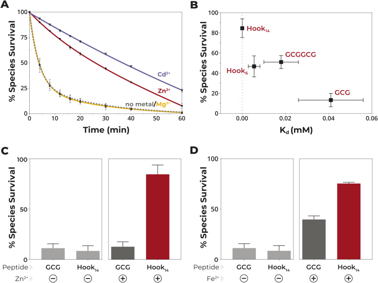
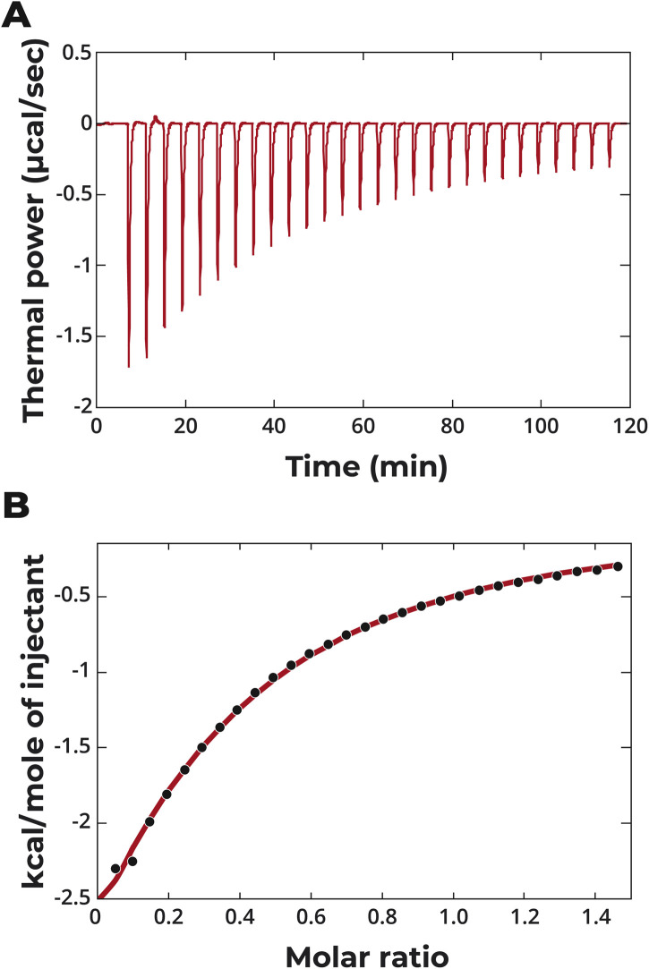
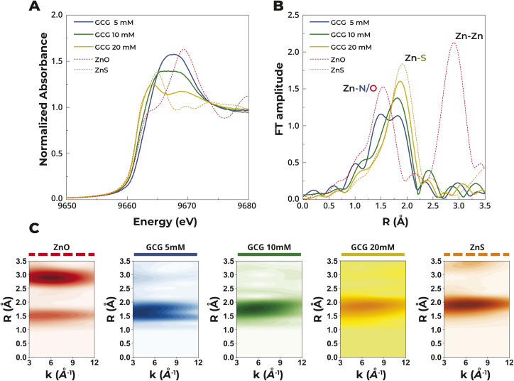
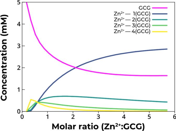
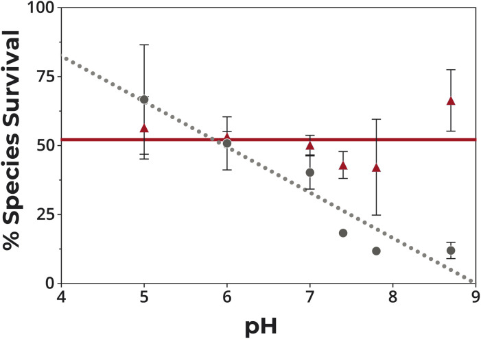
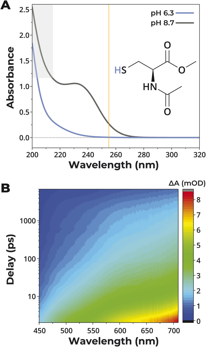
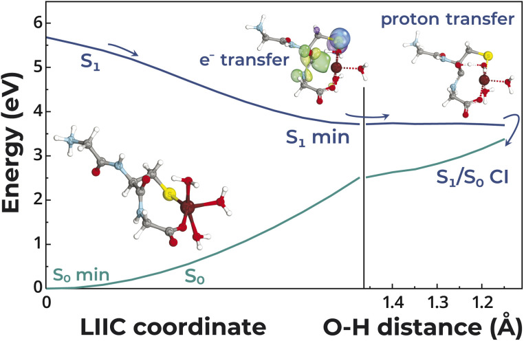
The transition from unregulated, prebiotic chemistry to metabolic-like systems capable of supporting an evolving protocell has remained difficult to explain. One hypothesis is that early catalysts began to prune the chemical landscape in a manner that facilitated the emergence of modern-day enzymes. As enzymes frequently rely on the intrinsic reactivity of metal ions, it follows that these early catalysts may have been metal ions coordinated to prebiotic peptides that have remained as core structures within extant proteins. Here, we demonstrate that UV light directly selects for the types of metal-binding peptide motifs found in biology. This is because bare cysteine is much more susceptible to photolysis than cysteine bound by a metal ion. Therefore, peptides with greater affinity for environmentally available metal ions, such as Fe2+ or Zn2+, are more stable. Our results are supported by mass spectrometry, calorimetry, X-ray absorption, NMR spectroscopy, transient absorption pump probe spectroscopy, and excited-state quantum-chemical calculations. Photostability arises from the ability of the metal ion to engage transiently generated reactive radical centers in a manner that prevents subsequent degradative processes. The data are consistent with the enrichment of a restricted set of high affinity, extant-like metallopeptides in surficial environments on the early Earth.


Conflict of interest statement

All authors declare they have no competing interests.

Figures

Fig. 1
Fig. 1. Metal binding protects against UV light and enables the selection of high-affinity peptides. (A) Degradation of GCG upon irradiation at 254 nm. Solutions of 5 mM GCG at pH 8.7 with or without 5 mM Zn2+, Cd2+, or Mg2+ were irradiated for 60 min. Quantification was performed by peak integration of HPLC chromatograms. Data were fit to a one-phase decay model. Data are mean ± SD; n = 3. (B) Peptide survival in the presence of 1.5 mM Zn2+vs. Kd of the peptide–Zn2+ complex. (C) Competition between Hook14 and GCG upon UV irradiation in the presence of Zn2+. (D) Competition between Hook14 and GCG in the presence of Fe2+ and UV light. Solution conditions for competition experiments were 1.25 mM Zn2+ or Fe2+, 5 mM GCG, and 2.5 mM Hook14 at pH 8.7. Irradiation was performed for 16 min at 254 nm. Data are mean ± SD; n = 3.
Fig. 2
Fig. 2. ITC of Zn2+ binding to GCG. Heat response (A) and integrated heat data as a function of the molar ratio of Zn2+/GCG (B) for injections of 2 mM ZnSO4 into 300 μM GCG. The continuous red line represents the best fit obtained with a single set of sites model and the “ligand in the cell” option.
Fig. 3
Fig. 3. XAS analysis of aqueous solutions of 5 mM Zn2+ with 5 mM, 10 mM, and 20 mM GCG compared to references ZnO and ZnS. (A) XANES rising edge spectra and (B) k2-weighted Fourier transformed spectra (3–12 Å−1k-range; Hanning window) highlighting the contributions from Zn–N/O and Zn–S scattering atoms. (C) Cauchy wavelet transforms showing the k-space and r-space dependence of Zn–N/O and Zn–S scattering alongside reference spectra of ZnO and ZnS. The colors used for each species are the same in each panel.
Fig. 4
Fig. 4. Concentration distribution of the different species present in solution during the titration of 5 mM GCG with Zn2+. At 5 mM GCG and 5 mM Zn2+, the calculated concentrations were 2.6 mM free GCG, 1.1 mM Zn–(GCG), 0.7 mM Zn–2(GCG), 0.4 mM Zn–3(GCG), and 0.2 mM Zn–4(GCG).
Fig. 5
Fig. 5. Survival of GCG in the presence of UV light at different pH with and without Zn2+. 5 mM GCG in 20 mM GG after exposure to light at 254 nm for 16 min in the absence (gray circles) and presence (red triangles) of 5 mM Zn2+.
Fig. 6
Fig. 6. UV-Vis and transient absorption spectra of the Cys analogue. (A) UV-Vis absorption spectra of 20 mM N-acetyl-l-cysteine methyl ester in water at 23 °C and pH 6.3 (blue line) and pH 8.7 (dark grey). The yellow line highlights the 255 nm excitation wavelength used in pump probe experiments. Absorbance above ∼1.2 (grey shaded area) may be less accurate due to the nonlinearity of UV-Vis detection at low photon flux. (B) 3D contour representation of the transient absorbance difference signal of N-acetyl-l-cysteine methyl ester after excitation at 255 nm. Spectra are shown on the x-axis. The delay time between pump and probe pulses is shown on a logarithmic scale on the y-axis (time resolution ∼1.5 ps), and the absorbance difference (ΔA) due to photoexcitation is color-coded and represented on the z-axis.
Fig. 7
Fig. 7. Potential energy profile of the photorelaxation mechanism of the GCG peptide coordinating Zn2+. The left side of the plot presents a linear interpolation in internal coordinates (LIIC) between the ground state structure of the Zn2+–peptide complex and the minimum-energy structure in the S1 state. The right side of the plot shows a relaxed scan along the CO⋯H distance associated with the proton transfer process from one of the water molecules coordinating the Zn2+ to the negatively charged carbonyl group. The excited-state and ground-state energies were calculated with the ADC(2) and MP2 methods, respectively, including the TZVP basis set.

Similar articles

References

    1. Copley S. D. Smith E. Morowitz H. J. Bioorg. Chem. 2007;35:430–443. doi: 10.1016/j.bioorg.2007.08.001. - DOI - PubMed
    1. Muchowska K. B. Varma S. J. Moran J. Nature. 2019;569:104–107. doi: 10.1038/s41586-019-1151-1. - DOI - PMC - PubMed
    1. Keller M. A. Turchyn A. V. Ralser M. Mol. Syst. Biol. 2014;10:725. doi: 10.1002/msb.20145228. - DOI - PMC - PubMed
    1. Bonfio C. Godino E. Corsini M. Fabrizi de Biani F. Guella G. Mansy S. S. Nat. Catal. 2018;1:616–623. doi: 10.1038/s41929-018-0116-3. - DOI
    1. Walton C. R. Rimmer P. Shorttle O. Front. Earth Sci. 2022;10:1011717. doi: 10.3389/feart.2022.1011717. - DOI

LinkOut - more resources