

Defect Size Dependent Reconstruction Strategy of the Nasal Tip Cutaneous Wounds: A Retrospective Study
- PMID: 40438275
- PMCID: PMC12117574
- DOI: 10.2147/CCID.S515533
Defect Size Dependent Reconstruction Strategy of the Nasal Tip Cutaneous Wounds: A Retrospective Study
Abstract
Objective: To achieve satisfying function and appearance of nasal tip after tumor resection.
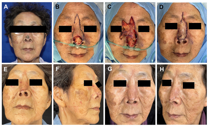
Methods: This retrospective study included patients with nasal tip tumors admitted between January 2010 and January 2024. A standardized data collection template was used to collect related variables. Tumors were all resected according to the guidelines, and the defects were repaired with different flaps. For nasal tip soft tissue defects smaller than 1 cm, a horn-shaped flap was used; for defects with a diameter of 1-2 cm, the bilobed flap was applied; for defects larger than 2 cm, frontonasal flap was designed.
Results: Twenty-eight patients were included in this study. All the skin defects were repaired according to the one-stage reconstruction strategy. All patients achieved primary healing without severe complications. Slight edema occurred in 5 patients, and slight infection occurred in 2 patients, all healed with dressing change in several days. Ideal aesthetic results achieved without distortion of the nasal tip or alar rim. During the follow-up period of 3-11 months, no tumor occurrence was observed.
Conclusion: The reconstruction strategy reported in this study is a promising way in the repair of nasal tip soft tissue defect with little complication and satisfying outcomes.
Keywords: bilobed flap; frontonasal flap; horn-shaped flap; nasal reconstruction; nasal tip defect.
© 2025 Wu et al.
Conflict of interest statement
Minliang Wu, Xinyi Zhang, and Ang Li are co-first authors for this study. All authors report no conflicts of interest in this work.
Figures


Similar articles
-
[Cosmetic outcome of nasal tip reconstruction with the frontonasal flap and other locoregional flaps - Cosmetic Outcome of Nasal Tip Reconstruction].Handchir Mikrochir Plast Chir. 2023 Aug;55(4):278-286. doi: 10.1055/a-2069-2246. Epub 2023 May 24. Handchir Mikrochir Plast Chir. 2023. PMID: 37224879 Free PMC article. German.
-
The Axial Frontonasal Flap for Nasal Tip Defect: A Single Centre Experience.Cureus. 2019 Oct 11;11(10):e5892. doi: 10.7759/cureus.5892. Cureus. 2019. PMID: 31772862 Free PMC article.
-
Supermicrosurgical reconstruction of nasal tip defects using the preauricular reversed superficial temporal artery flap.J Plast Reconstr Aesthet Surg. 2020 Jan;73(1):58-64. doi: 10.1016/j.bjps.2019.06.028. Epub 2019 Jul 16. J Plast Reconstr Aesthet Surg. 2020. PMID: 31466909
-
A Systematic Review and Overview of Flap Reconstructive Techniques for Nasal Skin Defects.Facial Plast Surg Aesthet Med. 2021 Dec;23(6):476-481. doi: 10.1089/fpsam.2020.0533. Epub 2021 Mar 1. Facial Plast Surg Aesthet Med. 2021. PMID: 33650884 Free PMC article.
-
Advancement flap for the reconstruction of nasal ala and lateral nasal tip defects.J Am Acad Dermatol. 2006 Dec;55(6):1032-5. doi: 10.1016/j.jaad.2006.08.049. J Am Acad Dermatol. 2006. PMID: 17110218 Review.
References
-
- Burget G, Menick F. Aesthetic Reconstruction of the Nose, 2nd Ed. St. Louis, MO, USA: Mosby; 1994.
LinkOut - more resources
Full Text Sources
