

Proximal-type Epithelioid Sarcoma of the Vulva: A Case Report
- PMID: 40438484
- PMCID: PMC12106100
- DOI: 10.47895/amp.vi0.10057
Proximal-type Epithelioid Sarcoma of the Vulva: A Case Report
Abstract
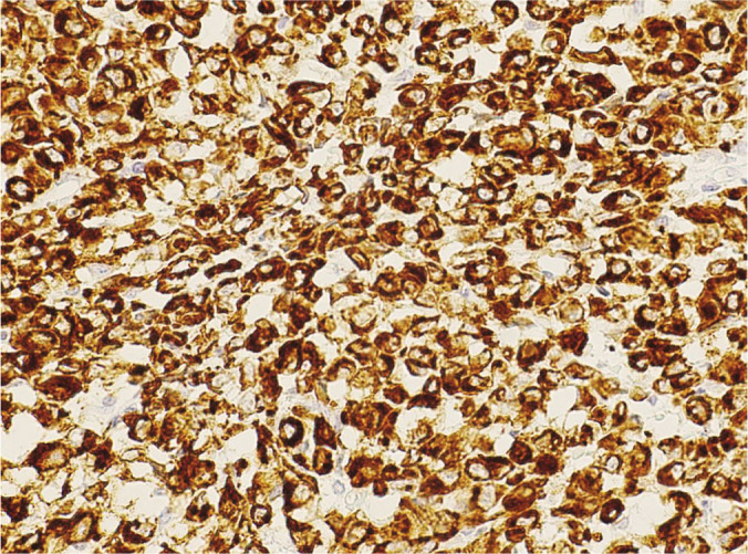
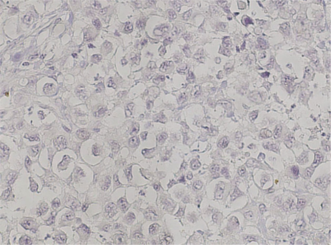
Epithelioid sarcoma is an uncommon mesenchymal malignancy which represents less than 1% of all sarcomas. Rarer still are reports of this tumor initially presenting in the vulva. We report a case of vulvar proximal-type epithelioid sarcoma. A 52-year-old had a 5-month history of slowly growing papule on the right labia majora. Excision of the mass revealed a tumor composed of large polygonal cells with abundant eosinophilic cytoplasm. An immunohistochemistry panel revealed cytokeratin AE1/AE3 positivity only. She underwent radical vulvectomy with bilateral groin node dissection. The specimen revealed a cream tan, firm, fairly defined mass at the right vulva. Microscopic examination showed a sheet-like growth pattern of large pleomorphic epithelioid cells with large vesicular nuclei and prominent nucleoli. The tumor showed loss of INI1 nuclear expression and absence of CD34 staining. EMA was positive. The case was signed out as proximal-type epithelioid sarcoma of the right vulva. Two months post-operatively, the patient was given concurrent chemotherapy with 5 cycles of cisplatin 40 mg/m2 and 6600 centigray vulvar intensity-modulated radiotherapy. She had no evidence of disease for five months until repeat workup showed tumor recurrence in the perineum. She was subsequently given 6 cycles of gemcitabine 900 mg/m2 and gemcitabine 900 mg/m2 with docetaxel 100 mg/m2. Two months after, repeat workup showed persistent progressive disease in the vulva. She was subsequently given 4 cycles of doxorubicin 60 mg/m2 and is for repeat workup. The immunohistomorphologic features of this tumor, in addition to its unusual location, present a diagnostic challenge. Clues to the diagnosis include an initial presentation as a soft tissue mass and microscopic features showing the presence of epithelioid to spindle cytomorphology with an infiltrative growth pattern. Immunohistochemistry studies revealing the loss of INI1 nuclear expression and expression of epithelial markers would ultimately establish the diagnosis of this rare clinical entity.
Keywords: epithelioid sarcoma; female urogenital diseases; vulvar neoplasms.
© The Author(s) 2025.
Conflict of interest statement
All authors declared no conflicts of interest.
Figures


References
-
- Oda Y, Nielsen TO, Dal Cin P, Le Loarer F. Epithelioid sarcoma. In: WHO Classification of Tumours Editorial Board. Soft tissue and bone tumours [Internet]. Lyon (France): International Agency for Research on Cancer; 2020 [cited 2024 April 2]. (WHO classification of tumours series, 5th ed.; vol. 3). Available from: https://tumourclassification.iarc.who.int/chapters/33.
-
- Yahiro S, Fujimoto T, Fujita I, Takai T, Sakuma T, Sudo T, et al. Proximaltype epithelioid sarcoma in pubic region expressing L-type amino acid transporter 1: a case report. SAGE Open Med Case Rep. 2022. Jan 8;10:2050313X211067917. doi: 10.1177/2050313X211067917. PMID: 35024147; PMCID: . - DOI - PMC - PubMed
Publication types
LinkOut - more resources
Full Text Sources
Molecular Biology Databases
Research Materials
Miscellaneous