Review
. 2025 Jul 31;121(8):1156-1172.
doi: 10.1093/cvr/cvaf089.

PRDM16, a new kid on the block in cardiovascular health and disease

Affiliations
Review

PRDM16, a new kid on the block in cardiovascular health and disease

Jore Van Wauwe et al. Cardiovasc Res. .

Abstract

Transcriptional regulation is essential for the development, homeostasis, and function of all organisms. Transcription factors and epigenetic modifiers play an indispensable role by direct or indirect interaction with DNA or chromatin. Although the role of transcription factor PRDM16 in adipose, haematopoietic, skeletal, and neural cell lineage specification is well-documented, its function within the cardiovascular system has only recently gained significant attention. Similar as in adipose tissue, PRDM16 displays an asymmetric expression pattern within the cardiovascular system, where it is exclusively expressed by ventricular cardiomyocytes and endothelial and smooth muscle cells of arteries while being absent in their atrial and venous counterparts. Concordantly, an increasing number of clinical and preclinical studies have identified PRDM16 as an important multi-modal regulator of cardiovascular development and function. Moreover, aberrant PRDM16 expression has now been linked to (cardio)vascular diseases, including left ventricular non-compaction, migraine, and coronary artery disease. In this review, we give a synopsis of PRDM16's expression and function within (developing) cardiovascular tissues and provide insights into how impaired PRDM16 signalling contributes to cardiovascular disease.

Keywords: Cardiomyopathy; Cell-fate decision; PRDM16; Transcription factors; Vascular disease.


Conflict of interest statement

Conflict of interest: None declared.

Figures

Graphical Abstract
Graphical Abstract
Figure 1
Figure 1
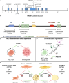
PRDM16 protein structure and mode of action outside the cardiovascular system. (A, B) The structure of the PRDM16 gene, consisting of 17 exons (A) and the corresponding protein structure (B). Pathogenic variants listed in Table 1 associated with cardiomyopathy are indicated. (C) PRDM16 is known for its transcriptional activator (green arrow) role in haematopoietic stem cell (HSC) maintenance and its repressor (red T-bar) role in cancer. (D) In the nervous system, PRDM16 functions as an epigenetic (epi) regulator to organise cells of the cerebral cortex. Hereto, it changes the epigenetic status of neural stem cells (NSCs, i.e. radial glia cells), thereby affecting intermediate progenitor formation and cortical neuron migration. (E) PRDM16 is best known for its role as binary cell-fate decision-maker in adipose tissue, where it drives the differentiation of brown adipose tissue (BAT), thereby repressing the transcriptional programme of white adipose tissue (WAT). Expression of PRDM16 in adipose progenitors or MYF5+ myocyte progenitors results in a BAT cell fate. Moreover, overexpression in myoblasts or WAT results in an identity switch of these cell types towards a more BAT-like phenotype. In this myogenic-adipocyte cell fate, PRDM16 functions as a transcription factor (TF), co-factor or epigenetic regulator. The figure was designed in BioRender and panel A was adapted from Ref.
Figure 2
Figure 2
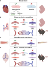
PRDM16 has an asymmetric expression pattern in the cardiovascular system. (A) In the developing mouse heart, PRDM16 expression is only detected in ventricular cardiomyocytes (CMs) while it is absent from atrial CMs. Moreover, within the ventricle, PRDM16 is more expressed in the compact myocardium compared to the trabecular zone. (B) In the systemic vasculature, we and others have shown that PRDM16 expression is restricted to arterial endothelial cells (aECs) and smooth muscle cells (aSMCs), whilst being absent from capillary (cap)ECs, venous (v)ECs, and vSMCs. (C) In accordance with its expression within the mouse vascular system, prdm16 is only expressed in aECs, but not in vECs of the tail axial vessels of zebrafish embryos. (D) In the cerebral vasculature of adult mice, PRDM16 expression is detected in aECs and aSMCs, and capECs, but is absent in vECs and vSMCs. The figure was designed in BioRender.
Figure 3
Figure 3
Heterogeneity in preclinical mouse models with myocardial PRDM16 deficiency. (A) Cardiomyocyte (CM)-specific deletion of Prdm16 using the Myh6 Cre-driver induced hypertrophic cardiomyopathy (HCM) and hypotension, respectively more pronounced or exclusively present in female mice. (B) Using the same Cre-driver, but a different Prdm16fl/fl mouse model, with LoxP sites flanking exon 6/7, dilated cardiomyopathy (DCM) was observed with reduced ejection fraction (EF), again more pronounced in female mice. (C) In a CM-specific KO model using Mesp1 as a Cre-driver, HCM was seen only upon metabolic stress or ageing. (D) An adult inducible (using tamoxifen) CM-specific Cre-recombinase model based on Myh6 resulted in HCM in one study but no functional abnormalities in another report. (E) In a CM-specific mouse model using Xmlc2 or cTnT as Cre-drivers, postnatal mortality was seen in the complete offspring by 7 days of age (P7). Embryonic hearts showed a left ventricular non-compaction (LVNC) phenotype. (F) CM-specific deletion of Prdm16 during cardiac development using a Sm22α-driven Cre-recombinase resulted in contractile dysfunction, hyperplasia of the ventricular conduction system (VCS), and hence abnormal electrophysiology of the postnatal heart, resulting in premature death by P21. (G, H) Ubiquitous homozygous Prdm16-KO mouse models are lethal postnatally and feature left ventricular hypoplasia. (I) Ubiquitous heterozygous deletion of Prdm16 (csp1 mutant) showed normal heart morphology but reduced EF and an altered metabolism more pronounced in females. (J, K) Overexpression of the nonsense variant Q187X was embryonic lethal in a homozygous setting and displayed LVNC in a heterozygous setting. Heterozygous females were slightly underrepresented in the offspring. The figure was designed in BioRender.
Figure 4
Figure 4
Preclinical mouse models with PRDM16 deficiency in the vascular system. (A) Ubiquitous heterozygous PRDM16-deficient mice demonstrated reduced arterial dimensions compared to wild-type (WT) littermates. (B) In the same model, after induction of femoral artery ligation (FAL) impaired flow recovery was seen. L: ligated; UL: unligated. (C) In an endothelial cell (EC)-specific knockout (KO) model using the inducible Cdh5 Cre-driver, this impaired flow recovery after FAL was recapitulated. (D) The same study showed that the impairment in blood flow recovery following FAL was not observed in mice with smooth muscle cell (SMC)-Prdm16 deletion. (E) PRDM16 overexpression (PRDM16OE) in ApoE−/− rats led to decreased atherosclerotic plaque formation. (F) Prdm16 expression was upregulated in perivascular adipose tissue of ApoE−/− mice, showing reduced atherosclerosis following all trans (t)-retinoic acid treatment. (G) Adipose-specific deletion of Prdm16 using the Adipoq Cre-driver or by locally silencing Prdm16 expression via Prdm16 siRNA resulted in exacerbated vascular inflammation and neointima formation following endovascular wire injury. (H) SMC-specific deletion of Prdm16 using Myh11-, Itga8- or Sm22α-Cre models showed reduced contractility, disrupted SMC homeostasis, and dysregulated blood pressure (BP). (I) Using the Myh11-Cre mouse model, elastase-induced abdominal aortic aneurysms (AAAs) were aggravated compared to challenged WT controls. (J) SMC-PRDM16 deletion using the Myh11-Cre or the Sm22α-Cre mouse model did not affect atherosclerotic burden (upon adeno-associated viral (AAV) transfer of a gain-of-function proprotein convertase subtilisin/kexin type 9 (PCSK9) mutant and exposure to Western diet) but altered atherosclerotic plaque composition. The figure was designed in BioRender.

References

    1. Casamassimi A, Rienzo M, Di Zazzo E, Sorrentino A, Fiore D, Proto MC, Moncharmont B, Gazzerro P, Bifulco M, Abbondanza C. Multifaceted role of PRDM proteins in human cancer. Int J Mol Sci 2020;21:2648. - PMC - PubMed
    1. Nishikata I, Sasaki H, Iga M, Tateno Y, Imayoshi S, Asou N, Nakamura T, Morishita K. A novel EVI1 gene family, MEL1, lacking a PR domain (MEL1S) is expressed mainly in t(1;3)(p36;q21)-positive AML and blocks G-CSF-induced myeloid differentiation. Blood 2003;102:3323–3332. - PubMed
    1. Di Zazzo E, De Rosa C, Abbondanza C, Moncharmont B. PRDM proteins: molecular mechanisms in signal transduction and transcriptional regulation. Biology (Basel) 2013;2:107–141. - PMC - PubMed
    1. Mochizuki N, Shimizu S, Nagasawa T, Tanaka H, Taniwaki M, Yokota J, Morishita K. A novel gene, MEL1, mapped to 1p36.3 is highly homologous to the MDS1/EVI1 gene and is transcriptionally activated in t(1;3)(p36;q21)-positive leukemia cells. Blood 2000;96:3209–3214. - PubMed
    1. Nishikata I, Nakahata S, Saito Y, Kaneda K, Ichihara E, Yamakawa N, Morishita K. Sumoylation of MEL1S at lysine 568 and its interaction with CtBP facilitates its repressor activity and the blockade of G-CSF-induced myeloid differentiation. Oncogene 2011;30:4194–4207. - PubMed

MeSH terms