. 2025 Aug;10(8):101260.
doi: 10.1016/j.jacbts.2025.02.019. Epub 2025 May 28.

Knockdown of PARM1 Alleviates Aortic Valve Calcification via the PRKCH-MAPK Signaling Pathway

Affiliations

Knockdown of PARM1 Alleviates Aortic Valve Calcification via the PRKCH-MAPK Signaling Pathway

Haochang Hu et al. JACC Basic Transl Sci. 2025 Aug.

Abstract

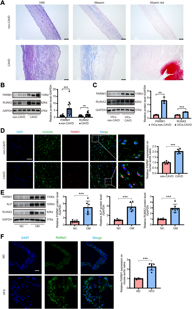
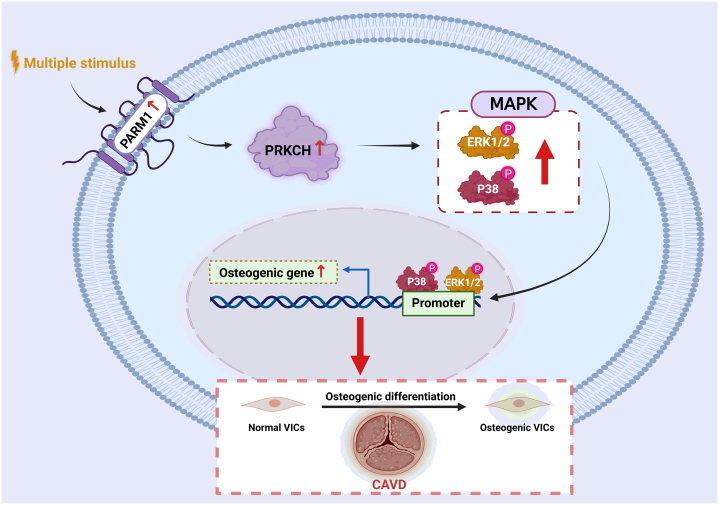
With the aging of the population, the prevalence of calcific aortic valve disease (CAVD) has increased yearly. However, effective means to delay or even reverse the progression of CAVD are still lacking. This study revealed that prostate androgen-regulated mucin-like protein 1 (PARM1) expression was significantly up-regulated in calcified aortic valve tissues. Functional investigations demonstrated that PARM1 knockdown effectively suppressed osteogenic differentiation of valvular interstitial cells (VICs) and mitigated pathological aortic valve calcification. Mechanically, PARM1 knockdown down-regulated PRKCH mRNA expression, consequently attenuating MAPK pathway activation during the osteogenic differentiation of VICs. In conclusion, PARM1 could be a feasible target for CAVD prevention.

Keywords: MAPK signaling pathway; PARM1; PRKCH; aortic valve calcification; osteogenic differentiation.


Conflict of interest statement

Funding Support and Author Disclosures This study was supported by State Key Laboratory of Transvascular Implantation Devices, the National Natural Science Foundation of China (82271606 to Dr X. Liu, 82200407 to Dr S. Cheng, 82301765 to Drs W. Hu, 82400436 to J. Chen, U22A20267 and 82030014 to Dr J. Wang), the National Key R and D Program of China (2019YFA0110400 to Dr J. Wang), the Key Program of Major Science and Technology Projects in Zhejiang Province (2021C03097 and 2024C03024 to Dr J. Wang, 2022C03063 to Dr X. Liu), Zhejiang Province Science and Technology Innovation Leading Talents Project (2023R5236 to Dr X. Liu), and the China Postdoctoral Science Foundation (2024M752874 to Dr J. Chen). All other authors have reported that they have no relationships relevant to the contents of this paper to disclose.

Figures

None
Graphical abstract
Figure 1
Figure 1
PARM1 Is Significantly Upregulated in Calcified Aortic Valves (A) Representative hematoxylin and eosin (H&E) staining, Masson’s staining, and Alizarin red staining of the noncalcified human aortic valves and calcified aortic valves (Scale bar: 100 μm). (B) The expression of prostate androgen-regulated mucin-like protein 1 (PARM1) were determined by Western blot in aortic valves from calcific aortic valve disease (CAVD) and non-CAVD patients. (C) PARM1 expression was detected in VICs isolated from noncalcified aortic valves and calcified aortic valves. (D) Representative immunofluorescence images of PARM1 localized on VICs in human aortic valve tissues (scale bar: 25 μm). (E) PARM1 protein levels in VICs stimulated with osteogenic medium (OM) determined by Western blot. (F) Representative immunofluorescence images of PARM1 in aortic valves from the Ldlr-/- mice fed a normal diet (ND) and a high-fat diet (HFD) (Scale bar: 25 μm). Values are mean ± SD. The unpaired Student's t-test was performed. ∗∗P < 0.01; ∗∗∗P < 0.001.
Figure 2
Figure 2
Knockdown or Overexpression of PARM1 Could Regulate Osteogenic Differentiation of VICs In Vitro (A and B) The expression levels of 2 osteogenic markers (ALP and RUNX2) were evaluated by qRT-PCR (A) and Western blot (B) after VICs transfected with si-PARM1. (C) ALP activity was detected after VICs transfected with si-PARM1 or si-Scr in OM. (D) Calcium content in VICs of each group was measured after 14 days of OM intervention. (E, F) Calcium deposits in VICs were detected by Alizarin red staining. (G, H) The expression levels of ALP and RUNX2 after PARM1 overexpression in VICs were evaluated by quantitative real-time polymerase chain reaction (G) and Western blot (H). (I) ALP activity was detected after PARM1 overexpression in VICs. (J) Calcium deposits in PARM1-overexpressed VICs were detected by Alizarin red staining. Values are mean ± SD. Analysis of variance with Tukey's post hoc test was applied for multiple pairwise comparisons. ∗P < 0.05; ∗∗P < 0.01; ∗∗∗P < 0.001. Abbreviations as in Figure 1.
Figure 3
Figure 3
PARM1 Knockdown Ameliorates HFD-Induced Aortic Valve Calcification in Ldlr−/− Mice (A) Representative echocardiographic images of ND+sh-control, HFD+sh-control, and HFD+sh-PARM1 mice. (B) Quantification of peak transvalvular jet velocity and mean transvalvular pressure gradient of ND+sh-control, HFD+sh-control, and HFD+sh-PARM1 mice. (C) Representative images of H&E, Sirius red, Masson’s, and von Kossa staining of the aortic valves in ND+sh-control, HFD+sh-control, and HFD+sh-PARM1 mice (Scale bar: 200 μm). (D-G) Quantification of aortic valve thickness, Sirius red positive area, fibrotic area, and total calcified area. (H) Immunofluorescence of BMP2 in ND+sh-control, HFD+sh-control, and HFD+sh-PARM1 mice (Scale bar: 25 μm). (I) Immunofluorescence of ALP in ND+sh-control, HFD+sh-control, and HFD+sh-PARM1 mice (Scale bar: 25 μm). Values are mean ± SD. Analysis of variance with Tukey's post hoc test was applied for multiple pairwise comparisons. ∗∗P < 0.01; ∗∗∗P < 0.001. Abbreviations as in Figure 1.
Figure 4
Figure 4
MAPK Signaling Pathway Is Downstream of PARM1 in Regulating the Osteogenic Differentiation of VICs (A) Gene Ontology analysis of RNA-seq for si-Scr and si-PARM1 groups. (B) Kyoto Encyclopedia of Genes and Genomes (KEGG) analysis of RNA-seq for si-Scr and si-PARM1 groups. (C) Gene Set Enrichment Analysis of RNA-seq for si-Scr and si-PARM1 groups. (D) Western blot was used to analyze the phosphorylation level of MAPK signaling pathway after knocking down PARM1. (E) Protein levels of ALP and RUNX2 were evaluated by Western blot after the treatment of P38 inhibitor and ERK inhibitor in VICs. (F) ALP activity was detected after the treatment of P38 inhibitor and ERK inhibitor in VICs. (G) Calcium deposits after the treatment of P38 inhibitor and ERK inhibitor were detected by Alizarin red staining. Values are mean ± SD. The unpaired Student's t-test was performed (D). Analysis of variance with Tukey's post hoc test was applied for multiple pairwise comparisons (E to G). ∗P < 0.05; ∗∗P < 0.01; ∗∗∗P < 0.001; ns P > 0.05. Abbreviations as in Figure 1.
Figure 5
Figure 5
PRKCH is a Key Downstream Target of PARM1 in the Osteogenic Differentiation of VICs (A) Simultaneous significant differences in protein kinases after OM stimulation and PARM1 knockdown. (B) The mRNA levels of PRKCH, ERBB3, and DCLK1 were detected after OM stimulation and PARM1 knockdown. (C) The protein levels of PRKCH were detected after OM stimulation and PARM1 knockdown. (D) The expression levels of ALP and RUNX2 were evaluated by Western blot after VICs transfected with si-PRKCH. (E) ALP activity was detected after VICs transfected with si-PRKCH or si-Scr in OM. (F) Calcium content in VICs of each group was measured after 14 days of OM intervention. (G) Calcium deposits in VICs transfected with si-PRKCH were detected by Alizarin red staining. (H) The protein kinase C (PKC) activity with the treatment of Sotrastaurin was detected by Human PKC ELISA kit (MEIMIAN). (I) The expression levels of ALP and RUNX2 were evaluated by Western blot after VICs treated with Sotrastaurin. (J) ALP activity was detected after VICs treated with Sotrastaurin in OM. (K) Calcium content in VICs of each group was measured after 14 days of OM intervention. (L) Calcium deposits in VICs treated with Sotrastaurin were detected by Alizarin red staining. Values are mean ± SD. Analysis of variance with Tukey's post hoc test was applied for multiple pairwise comparisons. ∗P < 0.05; ∗∗P < 0.01; ∗∗∗P < 0.001. Abbreviations as in Figure 1.
Figure 6
Figure 6
Overexpression of PRKCH Reversed the Antiosteogenic Differentiation Effect of PARM1 Knockdown on VICs (A) The expression levels of ALP and RUNX2 were detected by Western blot. (B) The protein expression of ALP and RUNX2 were evaluated after VICs transfected with si-PARM1 and overexpressed PRKCH. (C) ALP activity was detected after VICs transfected with si-PARM1 and overexpressed PRKCH in osteogenic medium. (D, E) The phosphorylation level of P38 and ERK1/2 was detected by Western blot after VICs transfected with si-PARM1 and overexpressed PRKCH. (F) Calcium deposits in VICs transfected with si-PARM1 and overexpressed PRKCH were detected by Alizarin red staining. Values are mean ± SD. Analysis of variance with Tukey's post hoc test was applied for multiple pairwise comparisons. ∗P < 0.05; ∗∗P < 0.01; ∗∗∗P < 0.001. Abbreviations as in Figure 1.
Figure 7
Figure 7
Schematic Illustration of the Involvement of PARM1 in CAVD Under multifactor stimulation, up-regulated PARM1 could promote the osteogenic differentiation of VICs and contribute to the initiation and progression of CAVD via the PRKCH-MAPK axis.

References

    1. Xu H., Liu Q., Cao K., et al. Distribution, characteristics, and management of older patients with valvular heart disease in China: China-DVD Study. JACC Asia. 2022;2:354–365. - PMC - PubMed
    1. Roth G.A., Mensah G.A., Johnson C.O., et al. Global burden of cardiovascular diseases and risk factors, 1990-2019: update from the GBD 2019 Study. J Am Coll Cardiol. 2020;76:2982–23021. - PMC - PubMed
    1. Lindman B.R., Sukul D., Dweck M.R., et al. Evaluating medical therapy for calcific aortic stenosis: JACC state-of-the-art review. J Am Coll Cardiol. 2021;78:2354–2376. - PMC - PubMed
    1. Li J.J., Ma C.S., Zhao D., Yan X.W., Beijing Heart Society and Expert Committee Lipoprotein(a) and cardiovascular disease in Chinese population: a Beijing Heart Society Expert Scientific Statement. JACC Asia. 2022;2:653–665. - PMC - PubMed
    1. Kraler S., Blaser M.C., Aikawa E., Camici G.G., Luscher T.F. Calcific aortic valve disease: from molecular and cellular mechanisms to medical therapy. Eur Heart J. 2022;43:683–697. - PMC - PubMed

LinkOut - more resources