. 2025 May 29;16(1):943.
doi: 10.1007/s12672-025-02788-6.

VSV-CHIKV activates antitumor immunity by inducing pyroptosis in a melanoma model

Affiliations

VSV-CHIKV activates antitumor immunity by inducing pyroptosis in a melanoma model

Fan Wu et al. Discov Oncol. .

Abstract

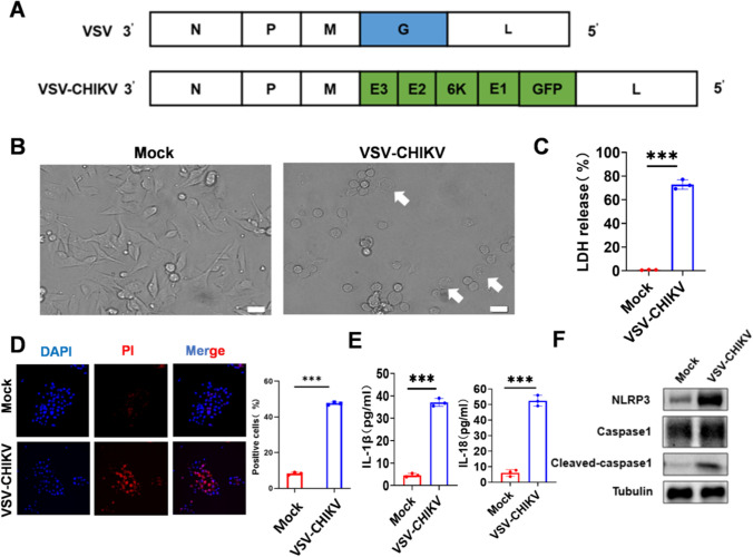
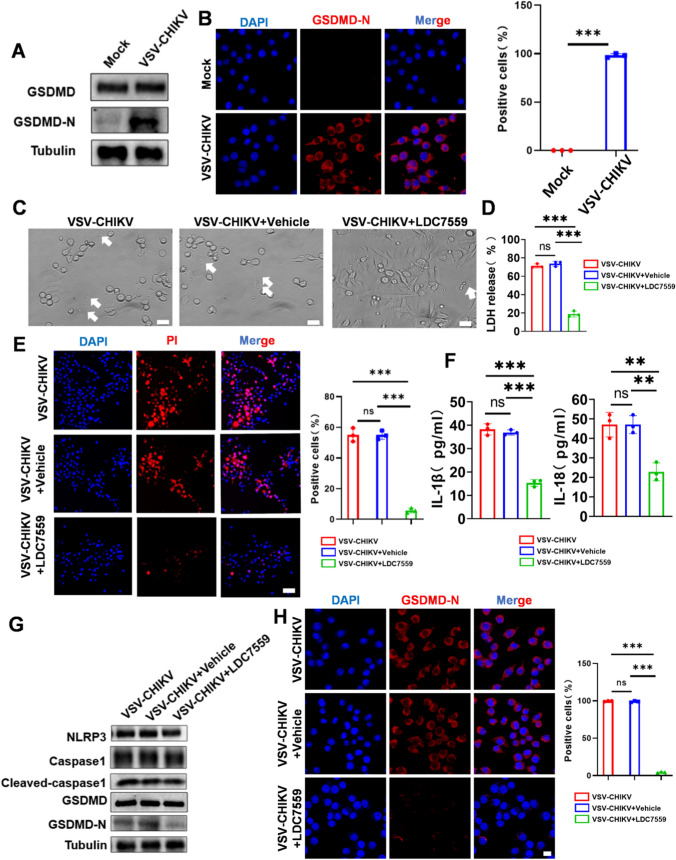
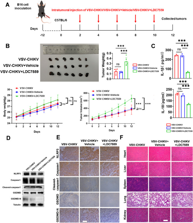
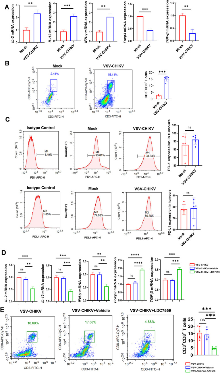
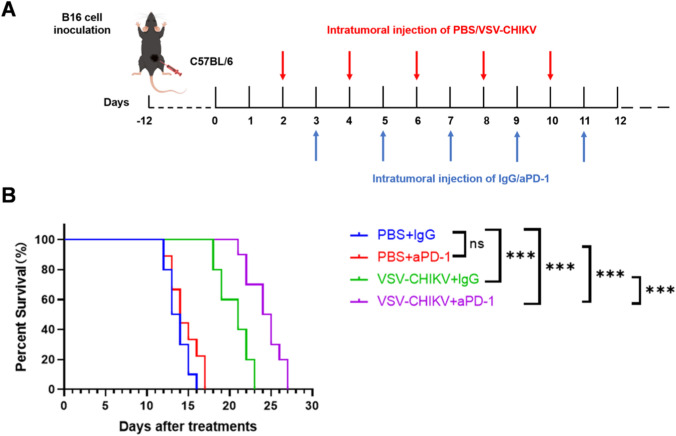
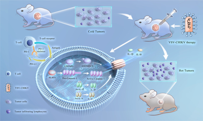
Melanoma is the most dangerous skin cancer due to its difficulty in treatment, high recurrence rate and metastatic ability. As a vector for oncolytic viruses (OVs), vesicular stomatitis virus (VSV) has been shown to be effective against malignant melanoma. However, the glycoprotein G protein of VSV has potential neurotoxicity. It has been shown that replacing glycoprotein G with E3-E2-6K-E1 of chikungunya virus (CHIKV) reduces its neurotoxicity and targets gliomas. Therefore, the aim of this study was to investigate the oncolytic effect of recombinant VSV-CHIKV on melanoma and the underlying mechanism. In this study, we found that recombinant VSV-CHIKV triggered GSDMD-mediated melanoma cell pyroptosis. Importantly, the NLRP3/Caspase-1/GSDMD axis was activated after VSV-CHIKV infection in melanoma cell lines and in a xenograft mouse model. Inhibition of GSDMD blocked cell pyroptosis, antitumor immunity and the tumor response in response to VSV-CHIKV treatment, suggesting that VSV-CHIKV act through the GSDMD pathway. VSV-CHIKV-triggered GSDMD-mediated tumor pyroptosis recruited cytotoxic T lymphocytes (CTLs) into the tumor microenvironment, which was accompanied by the release of inflammatory mediators. This remodeled the tumor microenvironment and turned immunologically "cold" tumors into "hot" tumors, thereby sensitized these tumors to checkpoint blockade. Finally, the combination therapy of VSV-CHIKV and an immune checkpoint inhibitor (anti-PD-1) prolonged the survival of mice. In conclusion, the VSV-CHIKV strategy is an attractive biologic therapy against melanoma.

Keywords: Melanoma; Oncolytic virus; Pyroptosis; Vesicular stomatitis virus.


Conflict of interest statement

Declarations. Ethical approval: Animal experiments were approved by the Animal care & Welfare Committee, School of Stomatology, Fourth Military Medical University (kq-2022–020) and followed the ARRIVE guidelines. All methods were performed in accordance with relevant guidelines and regulations. Consent to participate: Not applicable. Consent for publication: Not applicable. Competing interests: The authors declare no competing interests.

Figures

Fig. 1
Fig. 1
Oncolytic VSV-CHIKV induces melanoma cell death (MOI = 1, 24 h). A Schematic showing the wild-type VSV and chimeric VSV-CHIKV genomes, in which the VSV glycoprotein G gene (blue) is replaced by the chikungunya glycoprotein sequence in the CHIKV structural polyprotein (green). B Images obtained by microscopy showing changes in VSV-CHIKV infection of B16 cells (scale bar: 20 μm). C LDH release from the culture supernatant of B16 cells infected with VSV-CHIKV (n = 3). ***P < 0.001 by t test. D Fluorescence microscopy observation of PI uptake in B16 cells (scale bars: 50 μm). ***P < 0.001 by t test. E The levels of IL-1β and IL-18 in the cell culture supernatant (n = 3). ***P < 0.001 by t test. F Western blot analysis of NLRP3, Caspase1, and Cleaved-caspase1 protein expression after the addition of VSV-CHIKV
Fig. 2
Fig. 2
Melanoma cell pyroptosis triggered by oncolytic VSV-CHIKV is mediated by the NLRP3/Caspase-1/GSDMD axis (MOI = 1, 24 h). A Western blot analysis of GSDMD and GSDMD-N protein expression after the addition of VSV-CHIKV. B Expression of the GSDMD-N protein in the cytoplasm of cells after VSV-CHIKV infection of B16 cells. GSDMD-N, red; DAPI, blue (scale bar: 10 μm). ***P < 0.001 by t test. C Images obtained by microscopy showing changes in B16 cells after the addition of LDC7559 (scale bar: 20 μm). D LDH release from culture supernatants of B16 cells after the addition of LDC7559 (n = 3). ***P < 0.001 by One-way ANOVA. E Fluorescence microscopy of PI uptake in B16 cells after the addition of LDC7559 (scale bar: 50 μm). ***P < 0.001 by One-way ANOVA. F The levels of IL-1β and IL-18 in the cell culture supernatant after the addition of LDC7559 (n = 3). **P < 0.01, ***P < 0.001 by One-way ANOVA. G Western blot analysis of NLRP3, Caspase1, Cleaved-caspase1, GSDMD and GSDMD-N protein expression after the addition of LDC7559. H Expression of GSDMD-N protein in the cytoplasm of cells after the addition of LDC7559 to B16 cells. GSDMD-N, red; DAPI, blue (scale bar: 10 μm). ***P < 0.001 by One-way ANOVA
Fig. 3
Fig. 3
Oncolytic VSV-CHIKV causes pyroptosis in a melanoma cancer model. A Schematic illustration of the experimental design. B16 cell tumor-bearing C57BL/6 mice were intratumorally treated with VSV-CHIKV. B B16 tumors collected after VSV-CHIKV infection; images of the tumors, tumors weights (n = 6), body weight (n = 6), tumors growth curves are shown. **P < 0.01 by t test. ***P < 0.001 by t test. C IL-1β and IL-18 levels in tumor tissues (n = 3). ***P < 0.001 by t test. D NLRP3, Caspase1, Cleaved-caspase1, GSDMD and GSDMD-N proteins in the mock group and the VSV-CHIKV group determined by Western blotting. E Immunohistochemical staining for the NLRP3, Caspase1, Cleaved-caspase1, GSDMD and GSDMD-N proteins in the mock group and the VSV-CHIKV group (scale bar: 100 μm). F HE staining of major organs (scale bar: 100 μm)
Fig. 4
Fig. 4
Inhibition of GSDMD impairs VSV-CHIKV-induced melanoma tumor suppression. A Schematic illustration of the experimental design. B16 cell tumor-bearing C57BL/6 mice were intratumorally treated with VSV-CHIKV, VSV-CHIKV + Vehicle, or VSV-CHIKV + LDC7559. B B16 tumors from the VSV-CHIKV, VSV-CHIKV + Vehicle, and VSV-CHIKV + LDC7559 groups were collected after treatment; images of the tumors, tumors weights (n = 6), body weight (n = 6) and tumors growth curves are shown. ***P < 0.001 by One-way ANOVA. C The levels of IL-1β and IL-18 in tumor tissues (n = 3). ***P < 0.001 by One-way ANOVA. D Expression of the NLRP3, Caspase1, Cleaved-caspase1, GSDMD and GSDMD-N proteins in the VSV-CHIKV, VSV-CHIKV + Vehicle and VSV-CHIKV + LDC7559 groups determined by Western blotting. E Immunohistochemical staining for the NLRP3, Caspase1, Cleaved-caspase1, GSDMD and GSDMD-N proteins in the VSV-CHIKV, VSV-CHIKV + Vehicle and VSV-CHIKV + LDC7559 groups (scale bar: 100 μm). F HE staining of major organs (scale bar: 100 μm)
Fig. 5
Fig. 5
Inhibition of GSDMD impairs VSV-CHIKV-induced antitumor immunity against melanoma tumors. A mRNA expression analysis of IL-2, IL-12, IFN-γ, TGF-β and Foxp3 in mouse tumors (n = 3). **P < 0.01, ***P < 0.001 by t test. B Flow cytometry detection of CD3+CD8+ T cells expression in melanoma tissues. The results of the quantitative analysis are shown on the right (n = 6). ***P < 0.001 by t test. C Flow cytometry detection of PD-1 and PD-L1 expression in melanoma tissues. The results of the quantitative analysis are shown on the right (n = 5). D mRNA expression analysis of IL-2, IL-12, IFN-γ, TGF-β and Foxp3 in mouse tumors (n = 3). **P < 0.01, ***P < 0.001 by One-way ANOVA. E Detection of CD3+CD8+ T cells by flow cytometry. The results of the quantitative analysis are shown on the right (n = 6). ***P < 0.001 by One-way ANOVA
Fig. 6
Fig. 6
VSV-CHIKV-based virotherapy enhances the efficacy of immune checkpoint blockade. A Schematic illustration of the experimental design. B16 cell tumor-bearing C57BL/6 mice were intratumorally treated with VSV-CHIKV and intraperitoneally treated with aPD-1 (5 mg/kg). B Kaplan–Meier survival curves were generated (n = 10)
Fig. 7
Fig. 7
VSV-CHIKV induces GSDMD-mediated pyroptosis and activates antitumor immunity. The NLRP3/Caspase-1/GSDMD axis was activated after melanoma cell lines were infected with VSV-CHIKV. VSV-CHIKV-triggered GSDMD-mediated tumor pyroptosis recruits CTLs into the tumor microenvironment, which is accompanied by the release of inflammatory mediators. This remodels the tumor microenvironment and turns immunologically “cold” tumors into “hot” tumors, thereby sensitizes these tumors to checkpoint blockade

Similar articles

References

    1. Long GV, Swetter SM, Menzies AM, Gershenwald JE, Scolyer RA. Cutaneous melanoma. Lancet. 2023 - PubMed
    1. Centeno PP, Pavet V, Marais R. The journey from melanocytes to melanoma. Nat Rev Cancer. 2023;23(6):372–90. - PubMed
    1. Kalaora S, Nagler A, Wargo JA, Samuels Y. Mechanisms of immune activation and regulation: lessons from melanoma. Nat Rev Cancer. 2022;22(4):195–207. - PubMed
    1. Kaufman HL, Kohlhapp FJ, Zloza A. Oncolytic viruses: a new class of immunotherapy drugs. Nat Rev Drug Discov. 2015;14(9):642–62. - PMC - PubMed
    1. Fukuhara H, Ino Y, Todo T. Oncolytic virus therapy: a new era of cancer treatment at dawn. Cancer Sci. 2016;107(10):1373–9. - PMC - PubMed

LinkOut - more resources