. 2025 Jun 3;122(22):e2414166122.
doi: 10.1073/pnas.2414166122. Epub 2025 May 29.

Histone variant H2A.W7 represses meiotic crossover formation in Arabidopsis heterochromatin

Affiliations

Histone variant H2A.W7 represses meiotic crossover formation in Arabidopsis heterochromatin

Pallas Kuo et al. Proc Natl Acad Sci U S A. .

Abstract

In eukaryotic genomes, DNA is packaged into nucleosomes to form chromatin. The incorporation of canonical or variant histones into nucleosomes confers different properties and influences chromatin structure to regulate cellular processes, including recombination. During meiosis, DNA double-strand breaks (DSBs) are formed and repaired as interhomolog crossovers. Nucleosome occupancy is generally associated with low crossover frequency, but it remains unclear which histone variants are involved in this process. In Arabidopsis, three variants of H2A coexist: H2A.X, H2A.Z, and H2A.W. Here, we show that H2A.W7 has a suppressive role on meiotic recombination. Genome-wide mapping of the crossover landscape revealed increased centromere-proximal recombination in h2a.w7. Moreover, H2A.W7 can be recruited to the 3a crossover hotspot via 21-24-nucleotide siRNAs during RNA-directed DNA methylation, causing increased nucleosome occupancy and decreased crossover frequency. Cytological analysis reveals that H2A.W7 restricts heterochromatin clustering during meiosis, which can form a mechanism to limit interhomolog recombination. Conversely, the linker histone H1, of which its loading is known to be restricted by H2A.W, promotes heterochromatin clustering and crossover on a heterochromatic genetic interval. Our study reveals a role for H2A.W7 in repressing crossover formation in Arabidopsis.

Keywords: H2A.W; crossover; epigenetics; meiosis; nucleosomes.


Conflict of interest statement

Competing interests statement:The authors declare no competing interest.

Figures
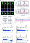
Fig. 1.
Fig. 1.
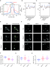
H2A.W6 and H2A.W7 localization on the chromatin. (A) Immunolocalization of H2A.W6 and H2A.W7 with ASY1 in prophase I. Chromatin is stained with DAPI. (Scale bar, 10 µm.) (B) Profiles of H2A.W6, H2A.W7 [log2(ChIP/input)], and SPO11-1-oligonucleotides [log2(oligos/gDNA)] in 10-kb cumulative windows along the chromosomes. Crossover frequency is presented in 300-kb cumulative windows. Vertical dotted lines represent the pericentromeres (PERI). (C) Mean coverage of SPO11-1-oligos [log2(oligos/gDNA)] within H2A.W6 or H2A.W7 peaks (vertical dashed lines) and 2-kb flanking regions. “S” and “E” represent the start and end of the peaks, respectively. Horizontal dashed lines represent 0-value. (D) A representative region from chromosome 3 showing H2A.W6, H2A.W7, SPO11-1-oligo profiles over genes and transposons. Orange dashed lines delineate an RNA LINE element and a DNA hAT element. Note that hAT contains H2A.W7 and SPO11-1-oligo (E) Bar graphs showing permutation test derived log2(observed/expected) overlap of the genome-wide H2A.W6 and H2A.W7 peaks with the indicated transposon families, SPO11-1-oligonucleotides and nucleosomes. Horizontal gray lines represent the significant threshold of α = 0.05.
Fig. 2.
Fig. 2.
H2A.W7 influences meiotic recombination and chromatin organization. (A) Profiles of crossover frequency in 300-kb cumulative windows and H2A.W6 and H2A.W7 [log2(ChIP/input)] in 10-kb cumulative windows along chromosome 3. Vertical dotted lines represent the PERI. Green and red dashed lines delineate the genetic intervals 420 and CEN3. (B and C) Crossover frequency (cM) within CEN3 (B) and 420 (C) intervals in the respective genotypes. Black points represent individual measurements and red points represent mean values. (D) Chromosome spreads of meiotic nuclei at pachytene stage in wild type (Col), h2a.w7, and h2a.w-2. Chromatin is stained with DAPI and chromocenter areas are defined as regions with higher intensity threshold of DAPI signal. (E) Immunostaining of MLH1 (white/red) on meiotic nuclei at diakinesis stage in wild type (Col), h2a.w7, and h2a.w-2. Chromatin is stained with DAPI (white). (F) Measurements of percentage chromocenter area/total chromatin area in wild type (Col), h2a.w7, and h2a.w-2. The mean value and SE of the means are shown. (G) Quantification of chromocenters in wild type (Col), h2a.w7, and h2a.w-2. Mean value and SE of the means are shown. (H) Quantification of MLH1 foci in wild type (Col), h2a.w7, and h2a.w-2. Mean value and SE of the means are shown. “n.s.” is abbreviated from no significance, ***P < 0.01). (Scale bar, 10 µm.)
Fig. 3.
Fig. 3.
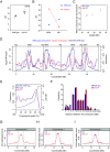
H2A.W7 suppresses crossover frequency in the heterochromatic pericentromeric regions. (A) Crossover frequency (cM) within the CEN3 interval in wild type and h2a.w7 ColxLer F1 hybrids. Black points represent individual measurements and red points represent mean values. (B) Crossover frequency within CEN3 and 420 intervals in wild type and h2a.w7 ColxLer F1 hybrids by genotyping-by-sequencing (GBS). (C) Mean crossover number for the chromosomes 1 to 4 in ColxLer hybrid of wild type and h2a.w7. (D) Crossover distribution along the chromosomes 1 to 4 using 300-kb adjacent windows in ColxLer hybrid of wild type and h2a.w7. Solid vertical lines represent the telomeres (TEL). Vertical dashed lines represent the PERI. (E) Crossover distribution along a telomere-to-centromere axis in ColxLer hybrid of wild type and h2a.w7. (F) Histogram showing frequency of cis-DCO distances in 2-Mb windows for wild type and h2a.w7 Col/Ler. (GI) Profile of DNA methylation in CG (G), CHG (H), and CHH (I) contexts on chromosome 1 from Col and h2a.w7.
Fig. 4.
Fig. 4.
Analysis of meiotic recombination in h2a.w7, cmt3, h1.1, and h1.2. (A) Crossover frequency (cM) within CEN3 interval in wild type (Col), h2a.w7, cmt3, and h2a.w7 cmt3. Black points represent individual measurements and red points represent mean values. (B and C) Crossover frequency (cM) within CEN3 (B) and 420 (C) intervals in wild type (Col), h1.1, h1.2, and h1.1 h1.2. Black points represent individual measurements and red points represent mean values. (“n.s.” is abbreviated from no significance, ***P < 0.01). (DF) Profile of DNA methylation in CG, (D), CHG (E), and CHH (F) contexts on chromosome 3 from Col and h1.1 h1.2. Vertical dashed lines represent the PERI. Red and green dashed lines delineate 420 an CEN3 genetic intervals. (G) Chromosome spreads of meiotic nuclei at pachytene stage in wild type (Col) and h1.1, h1.2. Chromatin is stained with DAPI and chromocenters are indicated by green arrows. (Scale bar, 10 µm.) (H) Quantification of chromocenters in wild type (Col) and h1.1 h1.2. Mean value and SE of the means are shown in bars. (I) Measurements of chromocenter percentage in wild type (Col) and h1.1 h1.2 using the ratio of chromocenter area: total area of chromatin. Mean value and SE of the means are shown in bars. ***P < 0.01 (MWW test).
Fig. 5.
Fig. 5.
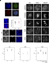
H2A.W7 and H2A.X are not essential for meiotic DSB repair. (A) Immunocolocalization of ASY1 and γH2AX on a meiotic nucleus at leptotene stage. Chromatin is stained with DAPI. (Scale bar, 10 µm.) (B) Zoom-in views of γH2AX (red) staining on DAPI-stained chromatin (blue). The white arrow shows that γH2AX foci are observed on chromocenters. (Scale bar, 1 µm.) (C) Quantification of MLH1 foci in Col and h2a.x at diakinesis stage. (D) MLH1 (white/red) immunostaining on meiotic chromosomes at diakinesis stage. Chromatin is stained with DAPI (white/blue). Mean values of MLH1 count are stated on the representative images. (Scale bar, 10 µm.) (E) Chromosome spread of Col, h2a.x, and h2a.w7 meiocytes at the specified stages. Chromatin is stained with DAPI. (Scale bar, 10 µm.) (F and G) Crossover frequency (cM) within 420 (F) and CEN3 (G) intervals in Col and h2a.x. Black points represent measurements from independent plants, and red points represent mean values. (H) Crossover frequency (cM) within CEN3 interval in the respective genotypes. Black points represent measurements from independent plants, and red points represent mean values. (“n.s.” means no significance, ***P < 0.05).
Fig. 6.
Fig. 6.
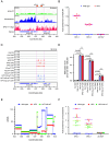
H2A.W7 suppresses crossover formation on 3a hotspot. (A) Representation of the 3a crossover hotspot locus showing H2A.W7, nucleosomes, SPO11-1-oligonucleotides, and genes. Pink vertical dashed lines represent the region targeted by the HP3 hairpin. Orange boxes represent the regions analyzed by qPCR assay. (B) Fold enrichment of H2A.W7 ChIP on HP3-1, HP3-2, and ACTIN7 relative to Ta2 in Col, HP3, and HP3 h2a.w7. (C) IGV browser view of DNA methylation with the 3a locus in Col, HP3, h2a.w7, and HP3 h2a.w7. Pink horizontal dashed lines represent the region targeted by the HP3 hairpin. (D) The bar graph shows the level of DNA methylation in CG, CHG, and CHH contexts within chromosome 3: 637 to 638.5 kb in Col, HP3, h2a.w7, and HP3 h2a.w7. t tests were performed to test for differences between genotypes. “n.s.” means not significant. (E) Recombination rate in cM/Mb within the 3a locus in ColxLer hybrid of wild type, HP3 and HP3 h2a.w7. (F) Fold enrichment of nucleosomes on HP3-1, HP3-2, and ACTIN7 relative to Ta2 in Col, HP3, and HP3 h2a.w7.

Similar articles

References

    1. Mercier R., Mézard C., Jenczewski E., Macaisne N., Grelon M., The molecular biology of meiosis in plants. Annu. Rev. Plant Biol. 66, 297–327 (2015). - PubMed
    1. Robert T., et al. , The TopoVIB-Like protein family is required for meiotic DNA double-strand break formation. Science. 351, 943–949 (2016). - PubMed
    1. Vrielynck N., et al. , A DNA topoisomerase VI-like complex initiates meiotic recombination. Science. 351, 939–943 (2016). - PubMed
    1. Hinch A. G., et al. , The configuration of RPA, RAD51, and DMC1 binding in meiosis reveals the nature of critical recombination intermediates. Mol. Cell. 79, 689–701 (2020). - PMC - PubMed
    1. Bishop D. K., Park D., Xu L., Kleckner N., DMC1: A meiosis-specific yeast homolog of E. coli recA required for recombination, synaptonemal complex formation, and cell cycle progression. Cell. 69, 439–456 (1992). - PubMed

LinkOut - more resources