. 2025 May 29;388(6750):eadt3971.
doi: 10.1126/science.adt3971. Epub 2025 May 29.

Conserved brain-wide emergence of emotional response from sensory experience in humans and mice

Affiliations

Conserved brain-wide emergence of emotional response from sensory experience in humans and mice

Isaac Kauvar et al. Science. .

Abstract

Emotional responses to sensory experience are central to the human condition in health and disease. We hypothesized that principles governing the emergence of emotion from sensation might be discoverable through their conservation across the mammalian lineage. We therefore designed a cross-species neural activity screen, applicable to humans and mice, combining precise affective behavioral measurements, clinical medication administration, and brain-wide intracranial electrophysiology. This screen revealed conserved biphasic dynamics in which emotionally salient sensory signals are swiftly broadcast throughout the brain and followed by a characteristic persistent activity pattern. Medication-based interventions that selectively blocked persistent dynamics while preserving fast broadcast selectively inhibited emotional responses in humans and mice. Mammalian emotion appears to emerge as a specifically distributed neural context, driven by persistent dynamics and shaped by a global intrinsic timescale.


Conflict of interest statement

Competing interests: B.D.H. is on the scientific advisory boards of Journey Clinical and Osmind and is a paid consultant to Arcadia Medicine, Inc. In the last three years, C.I.R. has been a consultant for Biohaven Inc., Osmind, and Biogen; has received research grant support from Biohaven Inc.; has received royalties from American Psychiatric Association Publishing; and also received a stipend from APA Publishing for her role as Deputy Editor at The American Journal of Psychiatry and a stipend for her role as Deputy Editor of Neuropsychopharmacology. K.D. is a founder and scientific advisor for Maplight Therapeutics and Stellaromics and a scientific advisor to RedTree LLC and Modulight. Other authors declare that they have no competing interests. Stanford University is submitting a patent application (63/761,036: Methods and Systems for Use in Connection With Psychiatric Disorders) to further facilitate therapeutic translation of the findings reported in this study. All protocols are freely available to nonprofit institutions and investigators.

Figures

Fig. 1.
Fig. 1.. A cross-species measurement of rapidly evoked negative emotion.
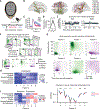
(A) Human eyepuff assay schematic. (B) Single-trial puff-aligned eye closure of example human participant during saline infusion or (C) ketamine infusion (0.5 mg/kg intravenous over 40 min) and (D) summarized across trials (mean ± SEM vertical dashed line, airpuff onset; horizontal bar, airpuff duration; dashed box, time window of interest corresponding to late eye closure, 0.3 to 0.8 s after airpuff onset). (E) Late eye closure, normalized by early eye closure (0.1 to 0.2 s after airpuff onset), decreases during ketamine (n = 4 human participants; each color represents a participant, mean ± SEM across trials). (F) Participants’ descriptions of experiences during eyepuff assay with saline or ketamine infusion. (G) Mouse eyepuff assay schematic. (H) Single-trial eye closure of example mouse during saline infusion and (I) ketamine infusion and (J) summarized across trials (mean ± SEM vertical dashed line, airpuff onset; horizontal bar, airpuff duration; dashed box, time window of interest corresponding to late eye closure, 0.3 to 0.8 s after airpuff onset). (K) Late eye closure, normalized by early eye closure (0.1 to 0.2 s afterpuff onset), decreases during ketamine (n = 5 mice; each color represents one participant, mean ± SEM across trials). (L) Effect of dissociative drugs ketamine (50 mg/kg) and PCP (20 mg/kg) on late, normalized by early, eye closure relative to preinfusion (n = 5 mice; bar height indicates mean). (M) Eye closure across sequence of eight closely spaced puffs, with administration of (M) saline, (N) ketamine, (O) PCP, and (P) general anesthetic dose of ketamine/xylazine cocktail (135 mg/kg ketamine with 15 mg/kg xylazine). (M) to (P) n = 5 mice, mean ± SEM (Q) Summary of ketamine’s cross-species impact on reflexive and affective components of the eyepuff eye closure response. ns (not significant), P-value ≥ 0.05, *P-value < 0.05. **P-value < 0.01. See table S3 for information on statistical analyses and sample sizes. [(A) and (G) partially created with BioRender.com.]
Fig. 2.
Fig. 2.. Human eyepuff-triggered intracranial electrical dynamics exhibit brain-spanning fast and slow components.
(A) Recording simultaneous brain-spanning iEEG with eyepuff assay before, during, and after ketamine infusion. (B) Locations of recording sites. (C) Eye closure response to airpuffs before, during, and after ketamine infusion (n = 7 participants, mean ± SEM). Vertical dashed line indicates time of airpuff onset. (D) Clinician-Administered Dissociative States Scale (CADSS) before, during, and after infusion (n = 7 participants, mean ± SEM). (E) Example preinfusion single-channel event-related potentials from insula (INS) and posteromedial cortex (PMC), with mean shown in teal and single trials shown in gray. (F) Schematic of analysis approach for identifying shared spectrotemporal neural response components across participants and channels, using matrix factorization. Each channel’s response can be represented as a linear combination of factor activations (per-factor weights are “loadings,” denoted as wfactor). (G) Spectrotemporal factors of neural eyepuff response, computed using pre- and post-infusion trials across participants (n = 7) and channels (n = 458), shown from onset of airpuff. (H) Loading of factors shown in G on Yeo7 cortical resting state networks (each row normalized by its sum). (I) Modulation of factor activity by ketamine. Factor loading shown before, during, and after infusion (mean ± SEM across channels), statistics compare infusion versus average of pre- and post-infusion. (J) Change in factor loading by ketamine for each Yeo7 resting state network. Blue indicates higher loading during preinfusion relative to during ketamine. ns, P-value ≥ 0.05. *P-value < 0.05. **P-value < 0.01. ***P-value < 0.001. ****P-value < 0.0001. See table S3 for information on statistical analyses and sample sizes.
Fig. 3.
Fig. 3.. Selective disruption of human regional eyepuff-triggered dynamics by ketamine.
(A) Schematic of permutation cluster test for identification of eyepuff-evoked spectrotemporal clusters that change significantly from preinfusion to ketamine. T-value (colormap) indicates size of change relative to variation across trials. Areas within black outlines indicate significantly changed spectrotemporal clusters. (B) Significantly changed clusters, by Yeo7 resting state network and (C) by brain region. See fig. S7C for full region names. Total number of trials across participants specified in parentheses; regions were sampled in different numbers of participants, yielding different numbers of trials per region. Blue signifies reduced power during ketamine. Vertical dashed lines indicate eyepuff onset. (D) Anatomical distribution of individual cortical contacts with significant changes, overlaid on Human Connectome Project (HCP) cortical parcellation. Representative fine-grained HCP areas with significantly changing contacts are indicated, with corresponding regions in parentheses. See table S3 for information on statistical analyses and sample sizes. ORB, Orbitofrontal Cortex; IFG, Inferior Frontal Gyrus; MFG, Middle Frontal Gyrus; INS, Insular Cortex; ACC, Anterior Cingulate Cortex; MCC, Mid-Cingulate Cortex; PMC, Posteromedial Cortex; MOT, Motor Cortex; SS, Somatosensory Cortex; SMG, Supramarginal Gyrus; PHG, Parahippocampal Gyrus; STG, Superior Temporal Gyrus; STS, Superior Temporal Sulcus; MTG, Middle Temporal Gyrus; ITG, Inferior Temporal Gyrus; TP, Temporal Pole; HIPP, Hippocampus; BG, Basal Ganglia; AMY, Amygdala; THAL ANT, Anterior Thalamus; THAL POS, Posterior Thalamus.
Fig. 4.
Fig. 4.. Brain-wide mouse eyepuff-triggered spiking dynamics have separable fast and slow components.
(A) Recording high-density cellular electrophysiology with eyepuff assay before, during, and after ketamine infusion. (B) Eye closure in response to airpuffs in recorded mice (n = 13 sessions, 10 mice; mean ± 95% CI). (C) Locations of recorded units, colored by the Allen Brain Atlas colormap. (D) Airpuff-triggered cellular activity, z-scored and organized by unsupervised clustering, combining neurons from 13 sessions and 10 mice. Left vertical colorbar, region locations of individual units, colored by Allen Brain Atlas colormap. Vertical dashed lines, airpuff onset and offset. Horizontal black lines, division between unsupervised cluster identities. (E) Average cluster responses to airpuff. (F) Hypothesized information flow from airpuff onset, based on anatomy. (G) Average cluster responses before and during ketamine (n = 13 sessions, 10 mice; mean ± 95% CI). Clusters are numbered following (D) and (E) from top to bottom. (H) Decay time of puff-triggered neural activity by region, ordered according to pre-ketamine decays. (I) Rise time of puff-triggered neural activity. (H) and (I) median ± 95% CI. See table S3 for information on statistical analyses and sample sizes, and table S2 for region names.
Fig. 5.
Fig. 5.. Mouse eyepuff-triggered neural activity accumulates to a persistent state with first order system dynamics.
(A) Eyepuff protocol during neural recording. Each trial consists of a series of eight regularly spaced puffs, with twenty trials preceding and twenty trials following remote ketamine bolus infusion. (B) Eye closure across puff series in recorded mice (n = 11 sessions, 9 mice; mean ± 95% CI). (C) Schematic of coding dimension analysis to identify emotion-like neural population encoding. (D) Emotion-like population activity across puff series (n = 11 sessions, 9 mice; mean ± 95% CI). Dashed lines, airpuffs. The discontinuity at t = 23 s corresponds to the time window used to calculate the coding dimension, whose projection is definitionally high. (E) Comparison of emotion-like activity during the first puff presentation before and during ketamine, quantifying rapid (0 to 0.25 s after onset) and late emotion-like activity (2 to 3 s after first airpuff onset) epochs. Dots indicate sessions (n = 11 sessions, 9 mice). Bars indicate mean ± 95% CI. (F) Two-phase first order model of emotion-like neural state dynamics, as a piecewise time-dependent system with three key components: (i) a step input signal corresponding to each 250-ms eyepuff, which inputs to the system with a saturating positive magnitude S, which is the maximum attainable state value of the system; (ii) a broadcast phase, in which the state is driven upwards by the input at rate τBROADCAST; and (iii) a persistence phase after input offset, in which the state decays at rate τPERSIST. x0 is the state value at each phase transition (input onset or offset), and (Sx0)+ = max(0, Sx0), which enforces that the driving input is non-negative and that the state saturates at magnitude S. (G) Model fit to the emotion-like neural population activity (fitting performed jointly across all 11 sessions, 9 mice). The only parameter set to differ between preinfusion and ketamine conditions is τPERSIST; other component values were fit jointly across preinfusion and ketamine. (H) and (I) Model fit to affective eye closure (masking out eye blinking in each 750-ms window post post puff-onset) for different doses of ketamine and PCP (n = 5 sessions and mice for each condition), with only τPERSIST as a free parameter. The other component values were fit jointly across doses of a given drug. ns, P-value ≥ 0.05. **P-value < 0.01. See table S3 for information on statistical analyses and sample sizes.
Fig. 6.
Fig. 6.. Persistent population dynamics disrupted by ketamine across species.
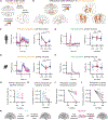
(A) Puff-triggered persistent population is identified for each participant using a coding dimension analysis with simultaneously recorded sites (channels for humans, neurons for mice). Participation in the persistent population is measured as loading on the persistent dimension. (B) Anatomical distribution of the persistent coding dimension across participants (n = 7 human sessions from 7 participants, n = 11 sessions from 9 mice). (C and D) Persistent population activity is decreased by ketamine and then recovers for both humans and mice. Mean ± 95% CI. See Methods for details on time windows and quantification. Vertical dashed lines, airpuff onset. (E and F) Fast population activity is not significantly altered by ketamine in humans (coding dimension and mean activity computed during 50 to 100 ms after puff onset) nor mice (coding dimension and mean activity computed during 0 to 70 ms after puff onset). Mean ± 95% CI. (G and H) Intrinsic timescale at baseline (outside of eyepuff assay) of persistent population is reduced during ketamine relative to preinfusion in humans and mice. (I and J) Coupling at baseline (measured as mean phase locking between channels in humans and mean correlation between neurons in mice) within persistent population is reduced during ketamine relative to preinfusion for humans and mice. See Methods for detail on quantification. (K) Summary of proposed transformation of a brief stimulus into affective state and ketamine’s impact. We use a human brain to illustrate, but the same applies to the mouse brain. ns, P-value ≥ 0.05. *P-value < 0.05. **P-value < 0.01. ***P-value < 0.001. ****P-value < 0.0001. See table S3 for information on statistical analyses and sample sizes.

Comment in

  • A wave of emotion.
    Karamihalev S, Gogolla N. Karamihalev S, et al. Science. 2025 May 29;388(6750):917-918. doi: 10.1126/science.adx8992. Epub 2025 May 29. Science. 2025. PMID: 40440399

References

    1. Bradley MM, Lang PJ, in Cognitive Neuroscience of Emotion (Oxford Univ. Press, 1999), pp. 242–276.
    1. Anderson DJ, Adolphs R, A framework for studying emotions across species. Cell 157, 187–200 (2014). doi: 10.1016/j.cell.2014.03.003 - DOI - PMC - PubMed
    1. Darwin C, The Expression of the Emotions in Man and Animals (John Murray, 1872). doi: 10.1037/10001-000 - DOI
    1. Erickson K, Schulkin J, Facial expressions of emotion: A cognitive neuroscience perspective. Brain Cogn. 52, 52–60 (2003). doi: 10.1016/S0278-2626(03)00008-3 - DOI - PubMed
    1. Panksepp J, The basic emotional circuits of mammalian brains: Do animals have affective lives? Neurosci. Biobehav. Rev 35, 1791–1804 (2011). doi: 10.1016/j.neubiorev.2011.08.003 - DOI - PubMed

LinkOut - more resources