. 2025 May 30;16(1):270.
doi: 10.1186/s13287-025-04394-3.

IFN-α and vitamin B6-induced galectin-9 promotes the immunomodulatory function of human clonal mesenchymal stem cells

Affiliations

IFN-α and vitamin B6-induced galectin-9 promotes the immunomodulatory function of human clonal mesenchymal stem cells

Jee-Hoon Nam et al. Stem Cell Res Ther. .

Abstract

Background: Mesenchymal stem cells (MSCs) possess a variety of immunomodulatory functions that can vary depending on the MSC line. Investigating priming strategies is essential for increasing the immunomodulatory potential of MSCs.

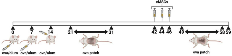
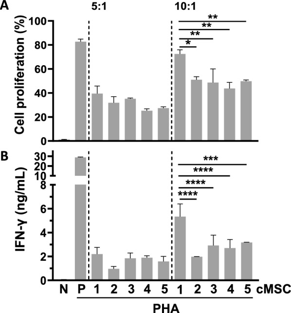
Methods: Human clonal MSCs (cMSCs) were primed with TNF-α, IFN-γ, IL-1β, IFN-α, and vitamin B6. Their immunomodulatory functions, including T-cell proliferation and cytokine production, were analyzed. The primed cMSCs were injected intravenously into a mouse model of ovalbumin-induced atopic dermatitis (AD), and their therapeutic effects were evaluated.

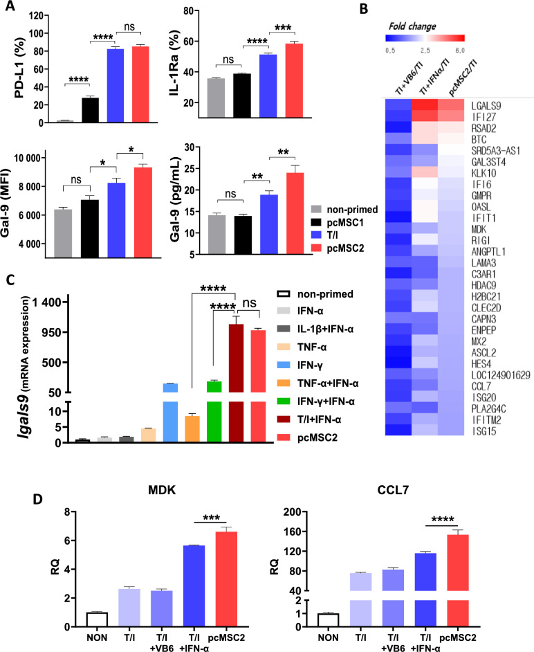
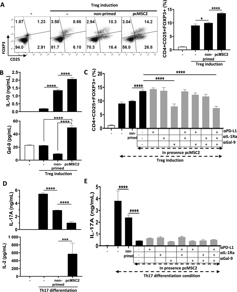
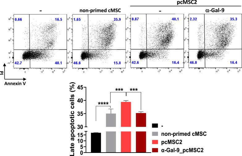
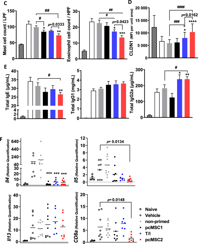
Results: We identified IFN-α and vitamin B6 as promising priming agents when they are combined with TNF-α and IFN-γ. The primed cMSCs showed expression of galectin-9 (Gal-9), IL-1Ra, and PDL-1. Gal-9 facilitates the induction of regulatory T cells (Tregs) and apoptosis. Treatment with primed cMSCs significantly alleviated pathological changes in an AD mouse model. Notable improvements included a reduction in epidermal thickness (p < 0.05), a decreased number of mast cells and eosinophils in the dermis (p < 0.01), restored expression of claudin-1 in the epidermis (p < 0.0001), and lower serum levels of IgE (p < 0.05).

Conclusions: This novel combination of priming factors significantly promotes the immunomodulatory functions of cMSCs by inducing Gal-9. Consequently, Gal-9 may serve as an excellent biomarker for screening primed cMSCs for their immunomodulatory capabilities, facilitating a more accurate assessment of their therapeutic effectiveness.

Keywords: Atopic dermatitis; Clonal mesenchymal stem cell; Galectin-9; IFN-α; Priming; Vitamin B6.


Conflict of interest statement

Declarations. Ethics approval and consent to participate: This study includes samples from both humans and animals. The isolation of human cMSC from bone marrow samples was approved by three institutions: 1. The study titled “Bone Marrow Donation for Harvesting, Clinical Trial Researching, and Culturing of Stem Cell Therapeutics Bone Marrow-Derived Adult Stem Cells from Healthy Donors” received approval from the IRB of the Catholic University of Korea, Seoul St. Mary’s Hospital, with the approval number IRB No. KC15CSSE0336, dated December 14, 2014. 2. The study titled “Bone Marrow Donation for Harvesting and Culturing Bone Marrow-Derived Adult Stem Cells from Healthy Donors” was approved by the IRB of Inha University Hospital, with the approval number IRB No. 10–51, dated October 11, 2010. 3. The study titled “Clinical Grade Bone Marrow Collection for Clinical Research and/or Future Manufacturing of Human Cell Therapy Products” was approved by AllCells (Alameda, CA, USA), with the approval number IRB No. 949–542-3882, dated December 14, 2020. The use of PBMC from healthy donors has been approved. The study titled “Blood Donation from Healthy Donors for Analyzing the Immune Mechanism of Adult Stem Cells” received approval from the IRB of Inha University Hospital, with approval number IRB No. 2014–01-082–019, dated November 14, 2022. Written informed consent was obtained from all study participants. The “Efficacy Evaluation of Cell Therapy for the Treatment of Atopic Dermatitis” study received approval from the IACUC of Gachon University for the mouse experiments. The approval number is IACUC No. LCDI-2023–0054, dated June 14, 2023. Consent for publication: All authors gave consent for publication. Competing interests: The authors have declared no competing interests.

Figures

Fig. 1
Fig. 1
Graphical schematic for the AD model. A schematic diagram illustrating the cMSC injection process following the induction of AD in a mouse model
Fig. 2
Fig. 2
Diversity of the immunomodulatory capacities of various cMSCs. A Flow cytometry analysis of T-cell proliferation. B IFN-γ levels measured by ELISA. N: negative control, P: positive control. The error bars indicate the standard deviation (SD). Two independent experiments were conducted. *p values were determined using one-way ANOVA. Significance levels: *p < 0.05, **p < 0.01, and ****p < 0.0001
Fig. 3
Fig. 3
Screening of priming conditions to promote the immunomodulatory functions of cMSCs. A Expression of immunomodulatory genes in cMSCs after treatment with various cytokines and vitamin B6. B T-cell proliferation by flow cytometry. C–F IFN-γ, TNF-α, IL-5, and IL-17A levels measured by ELISA. Error bars represent standard deviation (SD). *p and #p values represent comparisons with nonprimed and T/I-stimulated cMSCs, respectively. All p values were determined using one-way ANOVA. Significance levels: *p < 0.05, **p < 0.01, ***p < 0.001, ****p < 0.0001, #p < 0.05, ##p < 0.01, and ####p < 0.0001
Fig. 4
Fig. 4
Synergistic effects of IFN-α and Vit B6. A PD-L1, IL-1Ra, and Gal-9 protein levels in pcMSCs analyzed by flow cytometry and ELISA. Gal-9 MFI represents the median. B Heatmap of the 30 most differentially expressed genes in cMSCs under four priming conditions. C, D Gal-9, MDK, and CCL7 mRNA expression in pcMSCs. The error bars represent the standard deviation (SD). Three independent experiments were performed. Significance levels: *p < 0.05, **p < 0.01, ***p < 0.001 and ****p < 0.0001
Fig. 5
Fig. 5
The effects of Gal-9 secreted by pcMSCs on Treg and Th17 cells. A Flow cytometry analysis of CD25⁺Foxp3⁺ Treg cells after coculture with pcMSC2. B IL-10 and Gal-9 expression levels measured by ELISA. C Treg induction in the presence of neutralizing antibodies. D IL-17A and IL-2 levels in Th17 cell differentiation with pcMSC2. E IL-17A expression after treatment with neutralizing antibodies. The error bars represent the standard deviation (SD). Three independent experiments were performed. *p values were determined using one-way ANOVA. Significance levels: *p < 0.05, **p < 0.01, ***p < 0.001 and ****p < 0.0001
Fig. 6
Fig. 6
Effects of Gal-9 secreted by pcMSC2 on CD4 T-cell apoptosis. Flow cytometry analysis of late apoptotic CD4⁺ T cells after coculture with pcMSC2 under CD3/CD28 stimulation, with or without Gal-9 neutralization. The error bars represent the standard deviation (SD). Three independent experiments were performed. *p values were determined using one-way ANOVA. Significance levels: ***p < 0.001 and ****p < 0.0001
Fig. 7
Fig. 7
Primed cMSCs alleviated OVA-induced AD. A Histological analysis of skin tissues by H&E, toluidine blue, Congo red, and IF staining for CLDN1. B, C Quantification of skin thickness, mast cells, and eosinophils. D CLDN1 expression shown as MFI per unit area. E Serum levels of IgE, IgG1, and IgG2a measured by ELISA. F Relative mRNA expression in skin lesions. Data are presented as mean ± SEM. Unpaired t tests were used for statistical analysis: *p values indicate differences from the vehicle group, #p values from the nonprimed group, and the comparison with T/I-primed cMSCs is shown as a numerical p value. Significance levels: *p < 0.05, **p < 0.01, ***p < 0.001, ****p < 0.0001, #p < 0.05, ##p < 0.01, ###p < 0.001 and ####p < 0.0001
Fig. 7
Fig. 7
Primed cMSCs alleviated OVA-induced AD. A Histological analysis of skin tissues by H&E, toluidine blue, Congo red, and IF staining for CLDN1. B, C Quantification of skin thickness, mast cells, and eosinophils. D CLDN1 expression shown as MFI per unit area. E Serum levels of IgE, IgG1, and IgG2a measured by ELISA. F Relative mRNA expression in skin lesions. Data are presented as mean ± SEM. Unpaired t tests were used for statistical analysis: *p values indicate differences from the vehicle group, #p values from the nonprimed group, and the comparison with T/I-primed cMSCs is shown as a numerical p value. Significance levels: *p < 0.05, **p < 0.01, ***p < 0.001, ****p < 0.0001, #p < 0.05, ##p < 0.01, ###p < 0.001 and ####p < 0.0001

Similar articles

References

    1. Huang Y, Wu Q, Tam PKH. Immunomodulatory mechanisms of mesenchymal stem cells and their potential clinical applications. Int J Mol Sci. 2022;23(17):10023. 10.3390/ijms231710023. - PMC - PubMed
    1. Yang G, Fan X, Liu Y, Jie P, Mazhar M, Liu Y, et al. Immunomodulatory mechanisms and therapeutic potential of mesenchymal stem cells. Stem Cell Rev Rep. 2023;19(5):1214–31. - PMC - PubMed
    1. Sharan J, Barmada A, Band N, Liebman E, Prodromos C. First report in a human of successful treatment of asthma with mesenchymal stem cells: a case report with review of literature. Curr Stem Cell Res Ther. 2023;18(7):1026–9. - PubMed
    1. MacLoughlin R. Advancing mesenchymal stem cell therapy for asthma towards clinical translation. Ann Transl Med. 2022;10(20):1083. - PMC - PubMed
    1. Dalal J, Gandy K, Domen J. Role of mesenchymal stem cell therapy in Crohn’s disease. Pediatr Res. 2012;71(4 Pt 2):445–51. - PubMed

MeSH terms

LinkOut - more resources