

Acute Lateral Ankle Sprain Impairs Function and Strength Without Altering Muscle or Tendon Stiffness: A Controlled Observational Study
- PMID: 40442868
- PMCID: PMC12214425
- DOI: 10.1111/os.70082
Acute Lateral Ankle Sprain Impairs Function and Strength Without Altering Muscle or Tendon Stiffness: A Controlled Observational Study
Abstract
Introduction: Acute lateral ankle sprain (LAS) frequently results in persistent functional limitations. Understanding changes in calf muscle and Achilles tendon (AT) stiffness after LAS may shed light on mechanisms underlying impaired function.
Objective: To investigate the effects of acute LAS on the mechanical properties of the calf muscles and the Achilles tendon, ankle function, pain, edema, and strength.
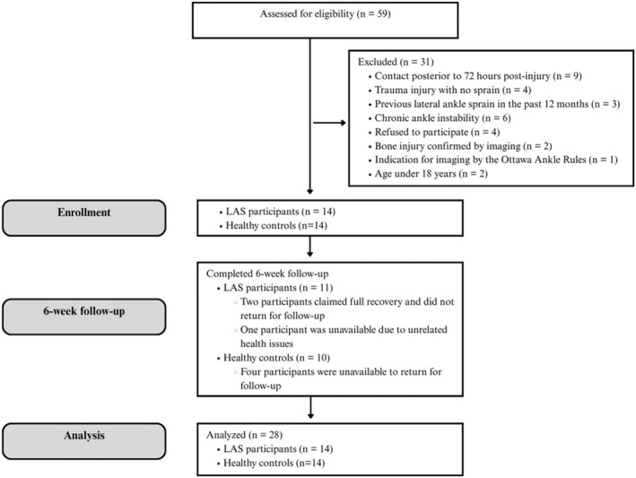
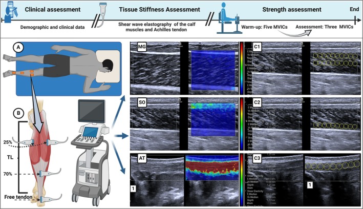
Methods: This controlled observational study was conducted from August 2023 to January 2025. Fourteen participants with acute LAS and 14 healthy controls were evaluated twice, 6 weeks apart. Shear wave elastography (SWE) assessed the stiffness of the triceps surae and AT. Ankle function, pain, and edema were evaluated using the Foot and Ankle Outcome Score, Visual Analog Scale, and figure-of-eight method. Plantar flexion strength was measured via isometric dynamometry.
Results: No significant differences in stiffness were found between or within groups (soleus: p = 0.932; MG: p = 0.760; LG: p = 0.800; AT: p = 0.070), although a time effect (p = 0.005, η2 = 0.269) indicated a general increase in AT stiffness over time (MD = -0.72, p = 0.05, d = 2.86). At baseline, the LAS group exhibited reduced ankle function (MD = 3.43, p < 0.001, d = 2.20), increased pain (MD = 1.88, p < 0.001, d = 1.86), and greater edema (MD = -51.27, p < 0.001, d = -3.58). Over time, improvements were noted in function (MD = -37.04, p < 0.001, d = 2.27), pain (MD = 2.66, p < 0.001, d = -1.31), and edema (MD = 1.07, p = 0.014, d = -0.95), but ankle function remained lower in the LAS group at follow-up (MD = -14.17, p < 0.001, d = -1.79). For plantar flexion strength, no group × time interaction was found (p = 0.745), but a group effect indicated lower peak torque in the LAS group (MD = -32.05, p = 0.012, d = -3.82). A time effect (p < 0.001, η2 = 0.622) showed increased torque across both groups (MD = -18.74, p < 0.001, d = 3.07).
Conclusion: LAS reduces ankle function and leads to pain and edema but does not induce notable changes in calf muscle or AT stiffness within 6 weeks.
Keywords: Achilles tendon; ankle sprains; elastography; functional performance; muscle strength.
© 2025 The Author(s). Orthopaedic Surgery published by Tianjin Hospital and John Wiley & Sons Australia, Ltd.
Conflict of interest statement
The authors declare no conflicts of interest.
Figures
Similar articles
-
Heel kicking exercise rapidly improves pain and function in patients with acute lateral ankle sprain: a randomized controlled trial.BMC Musculoskelet Disord. 2025 Jul 8;26(1):666. doi: 10.1186/s12891-025-08881-9. BMC Musculoskelet Disord. 2025. PMID: 40629319 Free PMC article. Clinical Trial.
-
Physical exercise training interventions for children and young adults during and after treatment for childhood cancer.Cochrane Database Syst Rev. 2016 Mar 31;3(3):CD008796. doi: 10.1002/14651858.CD008796.pub3. Cochrane Database Syst Rev. 2016. PMID: 27030386 Free PMC article.
-
Rehabilitation for ankle fractures in adults.Cochrane Database Syst Rev. 2024 Sep 23;9(9):CD005595. doi: 10.1002/14651858.CD005595.pub4. Cochrane Database Syst Rev. 2024. PMID: 39312389
-
Oral non-steroidal anti-inflammatory drugs versus other oral analgesic agents for acute soft tissue injury.Cochrane Database Syst Rev. 2015 Jul 1;(7):CD007789. doi: 10.1002/14651858.CD007789.pub2. Cochrane Database Syst Rev. 2015. Update in: Cochrane Database Syst Rev. 2020 Aug 12;8:CD007789. doi: 10.1002/14651858.CD007789.pub3. PMID: 26130144 Updated.
-
Braces and orthoses for treating osteoarthritis of the knee.Cochrane Database Syst Rev. 2015 Mar 16;2015(3):CD004020. doi: 10.1002/14651858.CD004020.pub3. Cochrane Database Syst Rev. 2015. PMID: 25773267 Free PMC article.
References
-
- Ivins D., “Acute Ankle Sprain: An Update,” American Family Physician 74, no. 10 (2006): 1714–1720. - PubMed
-
- Doherty C., Delahunt E., Caulfield B., Hertel J., Ryan J., and Bleakley C., “The Incidence and Prevalence of Ankle Sprain Injury: A Systematic Review and meta‐Analysis of Prospective Epidemiological Studies,” Sports Medicine 44, no. 1 (2014): 123–140. - PubMed
Publication types
MeSH terms
Grants and funding
- 000193-00002357/Fundação de Apoio à Pesquisa do Distrito Federal
- 00193-00001132/2024-88/Fundação de Apoio à Pesquisa do Distrito Federal
- 00193-00001261/Fundação de Apoio à Pesquisa do Distrito Federal
- 00193.00000773/2021-72/Fundação de Apoio à Pesquisa do Distrito Federal
- 00193.00000859/2021-3/Fundação de Apoio à Pesquisa do Distrito Federal
- 00193.00001222/2021-26/Fundação de Apoio à Pesquisa do Distrito Federal
- 00193-00001301/2024-80/Fundação de Apoio à Pesquisa do Distrito Federal
- Finance Code 001/Coordenação de Aperfeiçoamento de Pessoal de Nível Superior
- 131422/2023-5/Conselho Nacional de Desenvolvimento Científico e Tecnológico
- 141130/2023-7/Conselho Nacional de Desenvolvimento Científico e Tecnológico
- 309435/2020-0/Conselho Nacional de Desenvolvimento Científico e Tecnológico
- 310269/2021-0/Conselho Nacional de Desenvolvimento Científico e Tecnológico
LinkOut - more resources
Full Text Sources
Medical
