. 2024 Dec 20;3(5):301-309.
doi: 10.1021/cbmi.4c00076. eCollection 2025 May 26.

Screening Anti-Parkinson's Disease Drugs in Living Mouse Brains via a Peroxynitrite-Activated Fluorescent Probe

Affiliations

Screening Anti-Parkinson's Disease Drugs in Living Mouse Brains via a Peroxynitrite-Activated Fluorescent Probe

Jiao Wu et al. Chem Biomed Imaging. .

Abstract

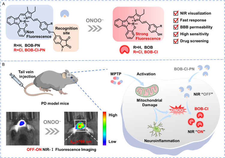
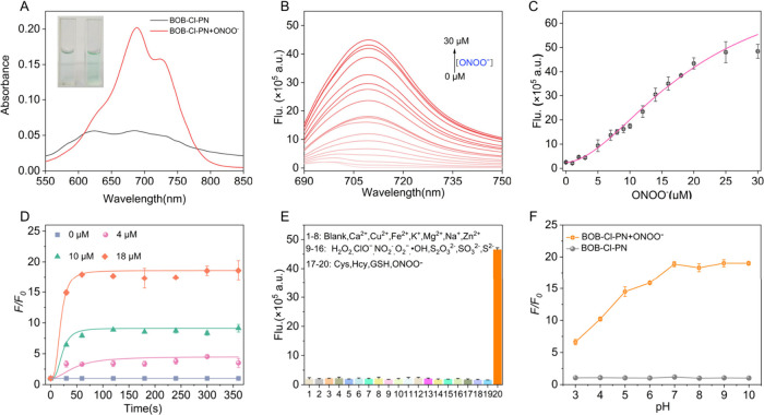
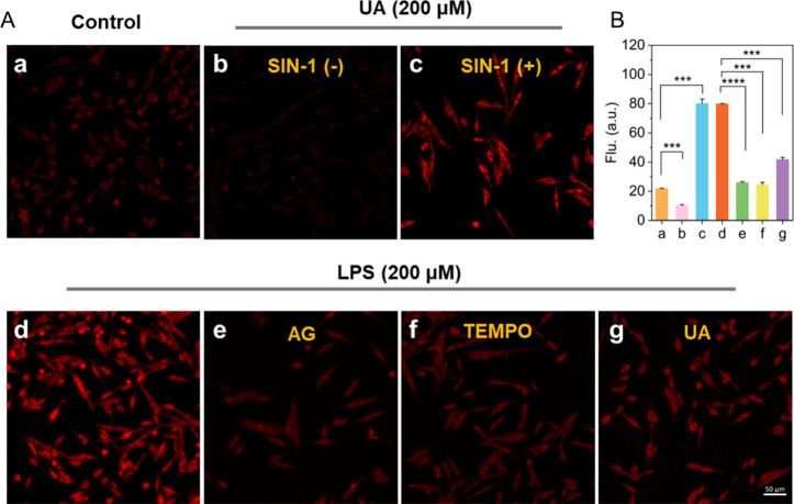
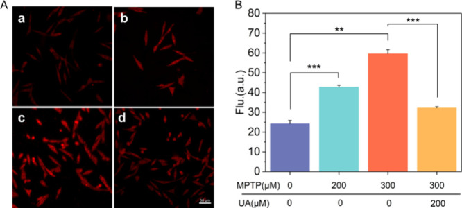
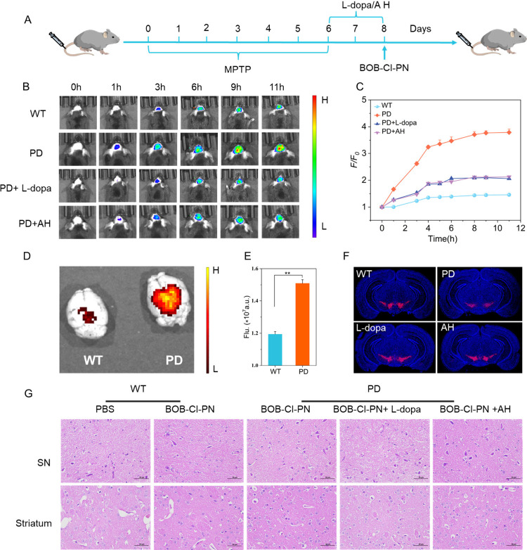
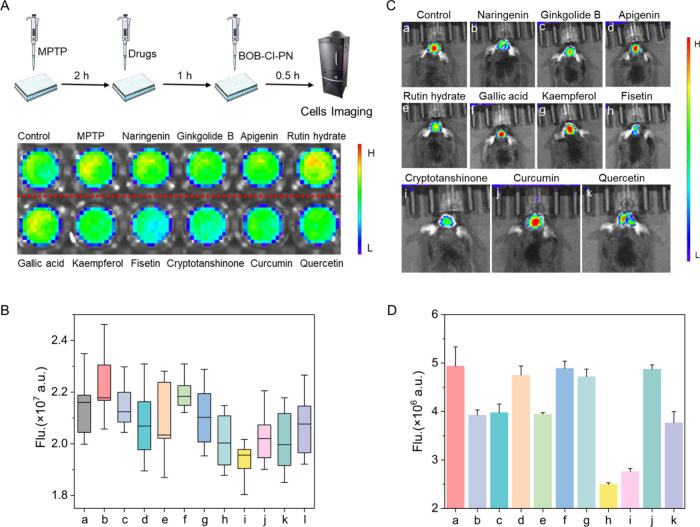
Screening anti-Parkinson's disease (PD) drugs at in vivo brain level is imperative for managing PD yet currently remains unaccomplished. Peroxynitrite (ONOO-) has been implicated in PD progression. Thus, developing in vivo ONOO--based imaging tools for anti-PD drug screening holds promise for early prognosis and treatment of PD. Consequently, a near-infrared (NIR) fluorescence probe, BOB-Cl-PN, with high specificity, good sensitivity (LOD = 24 nM), and rapid response (<60 s), was devised to investigate ONOO- and PD relationships. Utilizing NIR fluorescence imaging, BOB-Cl-PN effectively monitored ONOO- fluctuations in 1-methyl-4-phenyl-1,2,3,6-tetrahydropyridine (MPTP)-induced PD cell models, establishing a cellular high throughput screening (cHTS) system for anti-PD drugs. In live animal imaging, BOB-Cl-PN's ability to penetrate the blood-brain barrier enabled ONOO- flux imaging of PD mouse brains. Moreover, BOB-Cl-PN served as an imaging contrast for in vivo screening of potential traditional Chinese medicines for PD therapy, identifying fisetin as having the best therapeutic index among 10 Chinese medicines. This study constructs a sensitive, efficient imaging contrast for monitoring ONOO- dynamics in PD brains and provides a valuable platform for cellular and in vivo screening of anti-PD drugs.

Keywords: Chinese medicine; Parkinson’s disease; brain disease; fluorescent probe; peroxynitrite.


Figures

1
1. (A) Design Principles of Probe. (B) Mechanism of BOB-Cl-PN Response to ONOO in Parkinson’s Disease Mice
1
1
(A) Absorption spectra of BOB-Cl-PN before and after reacting with ONOO. Inset: color change of BOB-Cl-PN before (left) and after (right) reacting with ONOO. (B) Spectral response of 10 μM BOB-Cl-PN with 0–30 μM ONOO. (C) Relationship of fluorescence intensities and ONOO concentrations. (D) Time course of fluorescence responses of BOB-Cl-PN with 0, 4, 10, 18 μM ONOO. (E) Fluorescence responses of 10 μM BOB-Cl-PN to various biological species including 20 μM 28: Ca2+, Cu2+, Fe2+, K+, Mg2+, Na+, Zn2+; 50 μM 916: H2O2, ClO, NO2 , O2 , OH, S2O3 2–, S2–, SO3 2–, 100 μM 1719: Cys, Hcy, GSH, and 20 μM ONOO. (F) Influences of pH on the fluorescence response of BOB-Cl-PN to ONOO. λexem = 688/708 nm.
2
2
(A) (a) PC12 cells were incubated with BOB-Cl-PN (5 μM) for 0.5 h and then imaged. (b) PC12 cells were pretreated with UA (200 μM) for 0.5 h, then incubated with BOB-Cl-PN (5 μM) for 0.5h for imaging. (c) PC12 cells were pretreated with UA (200 μM) for 0.5 h, then incubated with BOB-Cl-PN (5 μM) and SIN-1 (800 μM) for 0.5 h for imaging. (d) PC12 cells were pretreated with UA (200 μM) for 0.5 h and incubated with LPS (2 μg/mL) for 4 h, then incubated with BOB-Cl-PN (5 μM) for 0.5 h for imaging. (e, f, g) PC12 ells were pretreated with (e) AG (200 μM), (f) TEMPO (200 μM), or (g) UA (200 μM) during stimulation with LPS (2 μg/mL) for 4 h, subsequently incubated with BOB-Cl-PN (5 μM) for 0.5 h and then imaged. (B) Mean intensities in a–g. Scale bar = 50 μm. λex = 633 nm, λem = 650–749 nm. **p < 0.01, ***p < 0.001, ****p < 0.0001.
3
3
(A) NIRF imaging of ONOO generated by the MPTP induced PD cells. 5 μM BOB-Cl-PN loaded cells were co-incubated with (a) 0 μM; (b) 200 μM; (c) 300 μM MPTP; (d) 300 μM MPTP + 200 μM UA. (B) Mean intensities in a–d. Scale bar = 50 μm. λex = 633 nm, λem = 650–749 nm. **p < 0.01, ***p < 0.001, ****p < 0.0001.
4
4
(A) Schematic diagram of the experimental timeline. (B) Fluorescence imaging of ONOO in WT, PD, levodopa (L-dopa)-treated and amantadine hydrochloride (AH)-treated PD mice brains during 11 h via intravenous injection of BOB-Cl-PN (100 μL,10 μM). (C) Quantification of fluorescence signals in (A). (D) Ex vivo imaging of the brain from WT or PD mice postimaging. (E) Ex vivo fluorescence intensities of PD and WT mouse brains. (F) Immunofluorescence staining of substantia nigra in WT and PD mice. Scale bars: 1000 μm. (G) Hematoxylin and eosin staining of the substantia nigra and striatum. Scale bar = 50 μm. NIRF imaging: λex = 680 nm, λem = 780 nm. N = 3, **p < 0.01.
5
5
(A) Fluorescence imaging of PC12 cells treated with ten kinds of traditional Chinese medicines. (B) Box-plot expression of fluorescence intensity in A diagram (a–l: control, MPTP, naringenin, ginkgolide B, apigenin, rutin hydrate, gallic acid, kaempferol, fisetin, cryptotanshinone, curcumin, quercetin). (C) Fluorescence imaging of PD mice treated with Chinese medicine and BOB-Cl-PN. (D) Corresponding fluorescence intensities from (a–k). NIRF imaging: λex = 680 nm, λem = 780 nm.

Similar articles

References

    1. Hou Y., Dan X., Babbar M., Wei Y., Hasselbalch S. G., Croteau D. L., Bohr V. A.. Ageing as a risk factor for neurodegenerative disease. Nat. Rev. Neurol. 2019;15:565–581. doi: 10.1038/s41582-019-0244-7. - DOI - PubMed
    1. Kalia L. V., Lang A. E.. Parkinson’s disease. Lancet. 2015;386:896–912. doi: 10.1016/S0140-6736(14)61393-3. - DOI - PubMed
    1. Kordower J. H., Olanow C. W., Dodiya H. B., Chu Y., Beach T. G., Adler C. H., Halliday G. M., Bartus R. T.. Disease duration and the integrity of the nigrostriatal system in Parkinson’s disease. Brain. 2013;136:2419–2431. doi: 10.1093/brain/awt192. - DOI - PMC - PubMed
    1. Dong-Chen X., Yong C., Yang X., Chen-Yu S., Li-Hua P.. Signaling pathways in Parkinson’s disease: molecular mechanisms and therapeutic interventions. Signal Transduction Targeted Ther. 2023;8:73. doi: 10.1038/s41392-023-01353-3. - DOI - PMC - PubMed
    1. Zhu Y., Ouyang Z., Du H., Wang M., Wang J., Sun H., Kong L., Xu Q., Ma H., Sun Y.. New opportunities and challenges of natural products research: When target identification meets single-cell multiomics. Acta Pharm. Sin. B. 2022;12:4011–4039. doi: 10.1016/j.apsb.2022.08.022. - DOI - PMC - PubMed

LinkOut - more resources