. 2025 May 8.
doi: 10.1039/d5md00316d. Online ahead of print.

Design, synthesis and evaluation of pyrrolobenzodiazepine (PBD)-based PROTAC conjugates for the selective degradation of the NF-κB RelA/p65 subunit

Affiliations

Design, synthesis and evaluation of pyrrolobenzodiazepine (PBD)-based PROTAC conjugates for the selective degradation of the NF-κB RelA/p65 subunit

Peiqin Jin et al. RSC Med Chem. .

Abstract

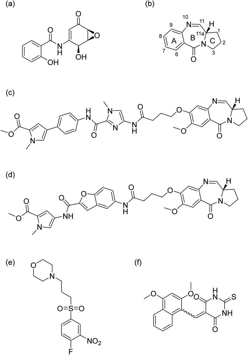
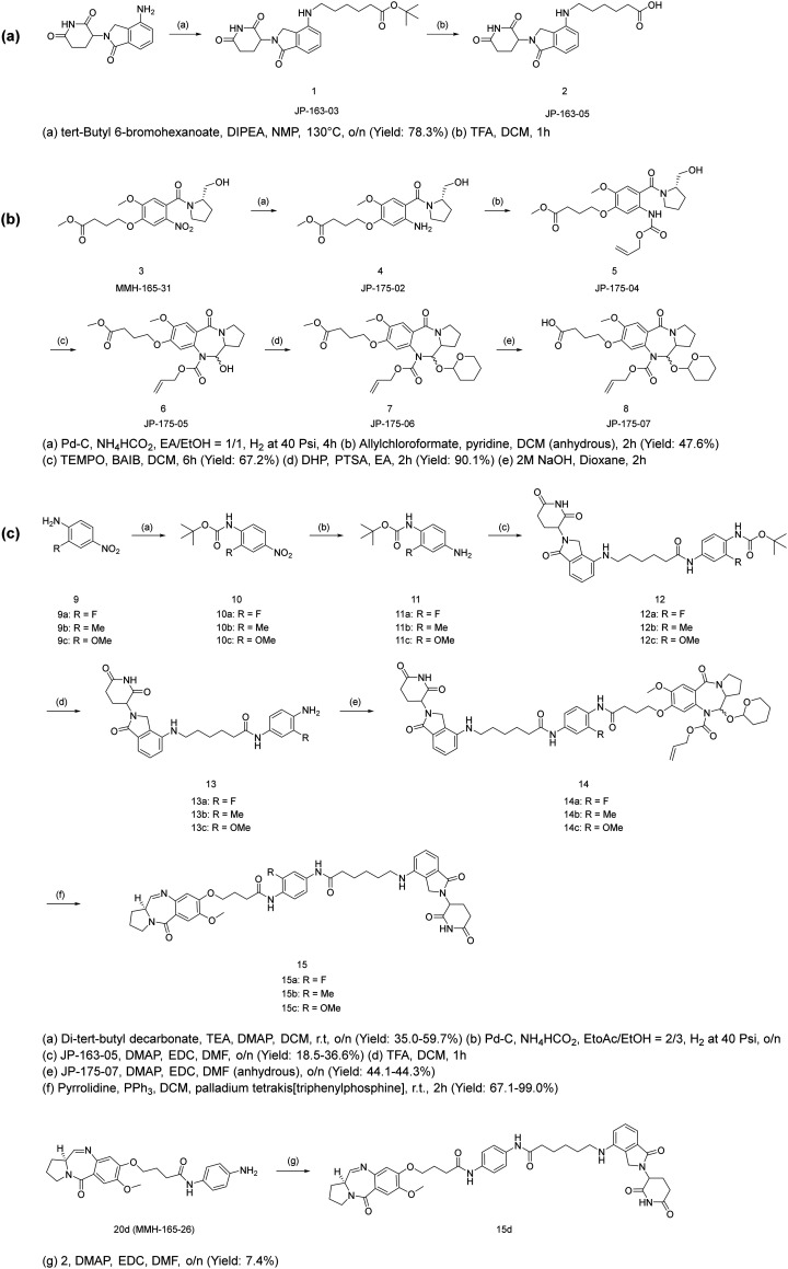
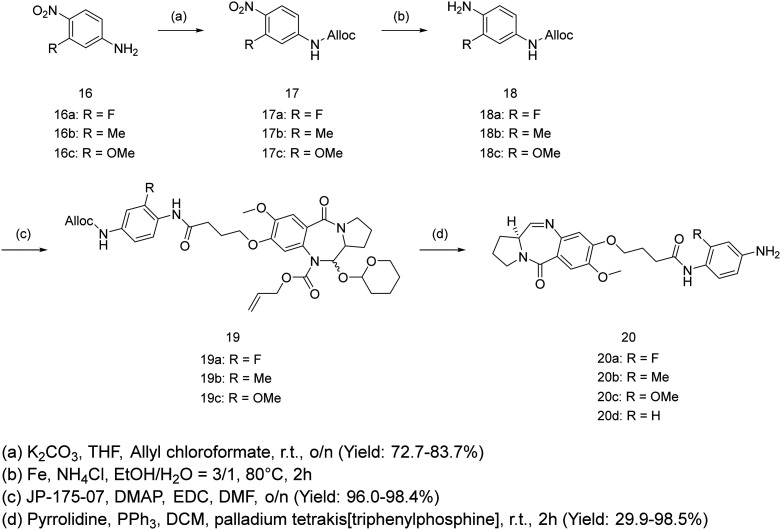
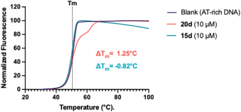
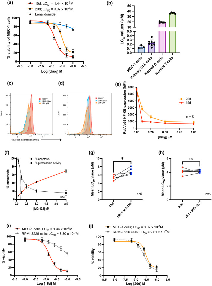
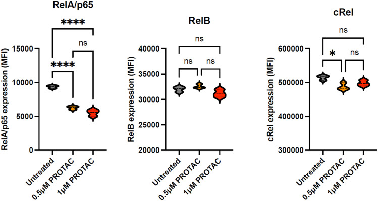
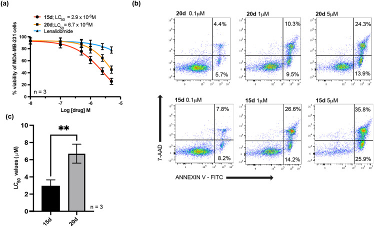
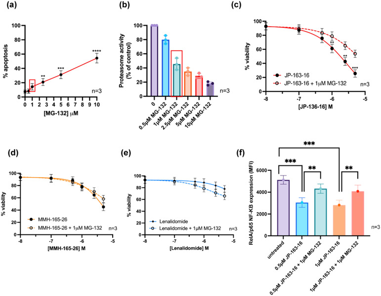
NF-κB signalling is frequently dysregulated in human cancers making it an attractive therapeutic target. Despite concerted efforts to generate NF-κB inhibitors, direct pharmacological inhibition of the kinases mediating canonical NF-κB has failed due to on-target toxicities in normal tissues. So, alternative strategies, designed to target specific components of the NF-κB signalling machinery, have the potential to selectively inhibit tumour cells whilst reducing the toxicities associated with broad inhibition of NF-κB in non-malignant cells. Here we present evidence that a C8-linked pyrrolobenzodiazepine (PBD) containing proteolysis-targeting chimera (PROTAC) selectively degrades the NF-κB subunit, RelA/p65, in a proteasome-dependent manner. Our lead PROTAC (JP-163-16, 15d) showed cytotoxicity with mean LC50 values of 2.9 μM in MDA-MB-231 cells, 0.14 μM in MEC-1 cells and 0.23 μM in primary chronic lymphocytic leukaemia cells. In contrast, 15d was two-logs less toxic in primary B- and T-lymphocytes (mean LD50 19.1 μM and 36.4 μM, respectively). Importantly, the development of 15d, by conjugating the C8-linked PBD with a cereblon-targeting ligand using a five-carbon linker, abolished the ability of the C8-linked PBD to bind to DNA, whilst demonstrating cytotoxicity in cancer cells associated with the degradation of RelA/p65. Mechanistically, 15d displayed PROTAC credentials through the selective degradation of NF-κB RelA/p65 in a proteasome-dependent manner and showed a five-fold reduction in potency in the cereblon deficient, lenalidomide resistant, myeloma cell line, RPMI-8226. To our knowledge, this work describes the first PROTAC capable of selective degradation of a single NF-κB subunit and highlights the therapeutic potential of our strategy for the treatment of RelA/p65-dependent tumours.


Conflict of interest statement

The authors have no relevant conflicts of interest.

Figures

Fig. 1
Fig. 1. Chemical structure of reported NF-κB targeting compounds (a) (−)-DHMEQ, (b) PBD core, (c) KMR-28-39 (TSG-1301), (d) Cpd 13, (e) CRL1101, and (f) IT-901.
Fig. 2
Fig. 2. Docking of PBD-PROTAC with the RelA/p65-p50-DNA complex (PDB:1VKX). (a) Molecular docking of the designed PBD-PROTAC (15d); (b) zoomed in figure showing the 15d binding pose in the RelA/p65-DNA binding interface; (c) overlapped docking results of PBD controls within the RelA/p65-DNA interface; (d) the typical 2D ligand-interface binding projection of PBD ligands. Here fluorine-substituted PBD (JP-193-12) was selected as an example, while the dashed lines highlight the protein–ligand interactions. (e) Chemical structure of the docked PBD building blocks.
Scheme 1
Scheme 1. Synthesis of (a) lenalidomide-based CRBN building block JP-163-05; (b) PBD core; (c) PBD-based PROTACs.
Scheme 2
Scheme 2. Synthesis of PBD controls.
Fig. 3
Fig. 3. FRET melting assay results reveal that the PROTAC (15d) does not significantly interact with DNA. Each compound was mixed with an AT-rich DNA sequence at 10 μM. 15d did not increase the melting temperature suggesting that it does not interact with DNA. In contrast, the PBD building block (20d) caused a marked increase in DNA melting temperature, confirming its ability to bind to and stabilise DNA.
Fig. 4
Fig. 4. (a) Overlaid dose–response curves for 15d, 20d, and lenalidomide in the MEC-1 cell line. Dose–response curves were generated using annexin V/7-AAD data following 48 h of exposure to each compound. The LD50 values were interpolated from each individual dose–response curve using GraphPad Prism 10, with all experiments performed in triplicate. (b) Shows the relative cytotoxic effect of 15d in MEC-1 cells, primary CLL cells and normal B- and T-lymphocytes. Normal lymphocytes were more than two logs less sensitive to the effects of 15d when compared with malignant B cells. RelA/p65 expression was significantly reduced in MEC-1 cells treated for 24 h with (c) 15d and (d) 20d. (e) In contrast to 20d, 15d did not show a dose-dependent reduction in RelA/p65, which suggests an event-driven mechanism of action consistent with other PROTACs. Furthermore, co-treatment with the proteasome inhibitor, MG-132, demonstrated a proteasome-dependent mechanism of action. (f) The cytotoxic and proteasome inhibitory effects of MG-132 were evaluated in MEC-1 cells and a concentration of 0.18 μM was chosen for the subsequent combination studies. (g) 15d shows a proteasome dependent mechanism of action. (h) In contrast, the PBD building block, 20d, showed a proteasome-independent mechanism of action. (i) The cytotoxic effects of 15d were shown to be dependent on CRBN expression by the five-fold reduction in potency in the CRBN deficient myeloma cell line, RPMI-8226. (j) In contrast, the PBD building block, 20d, showed similar potency in RPMI-8226 cells. All LC50 values were interpolated from individual dose–response curves using GraphPad Prism 10. Results are shown as the mean of five independent experiments carried out in duplicate. Statistical significance was determined using paired t-tests * p < 0.05.
Fig. 5
Fig. 5. Comparison the effect of 15d (PROTAC) on the expression of three NF-κB subunits RelA/p65, RelB and cRel. Each subunit was quantified using fluorescence-labelled antibodies; all experiments were performed three times in duplicate and data are presented as violin plots. Statistical significance was determined using the Kruskal–Wallis test with Dunn's multiple comparison post hoc correction. * p < 0.05, ** p < 0.01, *** p < 0.001, **** p < 0.0001.
Fig. 6
Fig. 6. Evaluation of 15d in MDA-MB-231 cells. (a) Dose–response curves for 15d, 20d, and lenalidomide in MDA-MB-231 cells. Cells were treated with a range of concentrations of the PROTAC (15d) and the individual constituent molecules (20d and lenalidomide). Dose–response curves were generated using annexin V/7-AAD data following 48 h of exposure to each compound. The LC50 values were interpolated from each individual dose–response curve using GraphPad Prism 10. All experiments were performed in triplicate. (b) Shows an example of the gating strategy used to identify viable and apoptotic MDA-MB-231 cells. The percentage of viable cells was defined by cells being annexin-V and 7AAD negative. (c) The mean LC50 values (+SD) for 15d and 20d are shown for three independent experiments carried out in triplicate. 15d was significantly more cytotoxic than 20d, ** p < 0.001.
Fig. 7
Fig. 7. The effects of the proteasome inhibitor, MG-132, in the MDA-MB-231 cell line. (a) MG-132 induced dose-dependent cytotoxicity which was (b) associated with a dose-inhibition of proteasomal activity. A dose of 1 μM MG-132 did cause a significant increase in cytotoxicity but induced a >50% reduction in proteasome activity. All experiments were performed in triplicate and statistical significance was determined using the Kruskal–Wallis test with Dunn's multiple comparison post hoc. ** p < 0.01, *** p < 0.001, **** p < 0.0001. (c) Overlaid dose–response curves of MDA-MB-231 cells treated with 15d with and without the addition of the proteasome inhibitor, MG-132. (d) 20d and (e) lenalidomide. The cytotoxic effect of 15d was significantly reduced by co-treatment with 1 μM MG-132. This was not the case for 20d or lenalidomide. All experiments were performed in triplicate and statistical significance was determined using the Kruskal–Wallis test with Dunn's multiple comparison post hoc correction. *** p < 0.001. (f) 15d mediated depletion of RelA/p65 expression in MDA-MB-231 cells was dependent on proteasome activity. Experiments were performed in triplicate and statistical significance was determined using the Kruskal–Wallis test with Dunn's multiple comparison post hoc correction. ** p < 0.01, *** p < 0.001.

Similar articles

References

    1. Liu T. Zhang L. Joo D. Sun S.-C. NF-κB Signaling in Inflammation. Signal Transduction Targeted Ther. 2017;2(1):17023. - PMC - PubMed
    1. Taniguchi K. Karin M. NF-KB, Inflammation, Immunity and Cancer: Coming of Age. Nat. Rev. Immunol. 2018;18(5):309–324. - PubMed
    1. Duran C. L. Karagiannis G. S. Chen X. Sharma V. P. Entenberg D. Condeelis J. S. Oktay M. H. Cooperative NF-KB and Notch1 Signaling Promotes Macrophage-Mediated MenaINV Expression in Breast Cancer. Breast Cancer Res. 2023;25(1):37. - PMC - PubMed
    1. Hewamana S. Alghazal S. Lin T. T. Clement M. Jenkins C. Guzman M. L. Jordan C. T. Neelakantan S. Crooks P. A. Burnett A. K. Pratt G. Fegan C. Rowntree C. Brennan P. Pepper C. The NF-KB Subunit Rel A Is Associated with in Vitro Survival and Clinical Disease Progression in Chronic Lymphocytic Leukemia and Represents a Promising Therapeutic Target. Blood. 2008;111(9):4681–4689. - PubMed
    1. Verzella D. Pescatore A. Capece D. Vecchiotti D. Ursini M. V. Franzoso G. Alesse E. Zazzeroni F. Life, Death, and Autophagy in Cancer: NF-KB Turns up Everywhere. Cell Death Dis. 2020;11(3):210. - PMC - PubMed