Review
. 2025 May 29;6(6):e70226.
doi: 10.1002/mco2.70226. eCollection 2025 Jun.

Lactylation: From Homeostasis to Pathological Implications and Therapeutic Strategies

Affiliations
Review

Lactylation: From Homeostasis to Pathological Implications and Therapeutic Strategies

Xi Chen et al. MedComm (2020). .

Abstract

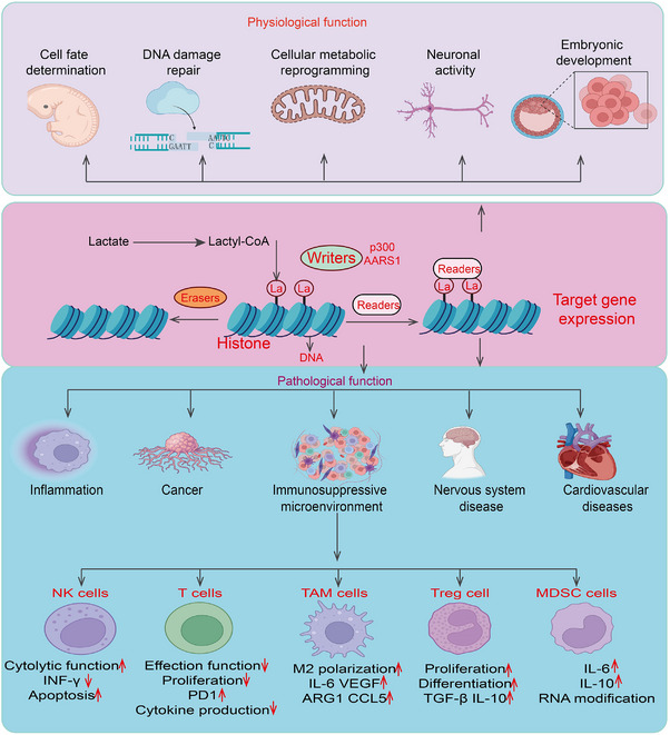
Lactylation, a recently identified post-translational modification, represents a groundbreaking addition to the epigenetic landscape, revealing its pivotal role in gene regulation and metabolic adaptation. Unlike traditional modifications, lactylation directly links metabolic intermediates, such as lactate, to protein function and cellular behavior. Emerging evidence highlights the critical involvement of lactylation in diverse biological processes, including immune response modulation, cellular differentiation, and tumor progression. However, its regulatory mechanisms, biological implications, and disease associations remain poorly understood. This review systematically explores the enzymatic and nonenzymatic mechanisms underlying protein lactylation, shedding light on the interplay between cellular metabolism and epigenetic control. We comprehensively analyze its biological functions in normal physiology, such as immune homeostasis and tissue repair, and its dysregulation in pathological contexts, including cancer, inflammation, and metabolic disorders. Moreover, we discuss advanced detection technologies and potential therapeutic interventions targeting lactylation pathways. By integrating these insights, this review aims to bridge critical knowledge gaps and propose future directions for research. Highlighting lactylation's multifaceted roles in health and disease, this review provides a timely resource for understanding its clinical implications, particularly as a novel target for precision medicine in metabolic and oncological therapies.

Keywords: biological significance; cancer immunotherapy; pathology; physiology; protein lactylation.


Conflict of interest statement

The authors declare no conflicts of interest.

Figures

FIGURE 1
FIGURE 1
Classification of post‐translational modifications of proteins. This schematic diagram illustrates various types of post‐translational modifications (PTMs) that proteins undergo, which significantly impact their structure, function, and interactions. At the center, “Protein post‐translation modification” is highlighted in a pink circle, with arrows pointing outward to different types of PTMs. Each modification is represented by a colored box containing a protein structure with an associated chemical tag: Acetylation (Ac): The addition of an acetyl group (‐COCH₃) to lysine residues, often regulating gene expression by modifying histones. Represented in a red box. SUMOylation (SUMO): The attachment of Small Ubiquitin‐like Modifier (SUMO) proteins, affecting nuclear transport, transcriptional regulation, and protein stability. Shown in an orange box. Lipidation: The addition of lipid moieties, such as fatty acids or prenyl groups, which facilitate membrane association and protein localization. Represented in a green box with a lipid tail symbol. Hydroxylation (‐OH): The enzymatic addition of hydroxyl groups, often occurring on proline or lysine residues, which is crucial for collagen stability and hypoxia signaling. Shown in a dark blue box. Glycosylation: The attachment of carbohydrate groups, influencing protein folding, stability, and cell−cell communication. Represented in a magenta box with a glycan structure. Disulfide bond (La): The formation of covalent bonds between cysteine residues, stabilizing protein tertiary and quaternary structures. Displayed in a cyan box. Ubiquitination (Ub): The covalent attachment of ubiquitin molecules, marking proteins for degradation via the proteasome or regulating cellular signaling. Shown in a brown box. Methylation (Me): The addition of methyl groups to lysine or arginine residues, influencing chromatin structure and transcriptional activity. Represented in a green box. Phosphorylation (P): The addition of phosphate groups to serine, threonine, or tyrosine residues, playing a key role in signal transduction and cellular regulation. Displayed in a light yellow box.
FIGURE 2
FIGURE 2
The role of nonhistone lactylation in cancer progression and chemoresistance. This schematic diagram illustrates the effects of nonhistone lactylation on key cancer‐related proteins, including p53, YAP, NBS1, and MRE11, highlighting its role in cancer progression and chemoresistance. The lactylation of p53 reduces phase separation, leading to decreased p53 activity. As a tumor suppressor, reduced p53 activity contributes to cancer progression. n: Lactylation of YAP enhances its protein stability, leading to increased YAP activity. YAP is a key effector in the Hippo signaling pathway, and its activation promotes cancer progression. The lactylation of NBS1 enhances its DNA‐binding ability, facilitating homologous recombination‐mediated DNA damage repair. This process contributes to cancer cell survival and chemoresistance. The lactylation of MRE11 enhances DNA end resection, promoting DNA damage repair through homologous recombination. This mechanism supports cancer cell survival under therapeutic stress, leading to chemoresistance.
FIGURE 3
FIGURE 3
The physiological functions and pathological function of lactylation in process. Histone Lactylation Mechanism: Lactate is converted to lactyl‐CoA, which serves as a substrate for histone lactylation. Writers (e.g., p300, AARS1) catalyze the addition of lactyl groups to histones, while erasers remove these modifications. Readers recognize lactylated histones, leading to altered gene expression. Physiological Functions: Histone lactylation plays essential roles in cell fate determination, DNA damage repair, cellular metabolic reprogramming, neuronal activity, and embryonic development. These functions are crucial for normal cellular and organismal homeostasis. Pathological Functions: Aberrant histone lactylation is implicated in various diseases, including inflammation, cancer, immunosuppressive microenvironments, nervous system diseases, and cardiovascular disorders. Immune Regulation: Histone lactylation influences immune cell function: NK Cells: Decreased cytolytic function, reduced IFN‐γ production, and increased apoptosis. T Cells: Reduced effector function, decreased proliferation and cytokine production, and upregulated PD‐1 expression. Tumor‐Associated Macrophages (TAMs): Promotes M2 polarization, upregulating IL‐6, VEGF, ARG1, and CCL5, which contribute to an immunosuppressive microenvironment. Regulatory T Cells (Tregs): Enhances proliferation, differentiation, and secretion of immunosuppressive cytokines (TGF‐β, IL‐10). Myeloid‐Derived Suppressor Cells (MDSCs): Increases IL‐6 and IL‐10 expression and promotes RNA modification, further suppressing anti‐tumor immunity.

Similar articles

References

    1. Ye L., Jiang Y., and Zhang M., “Crosstalk Between Glucose Metabolism, Lactate Production and Immune Response Modulation,” Cytokine & Growth Factor Reviews 68 (2022): 81–92. - PubMed
    1. Zhang D., Tang Z., Huang H., et al., “Metabolic Regulation of Gene Expression by Histone Lactylation,” Nature 574, no. 7779 (2019): 575–580. - PMC - PubMed
    1. Walton Z. E., Patel C. H., Brooks R. C., et al., “Acid Suspends the Circadian Clock in Hypoxia Through Inhibition of mTOR,” Cell 174, no. 1 (2018): 72–87.e32. - PMC - PubMed
    1. Xie Y., Hu H., Liu M., et al., “The Role and Mechanism of Histone Lactylation in Health and Diseases,” Frontiers in Genetics 13 (2022): 949252. - PMC - PubMed
    1. Li X., Yu T., Li X., He X., Zhang B., and Yang Y., “Role of Novel Protein Acylation Modifications in Immunity and Its Related Diseases,” Immunology 173, no. 1 (2024): 53–75. - PubMed

LinkOut - more resources