. 2025 Mar 3;3(5):261-271.
doi: 10.1021/prechem.4c00092. eCollection 2025 May 26.

3D Printed Microfluidic Devices for Integrated Immunoaffinity Extraction, Solid-Phase Extraction, and Fluorescent Labeling of Preterm Birth Biomarkers

Affiliations

3D Printed Microfluidic Devices for Integrated Immunoaffinity Extraction, Solid-Phase Extraction, and Fluorescent Labeling of Preterm Birth Biomarkers

James D Holladay et al. Precis Chem. .

Abstract

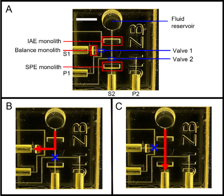
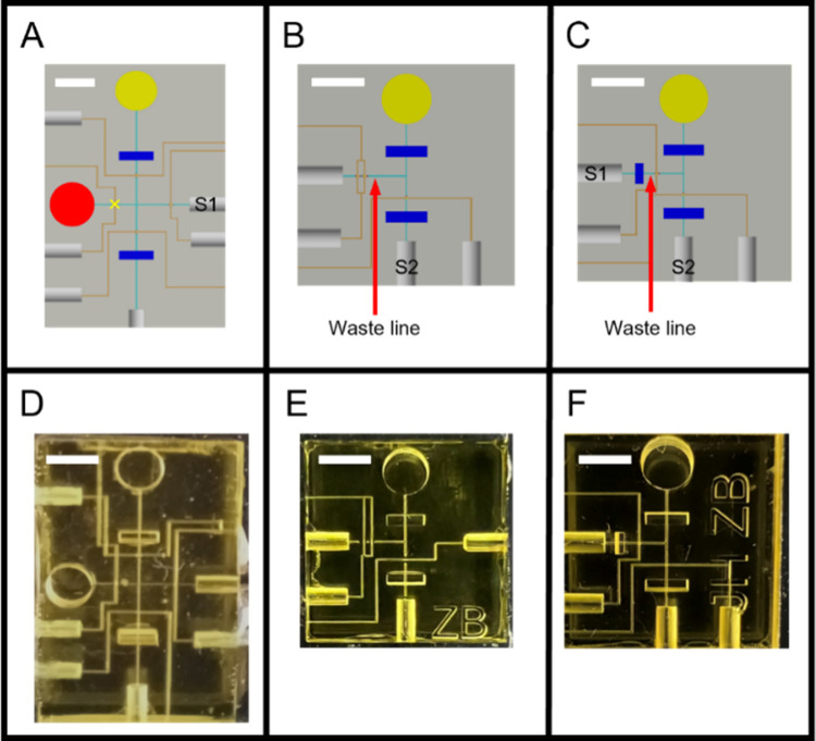
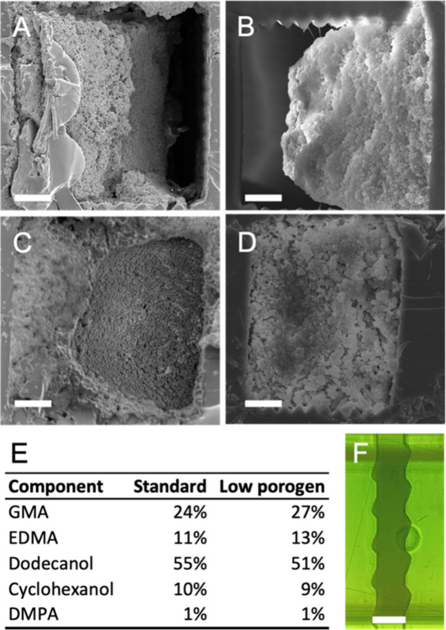
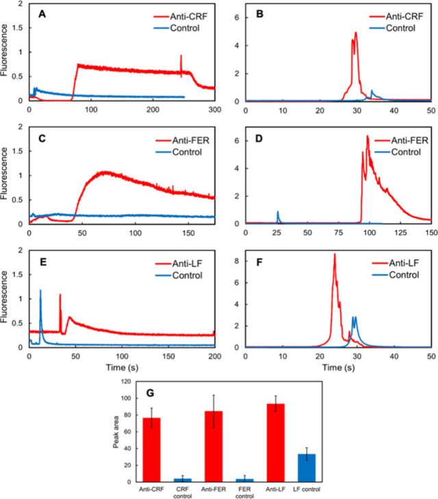
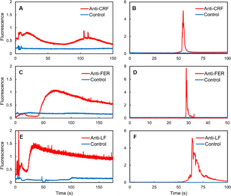
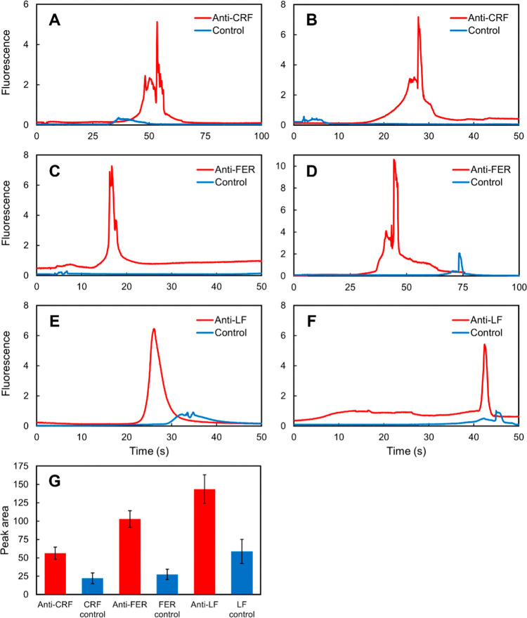
A miniaturized, biomarker-based diagnostic for preterm birth (PTB) risk will require multiple sample preparation steps to be integrated in a single platform. To this end, we created a 3D printed microfluidic device that combines immunoaffinity extraction (IAE), solid-phase extraction (SPE), and fluorescent labeling. This device uses an antibody-functionalized IAE monolith to selectively extract PTB biomarkers, a lauryl methacrylate reverse-phase SPE monolith to concentrate and facilitate fluorescent labeling of PTB biomarkers, and 3D printed valves to control flow through the monoliths. The advantageous iterative design process for arriving at a functional device is documented. The IAE/SPE device performed selective, reproducible extractions of three PTB biomarkers from buffer and depleted maternal blood serum, demonstrating its utility for single-biomarker and multiplexed extractions. After tandem extraction and fluorescent labeling, biomarkers eluted from the SPE monolith in a concentrated plug, facilitating future integration with downstream analysis techniques including microchip electrophoresis. This device effectively combines and automates orthogonal chromatographic extraction methods and constitutes a substantial step toward a complete microfluidic PTB prediction platform.

Keywords: 3D printing; lab-on-a-chip; microfluidics; molecular diagnostics; monoliths; point-of-care diagnostics.


Figures

1
1
Schematic of integrated IAE/SPE device. (A) Device features. S1, S2: vacuum (suction) ports 1 and 2. P1, P2: pneumatic ports 1 and 2. Scale bar is 3 mm. (B) Device set to direct flow through the IAE and balance monoliths. Pressure is applied to P2, closing valve 2. Vacuum applied through S1. (C) Directing flow through IAE and SPE monoliths. Valve 1 is closed by pressure through P1, vacuum applied through S2.
2
2
IAE/SPE integrated device design progression. (A–C) Top views of the CAD schematics for a few iterations of the integrated device. (D–F) Photographs of 3D printed devices. (A, D) Initial dual-reservoir, 4-valve design with 50 μm × 50 μm channels. SPE reservoir marked in red, valve 1 marked with a yellow “×”. (B, E) Compact, simplified design with 100 μm × 100 μm channels and two valves on the waste line. (C, F) Final design with 100 μm × 100 μm channels and a balance monolith on the waste line. Scale bars are 3 mm.
3
3
Optimizing monoliths for 100 μm × 100 μm channels. (A-D) SEM micrographs of monoliths polymerized in 100 μm × 100 μm channels taken at 800× magnification, 20 μm scale bars for reference. (A) IAE and (B) SPE monoliths created using formulations optimized for 50 μm × 50 μm channels, leading to incomplete monolith adhesion to channel walls. (C) IAE monolith created using low-porogen monomer mixture. (D) SPE monolith using previous formulation polymerized in a serrated monolith window. (E) Table of IAE monolith compositions. (F) Micrograph of SPE monolith in serrated channel, with 100 μm scale bar for reference. Monoliths were polymerized in a five-monolith test device (Figure S1).
4
4
IAE and SPE of prelabeled biomarkers in buffer using the integrated IAE/SPE device. IAE extractions were performed using either an IAE monolith functionalized with antibodies or a Tris-blocked control. (A, C, E) Fluorescence detection of biomarkers eluted from IAE monoliths after extraction from buffer. The eluted biomarkers were recaptured by the SPE monolith. (B, D, F) Fluorescence detection of biomarkers eluted from the SPE monolith. (G) SPE elution peak areas for antibody-functionalized and control devices, measured during the 90% ACN elution step after IAE and SPE. Error bars represent the standard deviation of three replicates. Prelabeled 375 nM CRF, 25 nM ferritin, or 12.5 nM lactoferrin was used for these experiments.
5
5
IAE and SPE of prelabeled biomarkers in depleted serum using the integrated IAE/SPE device. IAE extractions were performed using either an IAE monolith functionalized with antibodies or a Tris-blocked control. (A, C, E) Fluorescence detection of biomarkers eluted from IAE monoliths after extraction from depleted serum. The eluted biomarkers were recaptured by the SPE monolith. (B, D, F) Fluorescence detection of biomarkers eluted from the SPE monolith. Prelabeled 375 nM CRF, 25 nM ferritin, or 12.5 nM lactoferrin in depleted blood serum used for these experiments.
6
6
On-column labeling of biomarkers following IAE and SPE. IAE was performed using either an IAE monolith functionalized with antibodies or a Tris-blocked control. (A, C, E) Fluorescence detection of biomarkers eluted from the SPE monolith after IAE extraction from buffer, recapture on SPE monolith, and on-column labeling. (B, D, F) Fluorescence detection of biomarkers eluted from the SPE monolith after IAE extraction from depleted blood serum, recapture on SPE monolith, and on-column labeling. (G) SPE elution peak areas for antibody-functionalized and control devices, measured for extraction from buffer during the 90% ACN elution step after IAE and SPE. Error bars represent the standard deviation of three replicates. Unlabeled 375 nM CRF, 25 nM ferritin, or 12.5 nM lactoferrin used for these experiments.
7
7
Elution from an SPE monolith following multiplexed IAE extraction. IAE monoliths were functionalized with anti-CRF, antiferritin, and antilactoferrin antibodies. Fluorescence was measured during SPE elution following IAE/SPE of mixtures of biomarkers. Prelabeled 187.5 nM CRF, 12.5 nM ferritin, and 6.3 nM lactoferrin used for these experiments. The initial 50 s of baseline data before each peak were trimmed to allow for clearer comparisons between runs. Tests were performed in random order on a single device.

Similar articles

References

    1. Ohuma E. O., Moller A.-B., Bradley E., Chakwera S., Hussain-Alkhateeb L., Lewin A., Okwaraji Y. B., Mahanani W. R., Johansson E. W., Lavin T.. et al. National, regional, and global estimates of preterm birth in 2020, with trends from 2010: a systematic analysis. Lancet. 2023;402(10409):1261–1271. doi: 10.1016/S0140-6736(23)00878-4. - DOI - PubMed
    1. Patel S. S., Ludmir J.. Drugs for the Treatment and Prevention of Preterm Labor. Clin Perinatol. 2019;46(2):159–172. doi: 10.1016/j.clp.2019.02.001. - DOI - PubMed
    1. Khandre V., Potdar J., Keerti A.. Preterm Birth: An Overview. Cureus. 2022;14(12):e33006. doi: 10.7759/cureus.33006. - DOI - PMC - PubMed
    1. Luechathananon S., Songthamwat M., Chaiyarach S.. Uterocervical Angle and Cervical Length as a Tool to Predict Preterm Birth in Threatened Preterm Labor. Int. J. Womens Health. 2021;13:153–159. doi: 10.2147/IJWH.S283132. - DOI - PMC - PubMed
    1. Olarinoye A. O., Olaomo N. O., Adesina K. T., Ezeoke G. G., Aboyeji A. P.. Comparative diagnosis of premature rupture of membrane by nitrazine test, urea, and creatinine estimation. Int. J. Health Sci. (Qassim) 2021;15(6):16–22. - PMC - PubMed

LinkOut - more resources