. 2025 May 9;5(5):2172-2185.
doi: 10.1021/jacsau.5c00146. eCollection 2025 May 26.

Decoupling Global and Local Structural Changes in Self-aminoacylating Ribozymes Reveals the Critical Role of Local Structural Dynamics in Ribozyme Activity

Affiliations

Decoupling Global and Local Structural Changes in Self-aminoacylating Ribozymes Reveals the Critical Role of Local Structural Dynamics in Ribozyme Activity

Yu-Kai Cheng et al. JACS Au. .

Abstract

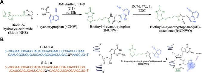
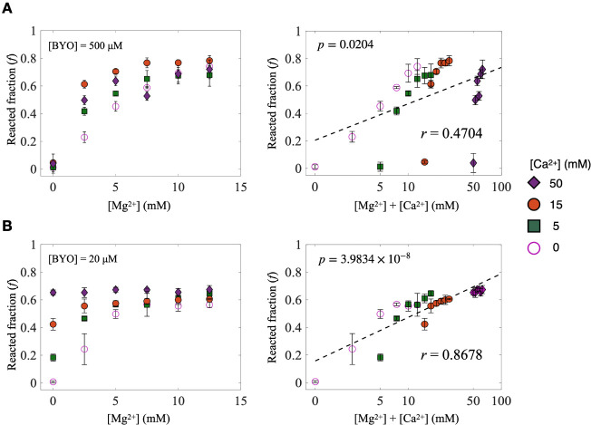
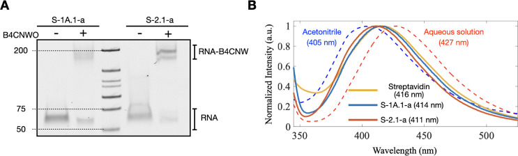
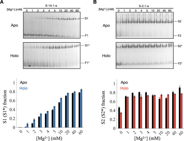
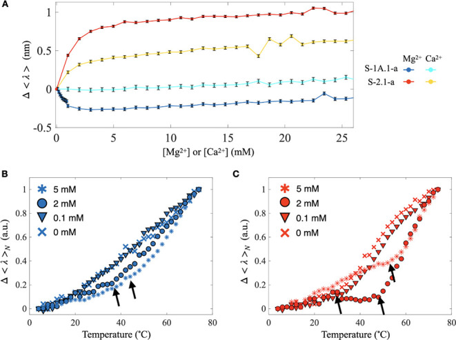
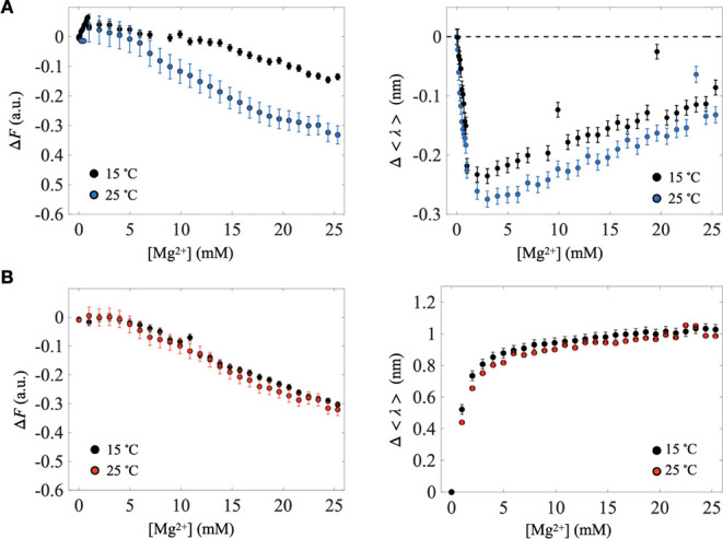
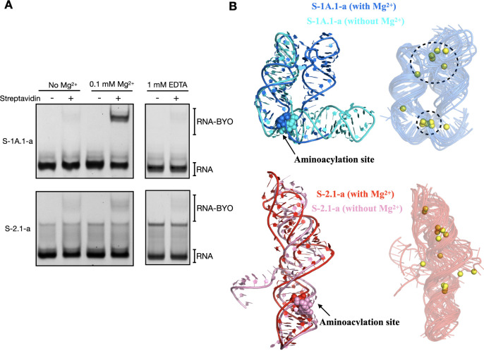
Self-aminoacylating ribozymes catalyze the attachment of amino acids to RNA, serving as pivotal models to investigate the catalytic roles of RNA in prebiotic evolution. In this study, we investigated how divalent metal ions (Mg2+ and Ca2+) modulate local and global structures in two such ribozymes, S-1A.1-a and S-2.1-a, using 4-cyanotryptophan (4CNW) fluorescence and native gel electrophoresis. By tracking 4CNW fluorescence changes at varying concentrations of Mg2+ and Ca2+ and temperatures, we determined how these ions influence the catalytic sites and overall conformations of the ribozymes. Our findings reveal that Mg2+ specifically binds to S-1A.1-a at low concentrations, stabilizing the local structure around the aminoacylation site and causing the site to become more buried, which is essential for catalytic activity. Although higher Mg2+ and Ca2+ concentrations induce global structural rearrangements, these shifts have minimal impact on the local environment of the aminoacylation site, underscoring the dominance of local structural stability in sustaining ribozyme function. In contrast, the activity of S-2.1-a effectively adapts to both Mg2+ and Ca2+, and its fluorescence results indicate a more solvent-exposed aminoacylation site. Overall, these data highlight that local structural changes in the ribozyme's catalytic core are more critical for its function than global conformational shifts. Our study highlights the importance of local environmental changes in ion-dependent ribozyme catalysis and provides insights into the molecular mechanisms of self-aminoacylating ribozymes.

Keywords: 4-cyanotryptophan (4CNW) fluorescence; divalent metal ions; local and global structural dynamics; ribozyme catalysis; self-aminoacylating ribozymes.


Figures

1
1
Synthesis and utilization of Biotinyl-4-cyanotryptophan-5­(4H)-oxazolone for probing the RNA aminoacylation site local environment. (A) Synthesis procedure of Biotinyl-4-cyanotryptophan-5­(4H)-oxazolone (B4CNWO). (B) Left: sequences of ribozymes S-1A.1-a and S-2.1-a, with labeling sites G65 and G54, respectively, highlighted in black. Right: B4CNWO binds to the 2′OH group of the RNA, yielding B4CNW conjugated RNA (RNA-B4CNW). The 4-cyanoindole group emits fluorescence upon excitation at 302 nm.
2
2
Dependence of S-1A.1-a and S-2.1-a activity on [Mg2+] and [Ca2+]. (A) Left: reacted fraction of S-1A.1-a with [BYO] = 500 μM as a function of [Mg2+] at different [Ca2+] (0, 5, 15, 50 mM). Right: the same data plotted with the sum of [Mg2+] and [Ca2+] concentrations on the x-axis in a logarithmic scale. (B) Left: reacted fraction of S-2.1-a with [BYO] = 20 μM as a function of [Mg2+] at different [Ca2+] (0, 5, 15, 50 mM). Right: the same data plotted with the sum of [Mg2+] and [Ca2+] concentrations on the x-axis in a logarithmic scale. The best linear fits (dashed line) are displayed. The calculated Pearson coefficients (r) and the p-value are shown. The error bars represent the standard deviation of the mean (n = 3).
3
3
Streptavidin gel-shift assay and fluorescence spectra of B4CNW-conjugated ribozymes. (A) Streptavidin gel-shift assay results confirm the conjugation of B4CNWO to S-1A.1-a and S-2.1-a. The shift in the RNA band upon conjugation with B4CNW (RNA-B4CNW) indicates successful labeling. (B) Fluorescence spectra of 4CNW in different environments. The dashed lines represent B4CNW in acetonitrile (blue) and aqueous solutions (red). The solid lines show the fluorescence spectra of B4CNW conjugated S-1A.1-a (blue), S-2.1-a (red), and B4CNW bound to streptavidin (yellow). The wavelength of the fluorescence peak for each condition is shown. The divalent ion concentration was zero for the B4CNW-conjugated ribozyme samples. The distinct spectra demonstrate the sensitivity of 4CNW to different environments.
4
4
Mg2+-induced shifts in the structural equilibrium of S-1A.1-a and S-2.1-a ribozymes conformations. Mg-native PAGE analysis of (A) S-1A.1-a and (B) S-2.1-a at varying Mg2+ concentrations (0–60 mM). The top gels show the unconjugated (apo) RNA sample, and the bottom gel shows the B4CNW conjugated (holo) RNA sample. The major bands are labeled as the fast-migrating (F1, F2, F1*, and F2*) and the slow-migrating bands (S1, S2, S1*, and S2*). The asterisk in the gel denotes unreacted F1 in the holo-form sample. The bar charts below quantify the fraction of the slow-migrating band within the total RNA in a lane, representing the proportion of this structural state relative to the total population. The error bars represent the standard deviation of the mean (n = 3). The data indicate that both ribozymes undergo structural transitions to bent conformations in response to Mg2+, suggesting a shift in structural equilibrium. The results show that the conjugation of B4CNW with ribozymes does not significantly perturb the overall structural equilibrium. The error bars represent the standard deviation among triplicate measurements.
5
5
Fluorescence analysis of the local environment of the aminoacylation site in B4CNW conjugated S-1A.1-a and S-2.1-a. (A) Change in average fluorescence wavelength, Δ⟨λ⟩, of B4CNW conjugated S-1A.1-a and S-2.1-a titrated with Mg2+ and Ca2+ at 25 °C. Increasing [Mg2+] results in a blueshift for S-1A.1-a (blue) and a redshift for S-2.1-a (red). In contrast, Ca2+ titration results in minimal change for S-1A.1-a (cyan) and a relatively minor redshift for S-2.1-a (yellow). The error bars represent the standard deviation of the mean (n = 5). (B,C) The normalized change in average fluorescence wavelength, Δ⟨λ⟩ N , for S-1A.1-a (red) and S-2.1-a (blue) at varying [Mg2+] (0, 0.1, 2, and 5 mM) with temperature changes. The change in slope is denoted by arrows, representing the onset of thermally induced unfolding temperature (T x ), determined as described in Materials and Methods. Detailed analysis is shown in Figure S14.
6
6
Temperature-dependent fluorescence analysis with Mg2+ titration. The relative peak fluorescence intensity (ΔF, left) and average fluorescence wavelength change (Δ⟨λ⟩, right) as a function of [Mg2+] at 15 and 25 °C for (A) S-1A.1-a and (B) S-2.1-a. For S-1A.1-a, the decrease in peak intensity indicates Mg2+-induced quenching, which is reduced at 15 °C, suggesting a dynamic quenching mechanism. The blue shift in Δ⟨λ⟩ implies the burial of the 4CNW fluorescence group within the RNA tertiary structure of S-1A.1-a supporting the dynamic quenching mechanism. For S-2.1-a, the reduction in relative peak fluorescence intensity with increasing [Mg2+] also indicates Mg2+-induced quenching but with no observable temperature dependence, suggesting a nondynamic quenching mechanism. The red shift in Δ⟨λ⟩ implies the exposure of the 4CNW fluorescence group for S-2.1-a. The trends of Δ⟨λ⟩ at two different temperatures are similar, indicating a consistent environmental change of the aminoacylation site to Mg2+ within this temperature range. The error bars represent the standard deviation of the mean (n = 5).
7
7
The activity of S-1A.1-a and S-2.1-a at low [Mg2+] and their structure predictions. (A) The streptavidin gel-shift assay shows the reacted fractions of S-1A.1-a (top) and S-2.1-a (bottom) with [BYO] = 500 μM at 4 °C in the absence of Mg2+, with 0.1 mM Mg2+, and with 1 mM EDTA. (B) AlphaFold 3 predicted structures of S-1A.1-a (top) and S-2.1-a (bottom) with (blue and red) and without (cyan and pink) Mg2+ are shown. The aminoacylation sites are highlighted in spheres. The predicted structures demonstrate the conformational changes induced by Mg2+ binding. The corresponding two-dimensional drawings created with RiboDraw are displayed in Figure S16. Ten predicted structures of each ribozyme with Mg2+ (yellow) from two different seeds (one with a single Mg2+ and the other with two Mg2+) were aligned. The clustered Mg2+ binding sites in S-1A.1-a are highlighted.

Similar articles

References

    1. Pressman A., Blanco C., Chen I. A.. The RNA World as a Model System to Study the Origin of Life. Curr. Biol. 2015;25(19):R953–R963. doi: 10.1016/j.cub.2015.06.016. - DOI - PubMed
    1. Robertson M. P., Joyce G. F.. The Origins of the RNA World. Cold Spring Harbor Perspect. Biol. 2012;4(5):a003608–a003608. doi: 10.1101/cshperspect.a003608. - DOI - PMC - PubMed
    1. Ishida S., Terasaka N., Katoh T., Suga H.. An aminoacylation ribozyme evolved from a natural tRNA-sensing T-box riboswitch. Nat. Chem. Biol. 2020;16(6):702–709. doi: 10.1038/s41589-020-0500-6. - DOI - PubMed
    1. Radakovic A., DasGupta S., Wright T. H., Aitken H. R. M., Szostak J. W.. Nonenzymatic assembly of active chimeric ribozymes from aminoacylated RNA oligonucleotides. Proc. Natl. Acad. Sci. U. S. A. 2022;119(7):e2116840119. doi: 10.1073/pnas.2116840119. - DOI - PMC - PubMed
    1. Su M., Schmitt C., Liu Z., Roberts S. J., Liu K. C., Röder K., Jäschke A., Wales D. J., Sutherland J. D.. Triplet-Encoded Prebiotic RNA Aminoacylation. J. Am. Chem. Soc. 2023;145(29):15971–15980. doi: 10.1021/jacs.3c03931. - DOI - PMC - PubMed

LinkOut - more resources