. 2025 Jan-Dec:21:17448069251348778.
doi: 10.1177/17448069251348778. Epub 2025 May 30.

Administration of 2-deoxy-D-glucose alleviates cancer-induced bone pain by suppressing microglial polarization to the M1 phenotype and neuroinflammation

Affiliations

Administration of 2-deoxy-D-glucose alleviates cancer-induced bone pain by suppressing microglial polarization to the M1 phenotype and neuroinflammation

Lin Liu et al. Mol Pain. 2025 Jan-Dec.

Abstract

Background: Cancer-induced bone pain (CIBP) is a debilitating complication with few effective treatments. Microglial activation contributes to the progression of CIBP. 2-deoxy-D-glucose (2-DG), a glycolytic inhibitor, could inhibit microglial activation. Its effect on CIBP remains unclear. This study aims to investigate the role of 2-DG in CIBP mice and underlying mechanisms.

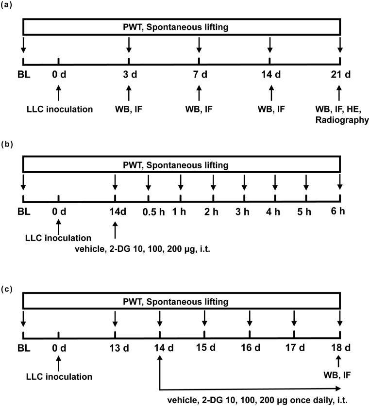
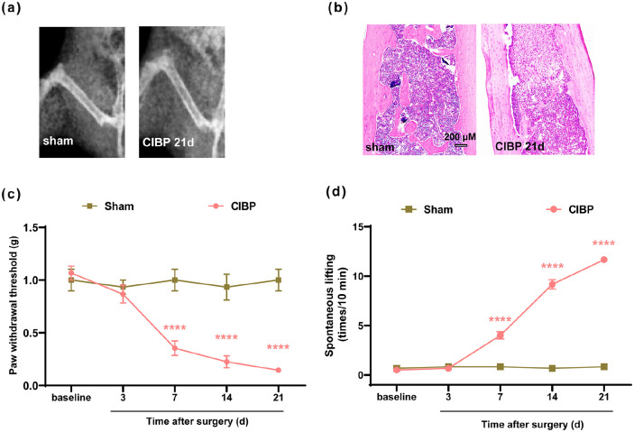
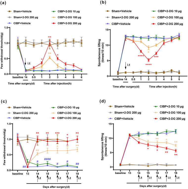
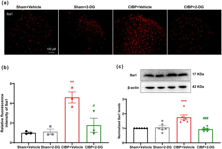
Methods: In this research, we established a CIBP mouse model by injecting Lewis lung cancer (LLC) cells into the bone marrow of the femur. Relevant pain behaviors were assessed by measuring the paw withdrawal threshold and spontaneous hind limb lifting. Additionally, the glycolysis inhibitor 2-DG was intrathecally administered to treat CIBP in mice. Western blotting and immunofluorescence techniques were employed to analyze microglial activation and M1/M2 phenotype markers in the spinal cord.

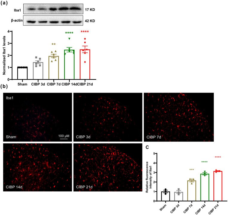
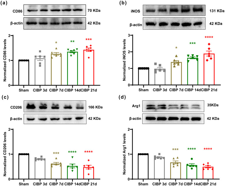
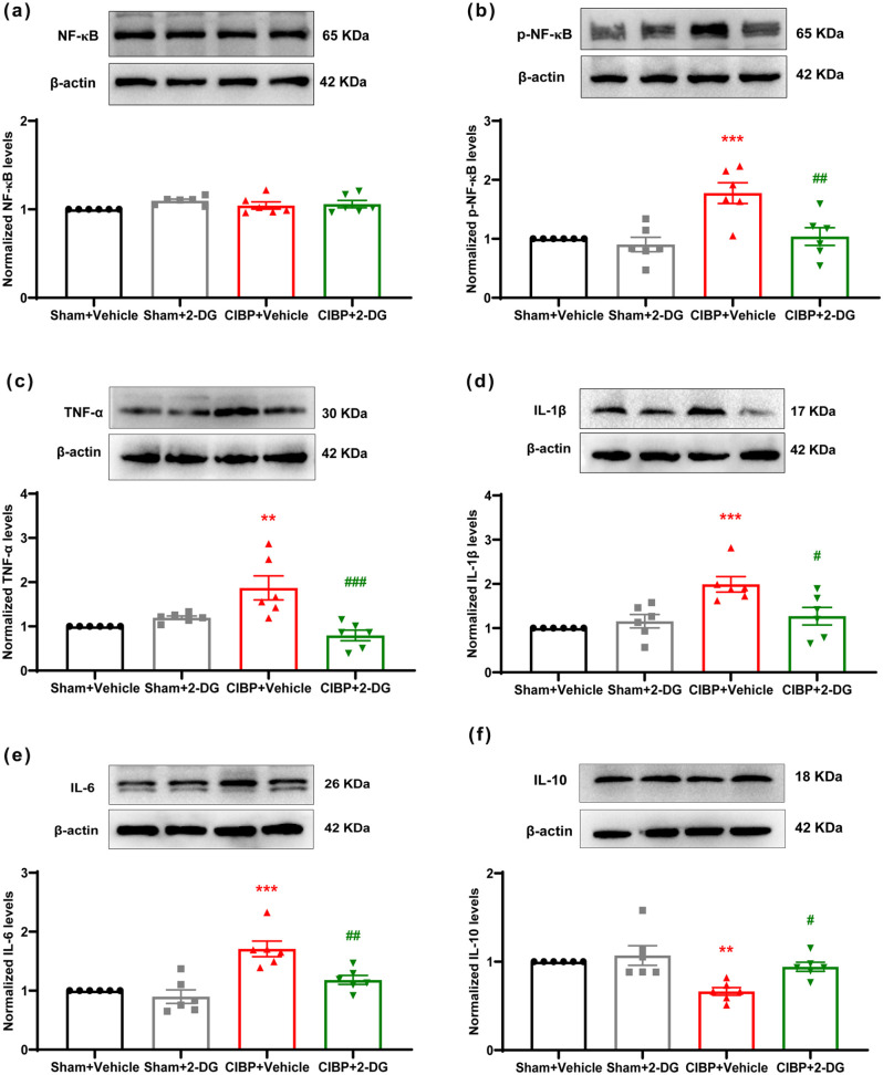
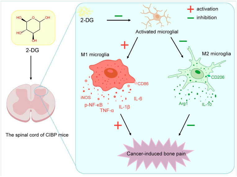
Results: Our findings demonstrated significant microglial activation and polarization toward the M1 phenotype in the spinal cord of CIBP mice. Intrathecal administration of 2-DG effectively alleviated pain-related behaviors in CIBP mice. Furthermore, this treatment suppressed microglial activation and M1 polarization, while significantly restoring levels of the M2 phenotype. Additionally, 2-DG attenuated the production of pro-inflammatory factors such as tumor necrosis factor-α (TNF-α), interleukin-1β (IL-1β), and interleukin-6 (IL-6), while boosting the secretion of the anti-inflammatory factor (IL-10) in the spinal cord of CIBP mice. Notably, 2-DG effectively suppresses microglia activation and M1 polarization in LPS + IFN-γ-induced BV-2 cells by downregulating CD86, iNOS expression, TNF-α, IL-1β, IL-6 levels while upregulating Arg-1, CD206 expression and IL-10 level.

Conclusion: These results suggest that 2-DG ameliorates mechanical allodynia, spontaneous pain and neuroinflammation in the spinal cord of CIBP mice by promoting the transition from the M1 phenotype to the M2 phenotype. This study may provide a novel strategy for the treatment of CIBP.

Keywords: 2-deoxy-D-glucose; Cancer-induced bone pain; microglia polarization; neuroinflammation.


Conflict of interest statement

Declaration of conflicting interestsThe author(s) declared no potential conflicts of interest with respect to the research, authorship, and/or publication of this article.

Figures

Figure 1.
Figure 1.
Schematic diagram of the experimental design: (a) changes in pain-related behaviors after LLC inoculation in mice. The spinal cord was collected at indicative days, (b) to determine whether a single administration of 2-DG can attenuate established CIBP in mice, 2-DG at doses of 10, 100, and 200 μg was intrathecally injected on day 14 following LLC inoculation, and (c) to clarify if continuous administration of 2-DG, can attenuate established CIBP in mice, 2-DG (10, 100, 200 μg) or vehicle was intrathecally administered once a day from day 14 for 5 days in a row. All behavioral tests were experienced 2 h after 2-DG administration or vehicle. The spinal cord was removed on day 18 for WB and IF after repeated 2-DG administration and behavioral tests. CIBP: cancer-induced bone pain; LLC: Lewis lung cancer.
Figure 2.
Figure 2.
Assessment of pain behaviors over time: (a) representative radiographs of the ipsilateral femur from sham and CIBP mice on day 21 after inoculation of PBS solution or LLC cells, (b) representative images of HE staining of femur sections from sham and CIBP mice, (c) the PWT of the left hind limb in sham mice and CIBP mice in response to von Frey filaments at the indicated time points, and (d) spontaneous lifting of the left hind limb at the indicated time points. ANOVA: analysis of variance; CIBP: cancer-induced bone pain; LLC: Lewis lung cancer; PWT: paw withdrawal threshold. The data are expressed as the mean ± SEM and analyzed using two-way repeated-measures ANOVA with Bonferroni’s post hoc test. Figure (c) ****p < 0.0001 versus the sham group. n = 6 per group. Figure (d) ****p < 0.01 versus the sham group. n = 6 per group.
Figure 3.
Figure 3.
Microglia is activated in the spinal cord of CIBP mice: (a) representative Western blot results reveal a marked upregulation of spinal Iba1 levels on days 3, 7, 14, and 21 after LLC cells inoculation, (b) and (c) representative immunofluorescence images show activated microglia in the spinal dorsal horn at the indicated time points. The histogram shows the immunofluorescence intensity analysis results. ANOVA: analysis of variance; LLC: Lewis lung cancer. The data are expressed as the mean ± SEM and analyzed using one-way repeated-measures ANOVA with Tukey post hoc test. ***p < 0.1, ****p < 0.001 versus the sham group. Scale bar = 100 μm. n = 3 per group.
Figure 4.
Figure 4.
The microglia in the spinal cord of CIBP mice polarized toward the M1 phenotype: (a)–(d) representative Western blotting bands and quantitative analysis show the expression levels of CD86, iNOS, CD206, and Arg1 protein in the Sham group and CIBP group on days 3, 7, 14, and 21 after LLC cells inoculation. β-actin was used as an internal control. ANOVA: analysis of variance; LLC: Lewis lung cancer. The data are expressed as the mean ± SEM and analyzed using one-way repeated-measures ANOVA with Tukey post hoc test. *p < 0.05, **p < 0.01, ***p < 0.001, ****p < 0.0001 versus the sham group. n = 6 per group.
Figure 5.
Figure 5.
Analgesic effect of intrathecal 2-DG on mechanical allodynia and spontaneous pain in CIBP mice: (a) a single dose of 2-DG (100 and 200 μg, i.t.) markedly increased the PWT in CIBP mice, starting at 1 h and lasting for at least 3 h, (b) a single dose of 2-DG (100 and 200 μg, i.t.) markedly decreased spontaneous lifting of the left hind limb in CIBP mice, starting at 1 h and lasting for at least 3 h, (c) and (d) the PWT and spontaneous lifting of the left hind limb was significantly decreased from day 14 to day 21 in 2-DG-treated CIBP mice compared with vehicle-treated CIBP mice.. ANOVA: analysis of variance; CIBP: cancer-induced bone pain; PWT: paw withdrawal threshold. The data are expressed as the mean ± SEM and analyzed using two-way repeated-measures ANOVA with Tukey post hoc test. Figure (a) *p < 0.05, **p < 0.01 compared with the CIBP + Vehicle group, #p < 0.05, ##p < 0.01 compared with the group treated with 2-DG (100 μg), n = 6 per group. Figure (b) **p < 0.05, ***p < 0.01, and ****p < 0.001 compared with the CIBP + Vehicle group, ##p < 0.01, ###p < 0.001 compared with the group treated with 2-DG (100 μg), n = 6 per group. Figure (c) and (d) **p < 0.01, ***p < 0.001, ****p < 0.0001 versus CIBP + Vehicle group, #p < 0.05, ##p < 0.01, ###p < 0.001 versus the group treated with 2-DG (100 μg). n = 6 per group.
Figure 6.
Figure 6.
Continuous administration of 2-DG inhibited microglial activation in the spinal cord of CIBP mice: (a) and (b) representative immunofluorescence images show that intrathecal 2-DG inhibited microglial activation in the spinal dorsal horn of CIBP mice. The histogram shows the immunofluorescence intensity analysis results, and (c) representative Western blotting bands and quantitative analysis show that 2-DG treatment reduced the expression levels of Iba1 in the spinal cord of CIBP mice. ANOVA: analysis of variance; CIBP: cancer-induced bone pain. The data are expressed as the mean ± SEM and analyzed using one-way repeated-measures ANOVA with Tukey post hoc test. Figure (a) and (b) **p < 0.001 versus Sham + Vehicle group, #p < 0.01 versus CIBP + Vehicle group. n = 3 per group. Figure (c) ***p < 0.001 versus Sham + Vehicle group, ###p < 0.01 versus CIBP + Vehicle group. n = 6 per group.
Figure 7.
Figure 7.
Continuous administration of 2-DG reversed the CIBP-induced increase in the M1 phenotype and the CIBP-induced decrease in the M2 phenotype: (a)–(d) representative Western blotting bands and quantitative analysis revealed that spinal M1 microglial markers (CD86, iNOS) were significantly upregulated in CIBP mice. Meanwhile, spinal M2 microglial markers (CD206, Arg-1) displayed an obvious decrease in CIBP mice. While, intrathecal 2-DG could reduce the levels of M1 microglial markers (CD86, iNOS) and increase those of M2 microglial markers (CD206, Arg-1) in the spinal cord in CIBP mice, and (e) representative immunofluorescence images show that Iba1 was colocalized with iNOS and CD206. ANOVA: analysis of variance; CIBP: cancer-induced bone pain. The data are expressed as the mean ± SEM and analyzed using one-way repeated-measures ANOVA with Tukey post hoc test. *p < 0.05, **p < 0.01, ****p < 0.0001 versus Sham + Vehicle group, #p < 0.05, ##p < 0.01, ####p < 0.0001 versus CIBP + Vehicle group. n = 6 per group.
Figure 8.
Figure 8.
Continuous administration of 2-DG inhibited pro-inflammatory cytokines and improved anti-inflammatory cytokines in CIBP mice: (a)–(f) representative Western blotting bands and quantitative analysis revealed that levels of inflammation-related factors p-NF-κB, pro-inflammatory cytokines (TNF-α, IL-1β, IL-6) were inhibited and anti-inflammatory cytokine (IL-10) was increased by 2-DG in CIBP mice. ANOVA: analysis of variance; CIBP: cancer-induced bone pain. The data are expressed as the mean ± SEM and analyzed using one-way repeated-measures ANOVA with Tukey post hoc test. **p < 0.01, ***p < 0.001 versus Sham + Vehicle group, #p < 0.05, ##p < 0.01, ###p < 0.001 versus CIBP + Vehicle group. n = 6 per group.
Figure 9.
Figure 9.
The schematic diagram illustrates that the potential mechanisms by which 2-DG treatment reduces spinal inflammation and alleviates cancer-induced bone pain. Intrathecal administration of 2-DG inhibits microglial polarization towards the M1 phenotype and suppresses neuroinflammation, thereby contributing to the relief of CIBP. −, inhibition; +, activation.

Similar articles

References

    1. Coleman RE. Clinical features of metastatic bone disease and risk of skeletal morbidity. Clin Cancer Res 2006; 12: 6243s–6249s. - PubMed
    1. Ji H, Jin X, Zhang Q, Zhou Y, Zhu C, Yang Y, Tang Z, Yu G, Wang C. A mouse model of cancer induced bone pain: from pain to movement. Front Behav Neurosci 2022; 16: 873750. - PMC - PubMed
    1. Paley CA, Johnson MI, Tashani OA, Bagnall AM. Acupuncture for cancer pain in adults. Cochrane Database Syst Rev 2015; 2015: CD007753. - PMC - PubMed
    1. Colosia A, Njue A, Bajwa Z, Dragon E, Robinson RL, Sheffield KM, Thakkar S, Richiemer SH. The burden of metastatic cancer-induced bone pain: a narrative review. J Pain Res 2022; 15: 3399–3412. - PMC - PubMed
    1. Tu H, Chu H, Guan S, Hao F, Xu N, Zhao Z, Liang Y. The role of the M1/M2 microglia in the process from cancer pain to morphine tolerance. Tissue Cell 2021; 68: 101438. - PubMed

MeSH terms