. 2025 Jun 25;10(6):e0001725.
doi: 10.1128/msphere.00017-25. Epub 2025 May 30.

A novel silver-ruthenium-based antimicrobial kills Gram-negative bacteria through oxidative stress-induced macromolecular damage

Affiliations

A novel silver-ruthenium-based antimicrobial kills Gram-negative bacteria through oxidative stress-induced macromolecular damage

Patrick Ofori Tawiah et al. mSphere. .

Abstract

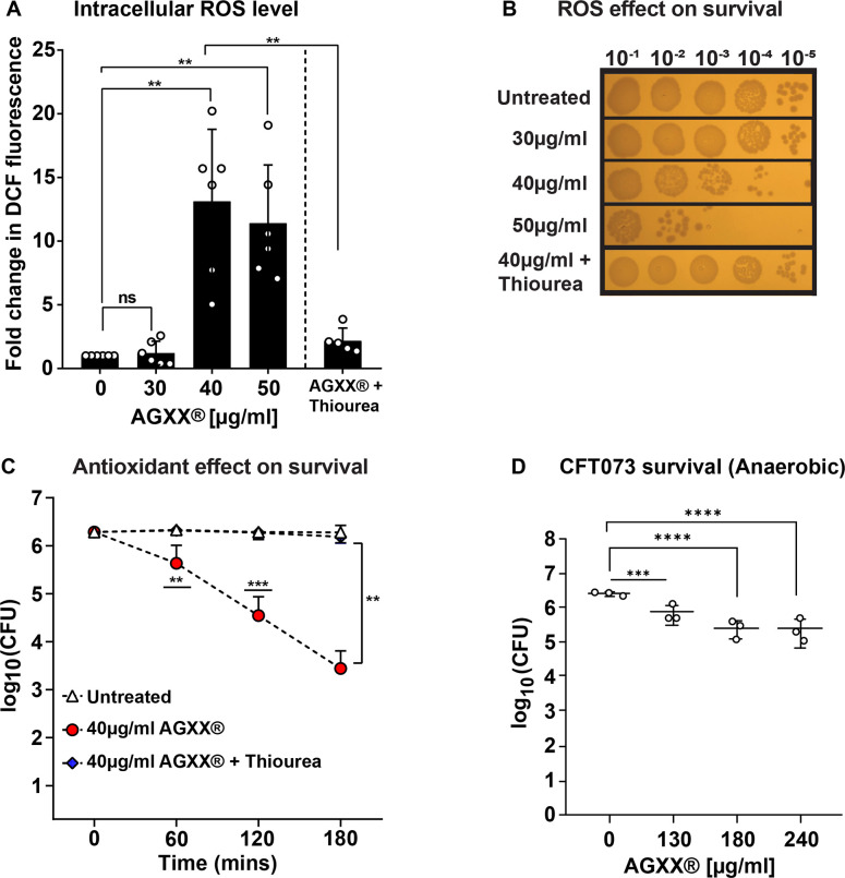
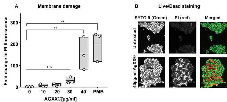
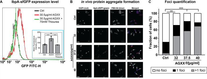
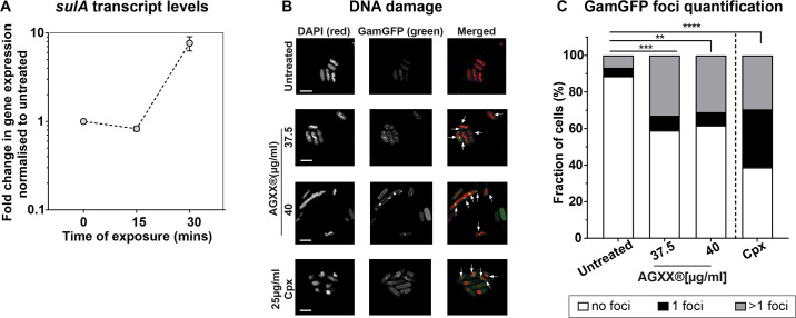
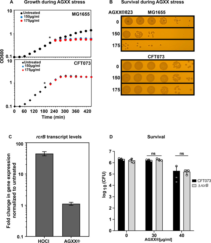
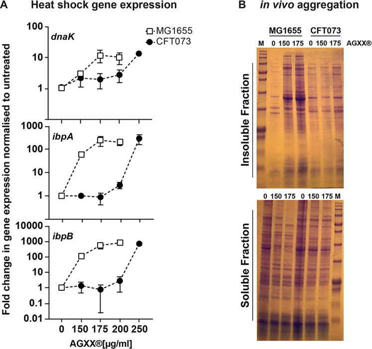
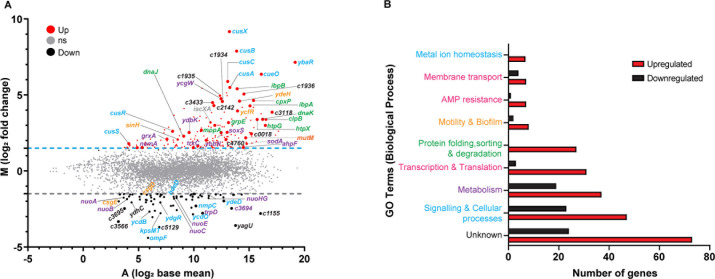
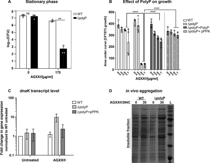
Amplified by the decline in antibiotic discovery, the rise of antibiotic resistance has become a significant global challenge in infectious disease control. Extraintestinal Escherichia coli (ExPEC), known to be the most common instigators of urinary tract infections (UTIs), represents such a global threat. Novel strategies for more efficient treatments are therefore desperately needed. These include silver nanoparticles, which have been used as antimicrobial surface coatings on catheters to eliminate biofilm-forming uropathogens and reduce the risk of nosocomial infections. AGXX is a promising silver-ruthenium coating that presumably kills bacteria through the generation of reactive oxygen species (ROS). However, neither AGXX's mode of action is fully understood, nor have its effects on Gram-negative bacteria or bacterial response and defense mechanisms toward AGXX been studied in detail. Here, we report that the bactericidal effects of AGXX are primarily based on ROS formation, as supplementation of the media with a ROS scavenger completely abolished AGXX-induced killing. We further show that AGXX impairs the integrity of the bacterial cell envelope and causes substantial protein aggregation and DNA damage already at sublethal concentrations. ExPEC strains appear to be more resistant to the proteotoxic effects of AGXX compared to non-pathogenic E. coli, indicating improved defense capabilities of the uropathogen. Global transcriptomic studies of AGXX-stressed ExPEC revealed a strong oxidative stress response, perturbations in metal homeostasis, as well as the activation of heat shock and DNA damage responses. Finally, we present evidence that ExPEC counteracts AGXX damage through the production of the chaperone polyphosphate, protecting cells from protein aggregation.IMPORTANCEThe rise in drug-resistant bacteria, together with the decline in antibiotic development, requires new strategies for infectious disease control. Gram-negative pathogens are particularly challenging to combat due to their outer membrane. This study highlights the effectiveness of the silver-containing antimicrobial AGXX against the Gram-negative bacterium Escherichia coli. AGXX effectively reduces bacterial survival by interfering with the membrane integrity and causing DNA damage and protein aggregation, which is likely a consequence of uncontrolled generation of oxidative stress. Our findings emphasize AGXX's potential as an antimicrobial surface coating and shed light on potential targets to reduce bacterial resistance to AGXX.

Keywords: antimicrobial agents; oxidative damage; oxidative stress; polyphosphate; silver; stress response.


Conflict of interest statement

The authors declare no conflict of interest.

Figures

Fig 1
Fig 1
AGXX-stressed bacteria accumulate large amounts of ROS, which contribute to the bactericidal effects of this antimicrobial. UPEC cells grown to mid-log phase were left untreated or treated with the indicated AGXX394C concentrations for 60 min before (A) intracellular ROS were quantified by H2DCFDA, and (B, C) survival was determined by serially diluting cells in PBS and spotting 5 µL onto LB agar for overnight incubation. 70 mM thiourea was used to quench ROS. (D) UPEC was grown under strictly anaerobic conditions in the presence of 1% potassium nitrate, treated with the indicated concentrations of AGXX394C for 4 hours, and survival monitored by CFU counts (n = 4–6, ±SD, one-way ANOVA, Dunnett and Sidak’s multiple comparison test; ns = P > 0.05, *P < 0.05, **P < 0.01, ***P < 0.001).
Fig 2
Fig 2
AGXX stress compromises bacterial membrane integrity. (A) CFT073 cells in the mid-log phase were treated with the indicated AGXX394C concentrations for 60 min, washed in PBS, and stained with 0.5 µM PI. PI fluorescence (λEx/Em: 535/617 nm) was measured by spectrophotometry and normalized to untreated cells (n = 3, ±SD). (B) Samples were washed in PBS after AGXX394C treatment, incubated with PI/Syto9 in the dark for 15 min at room temperature, and mounted on a glass slide with a 1% agarose pad for 63× imaging using inverted confocal microscopy. One representative image of four independent experiments is shown (one-way ANOVA, Sidak’s multiple comparison test; ns = P > 0.05, *P < 0.05, **P < 0.01).
Fig 3
Fig 3
AGXX causes extensive protein aggregation and increases the cellular demand for molecular chaperones. (A) Cellular IbpA-sfGFP fluorescence was monitored via flow cytometry after exposure of E. coli to sublethal AGXX394C treatment for 120 min (n = 5, ±SD). One representative image of five independent experiments is shown. Inset: quantified data from the five biological replicates. (B) Exponentially growing cells were either left untreated or treated with sublethal AGXX394C concentrations for 90 min. Samples were harvested, washed with PBS, and incubated with DAPI (nucleic acid stain) and FM4-64 (membrane stain) in the dark for 15 min. Cells were mounted on a glass slide with a 1% agarose pad for imaging at 63× via inverted confocal microscopy. Arrows illustrate foci formed when IbpA binds to protein aggregates in vivo. One representative image of four independent experiments (scale bar: 7.5 µm). (C) Confocal images were quantified by counting IbpA-sfGFP foci (n = 4) (one-way ANOVA, Sidak’s multiple comparison test; two-way ANOVA, Tukey’s multiple comparison test (compare total foci in untreated to AGXX treatment); ns = P > 0.05, *P < 0.05, **P < 0.01, ***P < 0.001, ****P < 0.0001).
Fig 4
Fig 4
AGXX causes DNA double-strand breaks. (A) sulA mRNA levels of AGXX394C-treated UPEC CFT073 cells were determined by qRT-PCR. Transcript levels were normalized to the housekeeping gene rrsD and calculated as fold changes based on the expression levels in the untreated control (n = 3, ±SD). (B) Cells expressing Gam-sfGFP were exposed to AGXX394C for 3 hours, washed, incubated with DAPI in the dark for 15 min, and visualized by confocal microscopy. Ciprofloxacin was used as a positive control. Arrows indicate Gam-sfGFP foci on the DAPI-stained DNA. One representative image of three independent experiments is shown (Scale bar: 5 µm). (C) Gam-sfGFP foci were quantified by counting the number of foci per cell (n = 3); (two-way ANOVA, Tukey’s multiple comparison test [compare total foci in untreated to AGXX treatment]; ns = P > 0.05, *P < 0.05, **P < 0.01, ***P < 0.001).
Fig 5
Fig 5
UPEC strain CFT073 shows increased tolerance to AGXX compared to K-12 E. coli strain MG1655, which is independent of RcrB. Overnight cultures of E. coli strains MG1655 and CFT073 were diluted into MOPSg to an OD600 of ~0.1 and grown to mid-log phase (OD600 ~0.5). Cells were either left untreated or exposed to increasing concentrations of AGXX. Growth (A) and survival (B) were recorded. (A) AGXX823 concentrations that resulted in a growth arrest of MG1655 had no effect on CFT073. (B) After 60 min of AGXX823 exposure, cells were serially diluted and spot-titered onto LB agar plates for overnight incubation at 37°C. One representative image of at least three independent experiments with similar outcomes. (C) rcrB mRNA levels of HOCl and AGXX394C-treated UPEC CFT073 cells were determined by qRT-PCR after 15 min. Transcript levels were normalized to the housekeeping gene rrsD and calculated as fold changes normalized to that of the untreated control (n = 3, ±SD). (D) Exponentially grown CFT073 WT and ΔrcrB cultures were either left untreated or treated with AGXX394C. After 60 min, cells were serially diluted and spot-titered onto LB agar plates for overnight incubation at 37°C (n = 3, ±SD).
Fig 6
Fig 6
AGXX-induced protein aggregation is less pronounced in members of the UPEC pathotype. Mid-log phase cultures of K-12 strain MG1655 and UPEC CFT073 were exposed to the indicated AGXX823 concentrations for 30 min. (A) Total RNA was extracted, genomic DNA removed, and mRNA reverse-transcribed into cDNA. qRT-PCR analysis was performed for differential expression analyses of genes ibpA, dnaK, and ibpB, which were normalized to the housekeeping gene rrsD and the untreated cells (n = 5–7, ±SD). (B) The extent of protein aggregation was determined after harvesting and cell lysis. Protein aggregates and soluble proteins were separated, extracted, separated by SDS-PAGE, and visualized by Coomassie staining. One representative image of five biological replicates is shown.
Fig 7
Fig 7
AGXX exposure of UPEC elicits significant changes in global gene expression. (A) Exponentially growing CFT073 cells were incubated with a sublethal concentration of AGXX394C for 30 min. Transcription was stopped by the addition of ice-cold methanol. Reads were aligned with the CFT073 reference genome (accession number: AE014075). Data are visualized as a ratio/intensity scatter plot (M/A-plot) of differentially expressed genes in AGXX-treated CFT073 cells. Statistically significantly upregulated genes are depicted above the blue dashed line, whereas statistically significantly downregulated genes are presented as black dots below the gray dashed line (M ≥ 1.5 or ≤−1.5, P ≤ 0.05). Light gray dots represent genes with no significant fold change in transcript level upon AGXX treatment (P > 0.5). Many of the upregulated genes can be categorized into metal ion homeostasis (blue IDs), protein homeostasis (green IDs), DNA damage (orange ID), and oxidative stress response (purple IDs), respectively. Transcriptome analysis was performed from three independent biological replicates. (B) Number of significantly differentially expressed genes of AGXX-stressed CFT073 grouped based on GO terms for biological processes. Genes with more than one biological process were assigned to their respective GO term from the KEGG pathway database.
Fig 8
Fig 8
Polyphosphate protects UPEC from AGXX stress. (A) The role of polyP for UPEC growth and survival during AGXX394C stress was determined in cells of the stationary phase. After 240 min, samples were serially diluted in PBS, spot-titered on LB agar, and incubated for 20 hours for CFU counts (n = 3, ±SD). (B) WT, ΔpolyP, ΔpolyP/pPPK, and ΔpolyP supplemented with 4 mM PolyP cultures were cultivated in MOPSg media in the presence of the indicated AGXX concentrations. Growth was monitored at 600nm for 16 hours and calculated as the area under the growth curve (n = 3–4, ±SD; Student t-test; ns = P > 0.05, *P < 0.05, **P < 0.01, ***P < 0.001, ****P < 0.0001). (C) Mid-log phase cultures of indicated strains were exposed to 30 µg/mL AGXX394C for 30 min before dnaK transcript levels were determined by qRT-PCR. Transcript levels were normalized to the housekeeping gene rrsD and calculated as fold changes based on the expression levels in the WT untreated control (n = 3, ±SD). (D) Mid-log phase cultures of UPEC CFT073 and ΔpolyP were exposed to the indicated AGXX394C concentration for 45 min. Protein aggregates were extracted, separated by SDS-PAGE, and visualized by Coomassie staining. One representative image of two biological replicates is shown.

Update of

Similar articles

Cited by

References

    1. Denamur E, Clermont O, Bonacorsi S, Gordon D. 2021. The population genetics of pathogenic Escherichia coli. Nat Rev Microbiol 19:37–54. doi: 10.1038/s41579-020-0416-x - DOI - PubMed
    1. Timm MR, Russell SK, Hultgren SJ. 2025. Urinary tract infections: pathogenesis, host susceptibility and emerging therapeutics. Nat Rev Microbiol 23:72–86. doi: 10.1038/s41579-024-01092-4 - DOI - PubMed
    1. Yang X, Chen H, Zheng Y, Qu S, Wang H, Yi F. 2022. Disease burden and long-term trends of urinary tract infections: a worldwide report. Front Public Health 10:888205. doi: 10.3389/fpubh.2022.888205 - DOI - PMC - PubMed
    1. Flores-Mireles AL, Walker JN, Caparon M, Hultgren SJ. 2015. Urinary tract infections: epidemiology, mechanisms of infection and treatment options. Nat Rev Microbiol 13:269–284. doi: 10.1038/nrmicro3432 - DOI - PMC - PubMed
    1. Vihta K-D, Stoesser N, Llewelyn MJ, Quan TP, Davies T, Fawcett NJ, Dunn L, Jeffery K, Butler CC, Hayward G, Andersson M, Morgan M, Oakley S, Mason A, Hopkins S, Wyllie DH, Crook DW, Wilcox MH, Johnson AP, Peto TEA, Walker AS. 2018. Trends over time in Escherichia coli bloodstream infections, urinary tract infections, and antibiotic susceptibilities in Oxfordshire, UK, 1998-2016: a study of electronic health records. Lancet Infect Dis 18:1138–1149. doi: 10.1016/S1473-3099(18)30353-0 - DOI - PMC - PubMed

MeSH terms

LinkOut - more resources