. 2025 May 30;16(1):952.
doi: 10.1007/s12672-025-02599-9.

Transglutaminase 2 nuclear localization enhances glioblastoma radiation resistance

Affiliations

Transglutaminase 2 nuclear localization enhances glioblastoma radiation resistance

Cuiping Sun et al. Discov Oncol. .

Abstract

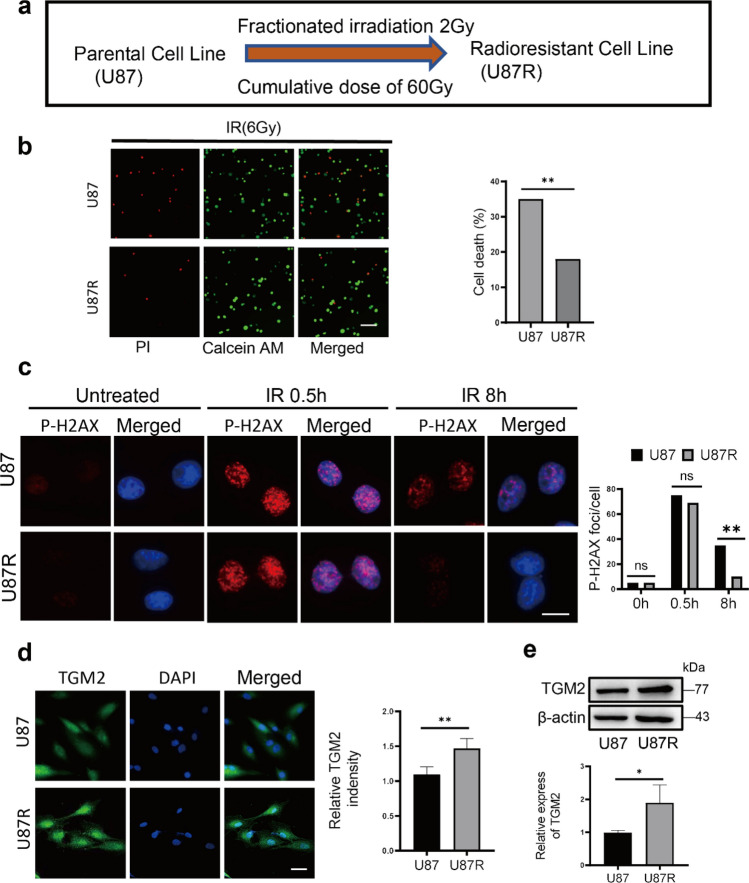
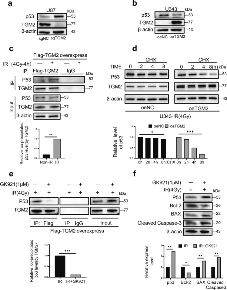
Radiotherapy remains the cornerstone of treatment for glioblastoma (GBM). However, the frequent occurrence of radiation resistance presents a significant therapeutic challenge. A comprehensive understanding of the mechanisms underlying this resistance is essential for improving GBM treatment strategies. In the present study, live-dead cell staining and immunofluorescence staining were employed, and irradiation-resistant cell lines were established. It was observed that transglutaminase 2 (TGM2) plays a pivotal role in enhancing radiation resistance in GBM, facilitating cell proliferation, and promoting DNA damage repair following irradiation. Moreover, immunofluorescence and nucleoplasmic protein extraction assays revealed that TGM2 in GBM rapidly translocates into the nucleus upon irradiation. Through co-immunoprecipitation assays, TGM2 was identified as binding to an increased amount of p53 proteins, thereby promoting p53 degradation post-irradiation. Notably, inhibition of this interaction resulted in a reduction of radiation resistance in GBM. In summary, this study underscores the significance of TGM2 nuclear translocation in radiation resistance and suggests that disrupting TGM2 binding to p53 may offer novel therapeutic insights for overcoming radiation resistance in GBM.

Keywords: DNA damage repair; Glioblastoma; Radiation resistance; Transglutaminase 2; Translocation; p53.


Conflict of interest statement

Declarations. Competing interests: The authors declare no competing interests.

Figures

Fig. 1
Fig. 1
TGM2 is associated with glioblastoma radiation resistance. (a) The correlations of TGM2 gene mRNA expression with survival of all WHO grade primary glioma (P < 0.0001). (b) TGM2 expression positively correlates with glioblastoma grade (P = 4.3e-08). (c) IHC to detect protein levels of TGM2 in different tumor grades. Quantification of IHC H-score for TGM2. Scale bar: 400 μm. (d) Representative IF images of TGM2 immunostaining in GBM cells. Green, TGM2; blue, DAPI. Scale bar: 10 μm. (e) WB analysis and quantification of TGM2 protein expression levels in GBM cells. (f) Death rates of U87 and T98G cells treated with different IR doses using inhibitor Cys-D. Cys-D is an inhibitor of TGM2. * P < 0.05 and ** P < 0.01
Fig. 2
Fig. 2
Radioresistant U87R cells demonstrate upregulated TGM2 protein expression. (a) Pattern of the establishment of U87R radioresistant cell line. (b) Representative IF images (left) and quantification (right) of PI and Calcein AM immunostaining in U87 and U87R cells. Green, PI; Red, Calcein AM. Scale bar: 100 μm. (c) Representative IF images of the levels of P-H2 AX in U87R and U87 cells after treated 6 Gy of RT. Red, P-H2 AX; blue, DAPI. Scale bar: 10 μm. (d) Representative IF images and quantification of TGM2 protein immunostaining in U87 and U87R cells. Green, TGM2; blue, DAPI. Scale bar: 20 μm. (e) WB analysis and quantification of TGM2 protein expression levels in U87 and U87R cells. * P < 0.05 and ** P < 0.01
Fig. 3
Fig. 3
TGM2 enhances proliferation after IR in GBM. (a) WB and quantification detected expression of TGM2 protein knockdown in U87 cells. (b) WB and quantification detected expression of TGM2 protein overexpressed in U343 cells. (c) CCK8 assay and quantification evaluated the proliferation capacity of GBM cells after knockdown or overexpression of TGM2 protein under IR. (d) Colony-formation assay and quantification detected the proliferation capacity of TGM2 protein in GBM cells after irradiation. (e) EdU assay and quantification evaluated the proliferation capacity of TGM2 protein in GBM cells after IR. Red, EdU; blue, DAPI. Scale bar: 100 μm. * P < 0.05, ** P < 0.01 and *** P < 0.001
Fig. 4
Fig. 4
TGM2 promotes DNA damage repair after irradiation in GBM. (a-b) Representative IF images and quantification of PI and Calcein AM immunostaining in U87 and U343 cells. Green, PI; Red, Calcein AM. Scale bar: 100 μm. (c) Representative IF images and quantification of the levels of P-H2 AX in U87 cells and treated 6 Gy of RT. Red, P-H2 AX; blue, DAPI. Scale bar: 10 μm. (d) Representative IF images and quantification of the levels of P-H2 AX in U343 cells and treated 6 Gy of RT. Red, P-H2 AX; blue, DAPI. Scale bar: 10 μm. (e) Representative images for TGM2 knockdown in U87 cells were examined with a flow cytometer to determine the cell cycle. (f) WB analysis and quantification of the key cell cycle regulators expression levels in U87 cells knockdown TGM2 and treated 4 Gy of RT.* P < 0.05 and ** P < 0.01
Fig. 5
Fig. 5
TGM2 was translocated into the nucleus after irradiation. (a) IHC staining of TGM2 protein in normal brain tissues and glioma tissues. Quantification of IHC H-score for nuclear TGM2. Scale bar: 100 μm. (b) Representative images for IF staining and quantification of TGM2 in u87 and U87R cells. Green, TGM2; blue, DAPI. Scale bar: 10 μm. (c) Representative images for IF staining of TGM2-GFP in u87 cells at different times after exposure to 6 Gy IR. Green, TGM2; blue, DAPI. Scale bar: 10 μm. (d) WB of TGM2 in both nucleus and cytoplasm lysates isolated from irradiated U87 cells. The levels of TGM2 protein expressions in both nuclear and cytoplasm were quantified with Image J software. ** P < 0.01 and *** P < 0.001
Fig. 6
Fig. 6
TGM2 promotes P53 degradation after irradiation in GBM cells. (a) WB analysis of TGM2 and p53 protein expression levels in original U87 cells and U87 cells knockdown TGM2 protein. (b) WB detected expression of TGM2 and p53 proteins in U343 cells with TGM2 overexpressed. (c) Co-IP assay of p53 bond withTGM2 in the Flag-TGM2 overexpressing cells after 4 Gy IR. The levels of co-precipitated p53 were normalized to the corresponding TGM2. (d) WB and quantification detect TGM2 in GBM cells after being treated with 4 Gy of RT and CHX (a translation inhibitor) at indicated time points. (e) Co-IP assay of p53 bond with TGM2 in the Flag-TGM2 overexpressing cells treated with 4 Gy of RT in the presence or absence of GK921 (1 μM). (f) WB analysis and quantification of p53 and apoptosis-related protein expression levels in GBM cells treated with or without GK921. ** P < 0.01 and *** P < 0.001

References

    1. Ostrom QT, Cioffi G, Waite K, Kruchko C, Barnholtz-Sloan JS. CBTRUS statistical report: primary brain and other central nervous system tumors diagnosed in the United States in 2014–2018. Neuro-Oncol. 2021. 10.1093/neuonc/noab200. - PMC - PubMed
    1. Chandra RA, Keane FK, Voncken F, Thomas CJ. Contemporary radiotherapy: present and future. Lancet. 2021;398(10295):171–84. 10.1016/S0140-6736(21)00233-6. - PubMed
    1. Ahmed SU, Carruthers R, Gilmour L, Yildirim S, Watts C, Chalmers AJ. Selective inhibition of parallel DNA damage response pathways optimizes radiosensitization of glioblastoma stem-like cells. Cancer Res. 2015;75(20):4416–28. 10.1158/0008-5472.CAN-14-3790. - PubMed
    1. Stupp R, Hegi ME, Mason WP, van den Bent MJ, Taphoorn MJ, Janzer RC, Ludwin SK, Allgeier A, Fisher B, Belanger K, Hau P, Brandes AA, Gijtenbeek J, Marosi C, Vecht CJ, Mokhtari K, Wesseling P, Villa S, Eisenhauer E, Gorlia T, Weller M, Lacombe D, Cairncross JG, Mirimanoff RO. Effects of radiotherapy with concomitant and adjuvant temozolomide versus radiotherapy alone on survival in glioblastoma in a randomised phase III study: 5-year analysis of the EORTC-NCIC trial. Lancet Oncol. 2009;10(5):459–66. 10.1016/S1470-2045(09)70025-7. - PubMed
    1. Yard BD, Adams DJ, Chie EK, Tamayo P, Battaglia JS, Gopal P, Rogacki K, Pearson BE, Phillips J, Raymond DP, Pennell NA, Almeida F, Cheah JH, Clemons PA, Shamji A, Peacock CD, Schreiber SL, Hammerman PS, Abazeed ME. A genetic basis for the variation in the vulnerability of cancer to DNA damage. Nat Commun. 2016;7:11428. 10.1038/ncomms11428. - PMC - PubMed

LinkOut - more resources