. 2025 May 30;11(22):eadr2753.
doi: 10.1126/sciadv.adr2753. Epub 2025 May 30.

High-affinity PQQ import is widespread in Gram-negative bacteria

Affiliations

High-affinity PQQ import is widespread in Gram-negative bacteria

Fabian Munder et al. Sci Adv. .

Abstract

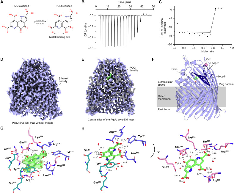
Pyrroloquinoline quinone (PQQ) is a soluble redox cofactor used by diverse bacteria. Many Gram-negative bacteria that encode PQQ-dependent enzymes do not produce it and instead obtain it from the environment. To achieve this, Escherichia coli uses the TonB-dependent transporter PqqU as a high-affinity PQQ importer. Here, we show that PqqU binds PQQ with high affinity and determine the high-resolution structure of the PqqU-PQQ complex, revealing that PqqU undergoes conformational changes in PQQ binding to capture the cofactor in an internal cavity. We show that these conformational changes preclude the binding of a bacteriophage, which targets PqqU as a cell surface receptor. Guided by the PqqU-PQQ structure, we identify amino acids essential for PQQ import and leverage this information to map the presence of PqqU across Gram-negative bacteria. This reveals that PqqU is encoded by Gram-negative bacteria from at least 22 phyla occupying diverse habitats, indicating that PQQ is an important cofactor for bacteria that adopt diverse lifestyles and metabolic strategies.


Figures

Fig. 1.
Fig. 1.. PqqU binds PQQ with a high affinity by enclosing it in an internal binding cavity.
(A) PQQ molecule visualized via a 2D stick model. PQQ changes from the oxidized form to the reduced form upon the two-electron transfer to both hydroxyl groups. The first aromatic ring contains nitrogen that, together with two carbonyl groups, coordinates a metal ion (Ca2+ or lanthanide) in PQQ-dependent enzymes (quinoproteins). (B) Representative thermogram of 100 μM PQQ titrated into 20 μM PqqU over ~50 min in 19 injections of 2 μl. The first injection (not included in the graph) is 0.4 μl. DP, differential power. (C) Negative peaks of injections integrated and plotted against the PqqU-PQQ molar ratio, excluding the first injection. (D) The PqqU coulomb potential map reveals distinct structures of PqqU such as the characteristic β barrel made of 22 individual β strands. The LMNG micelle has been removed for clarity. (E) Vertical cut of the PqqU coulomb potential map to reveal the density of a single PQQ molecule (green). (F) Cryo-EM structure of PqqU with bound PQQ (green) in the closed conformational state at a global resolution of 1.99 Å. Extracellular loops 7 and 8 of PqqU undergo a conformational change that encloses the substrate binding pocket (dark blue). Visualized as a side view with a slight tilt to expose the PQQ molecule. (G) PQQ (green) with map density surrounded by the 12 binding site residues colored by location within PqqU (β barrel, pink; loops 7 and 8, blue; plug domain, green). Interaction distances between PQQ and residues/waters visualized by dotted lines. (H) Rotational view of PQQ with the 11 binding site residues and bond lengths. Split into two parts for visual clarity based on location within the binding pocket.
Fig. 2.
Fig. 2.. PQQ capture by PqqU prevents binding of the PqqU-targeting phage.
(A) Close-up comparison of the surfaces of PqqU in open apo (red) and closed PQQ-bound (blue) states (left). In the closed PQQ-bound state, PQQ is obscured by loop 8. Close-up cartoon of PqqU in the closed conformational state (blue) compared to the open conformational state (red) visualizing the movement of loops 7 and 8 upon PQQ binding (right). (B) AlphaFold2 prediction of the extracellular side of PqqU in complex with two predicted IsaakIselin phage receptor binding proteins RBP-1 (pink) and RBP-2 (cyan). Cartoon view of the predicted complex (left) and zoomed cross-eye stereo view of the interface of the complex shown with RBP-1 and RBP-2 as cartoon and PqqU as a molecular surface (right). (C) AlphaFold2 prediction of the periplasmic side of PqqU in complex with the putative superinfection blocking protein SIP-1 (yellow), shown as in (B). (D) The structure prediction indicates that loops 7 and 8 are open during RBP-1 and RBP-2 binding, with clashes observed with loop 8 in the closed state.
Fig. 3.
Fig. 3.. Binding site mutations of PqqU reduce the ability to perform Entner-Doudoroff pathway glycolysis.
(A) Representative growth curve of E. coli ΔptsΔpqqU cultures grown in M63 minimal media containing 0.1% glucose and supplemented with 10 nM PQQ, unless indicated. The cells were transformed with plasmids containing mutated binding site variants of pqqU. h, hours. (B) The doubling time of cells was individually calculated from the exponential growth phase of each culture (n = 5, biological replicates). Statistical differences between PqqU and mutated PqqU were determined by performing paired t tests (ns, not significant; *P < 0.05 and **P < 0.01). (C) Rotational view of PQQ with the 12 binding site residues of PqqU. The mutated residues and their effect on culture growth are colored on the basis of severity.
Fig. 4.
Fig. 4.. Phylogeny and origin of PqqU-containing bacteria.
(A) Phylogenetic tree of bacteria encoding PqqU identified by our homology search of bacterial genomes. The tree was constructed on the basis of 16 ribosomal proteins. The classes of bacteria encoding the various clades of PqqU are shown, as well as the presence of genes encoding PQQ biosynthetic capacity or quinoproteins (PQQ-dependent dehydrogenases) in at least some of the members of that clade. Bacteria containing PqqU sequences used for structural modeling in complex with PQQ are numbered; see table S3 for sequence IDs. A sequence logo showing the conservation of PQQ-interacting residues across all identified PqqU homologs is shown as an inset; see fig. S8 for a full consensus logo and fig. S4 for a more detailed view of this tree. (B) Sunburst plot showing the environment of origin/isolation for bacteria encoding PqqU.
Fig. 5.
Fig. 5.. Chai-1 modeling indicates that diverse PqqU homologs bind PQQ.
(A) Comparison of the experimental structure of the PqqU-PQQ complex from E. coli with a selection of Chai-1 models of diverse PqqU-PQQ complexes (see data S4 for coordinate files for all models). (B) Comparison of models with glutamate and arginine substitutions as the position equivalent to K274 in E. coli PqqU. When glutamate substitutes lysine, a predicted metal ion binding site is formed, coordinated by PQQ and the glutamate. (C) A comparison of models lacking the semiconserved R536 in E. coli PqqU shows that the interaction between this residue and PQQ either is compensated for by arginine at another position in PqqU or is absent.
Fig. 6.
Fig. 6.. Model of the different strategies used by Gram-negative bacteria for obtaining PQQ for use in quinoproteins.
This diagram is a composite of the different strategies used by Gram-negative bacteria to obtain PQQ. On the basis of our genomic analysis, some strains use both PQQ biosynthesis and scavenging by PqqU to obtain PQQ, while others rely on one of these strategies.

Similar articles

References

    1. Anthony C., Pyrroloquinoline quinone (PQQ) and quinoprotein enzymes. Antioxid. Redox Signal. 3, 757–774 (2001). - PubMed
    1. Wehrmann M., Billard P., Martin-Meriadec A., Zegeye A., Klebensberger J., Functional role of lanthanides in enzymatic activity and transcriptional regulation of pyrroloquinoline quinone-dependent alcohol dehydrogenases in Pseudomonas putida KT2440. MBio 8, e00570-17 (2017). - PMC - PubMed
    1. Duine J. A., The PQQ story. J. Biosci. Bioeng. 88, 231–236 (1999). - PubMed
    1. Matsushita K., Toyama H., Yamada M., Adachi O., Quinoproteins: Structure, function, and biotechnological applications. Appl. Microbiol. Biotechnol. 58, 13–22 (2002). - PubMed
    1. Adachi O., Kubota T., Hacisalihoglu A., Toyama H., Shinagawa E., Duine J. A., Matsushita K., Characterization of quinohemoprotein amine dehydrogenase from Pseudomonas putida. Biosci. Biotechnol. Biochem. 62, 469–478 (1998). - PubMed

MeSH terms

LinkOut - more resources