. 2025 Aug;104(8):105336.
doi: 10.1016/j.psj.2025.105336. Epub 2025 May 26.

Duck plague virus US3 kinase phosphorylates and induces STING degradation to inhibit innate immune responses

Affiliations

Duck plague virus US3 kinase phosphorylates and induces STING degradation to inhibit innate immune responses

Bin Tian et al. Poult Sci. 2025 Aug.

Abstract

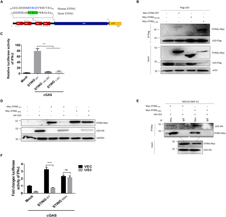
Duck plague virus (DPV) causes the highest mortality rate among aquatic birds; however, its antago nistic mechanism against antiviral innate immune responses remains elusive. In this study, we systematically screened and found that most DPV genes have inhibitory potential for duck cyclic guanosine monophosphate-adenosine monophosphate synthetase (cGAS)/stimulator of interferon (IFN) gene (STING) pathway-mediated antiviral responses, with the DPV US3 kinase showing the strongest inhibitory activity. Co-immunoprecipitation and immunoblotting assays demonstrated that DPV US3 interacted with STING and induced its degradation. Further mutagenesis experiments revealed that DPV US3 kinase activity was essential for phosphorylating STING, reducing STING dimerization, and inhibiting STING-mediated antiviral responses. Sequence alignment and mutagenesis studies have demonstrated that DPV US3 phosphorylates STING at serine 86, near the Endoplasmic reticulum (ER) retention sequence (R82YRGS86), disrupting its association with tank-binding kinase 1 (TBK1) and inducing STING degradation. Finally, US3 knockout attenuated DPV replication by activating higher levels of IFN and ISGs in vitro and in vivo. These results demonstrate that DPV promotes viral infection and pathogenicity by inducing STING degradation through the encoded US3 kinase, providing new insights into the mechanism of DPV immune evasion.

Keywords: Alpha herpesvirus; Immune evasion; Phosphorylation; STING; US3 kinase.


Conflict of interest statement

Declaration of competing interest The authors declare that they have no known competing financial interests or personal relationships that could have appeared to influence the work reported in this paper.

Figures

Fig 1
Fig. 1
DPV US3 inhibits the cGAS/STING signaling pathway. (A) The DEF cells were infected with 1 MOI of DPV CHv strain, the IFN-β mRNA was determined through Q-PCR method. (B) The DEF cells were transfected with duck IFN-β luciferase reporter gene, phRL-TK Renilla luciferase plasmid, cGAS and STING expression plasmids, together with DPV proteins expression plasmids, the luciferase activity was measured 36 hours after transfection and normalized to renilla luciferase activity. (C and D) The DEF cells were transfected with cGAS and STING expression plasmids, together with DPV US3 expression plasmid, the mRNA of IFN-β and MX were measured 24 hours after transfection through Q-PCR method. Graphs are presented as means ± SEM, data are representative of three independent experiments, *P < 0.05; **P < 0.01; ***P < 0.001; ****P < 0.0001 (t test or One-way ANOVAs followed by Tukey’s post hoc test for A, B, C, and D).
Fig 2
Fig. 2
DPV US3 protein kinase interacts with duck STING. (A) HEK293T cells were transfected with duck Flag-STING or Flag-IRF7 expression plasmids, together with DPV HA-US3 expression plasmid for 36 hours, then the whole cell lysates (WCLs) were collected and part of WCLs were used for CoIP against HA and immunoblot assays. (B and C) HEK293T cells were transfected with duck HA-cGAS, or HA-TBK1 expression plasmids, together with Flag-US3 expression plasmid for 36 hours. The WCLs were used for CoIP against Flag and immunoblot assays. (D) DEF cells were transfected with HA-US3 and Flag-STING expression plasmids for 36 hours. The WCLs were used for CoIP against Flag and immunoblot assays. (E and F) DEF cells were transfected with Myc-STING expression plasmid for 12 hours, then the DEF cells were infected with 1 MOI of DPV-CHv-US3-Flag for 36 hours, then the WCLs were used for Co-IP against Flag or Myc and immunoblot assays.
Fig 3
Fig. 3
DPV US3 reduces STING dimerization and induces STING degradation. (A) DEF cells were transfected with duck Myc-STING, and Myc-TBK1 expression plasmids, together with pcaggs or pcaggs-HA-US3 plasmids for 36 hours. The proteins level of duck STING, TBK1, US3, and actin were characterized through immunoblot assays. (B) DEF cells were transfected with duck Myc-STING expression plasmid, alone or together with DPV HA-US3 expression plasmid for 24 hours, respectively. Then the CHX (20 μM) was added and WCLs were collected at the indicated time and used for immunoblot assays. (C) DEF cells were transfected with duck Myc-STING expression plasmid, alone or together with DPV HA-US3 expression plasmid for 36 hours, respectively. Then MG132 (10 μM) and BAF A1 (20 μM) were added for 6 hours and the WCLs were extracted and used for immunoblot assays. (D) The DEF cells were transfected with duck Myc-STING expression plasmid for 12 hours and infected with 1 MOI of DPV-CHv-WT-GFP strain or DPV-CHv-dUS3-GFP strain for 36 hours, then the WCLs were collected for immunoblot assays. (E) HEK293T cells were transfected with indicated plasmids for 36 hours and MG132 and BAF A1 were added for 6 hours, then the WCLs were used for CoIP against Flag and immunoblot assays.
Fig 4
Fig. 4
The kinase activity of DPV US3 is indispensable for STING degradation and inhibition of STING mediated antiviral immune responses. (A) DEF cells were transfected with indicated plasmids for 36 hours and the WCLs were extracted and used for immunoblot assays with Phospho- (Ser/Thr) PKA substrate antibody, HA and GAPDH primary antibodies. (B) DEF cells were transfected for 36 hours and added MG132 and BAF A1 for 6 hours, then the WCLs were used for CoIP against Myc and immunoblot assays. (C and D) HEK293T cells and DEF cells were transfected with indicated plasmids for 36 hours and the WCLs were extracted and detected via immunoblot assays. (E) HEK293T cells were transfected with duck Myc-STING expression plasmid, alone or together with DPV HA-US3 and its kinase activity mutants expression plasmids for 36 hours, and added MG132 and BAF A1 for 6 hours, then the WCLs were used for CoIP against Flag and immunoblot assays. (F) The DEF cells were transfected, and the luciferase activity was measured 36 hours after transfection and normalized to renilla luciferase activity. (G-I) The DEF cells were transfected with the indicated plasmids for 24 hours and the IFN-β, MX, and OASL mRNA level were measured by Q-PCR assays. Graphs are presented as means ± SEM, data are representative of three independent experiments, *P < 0.05; **P < 0.01; ***P < 0.001; ****P < 0.0001 (One-way ANOVAs followed by Tukey’s post hoc test for the rest for F-I).
Fig 5
Fig. 5
STING S86A mutant resistant to US3 kinase induced degradation. (A) Schematic and sequence align of duck STING. (B) HEK293T cells were transfected with indicated plasmids for 36 hours and added MG132 and BAF A1 for 6 hours and the WCLs were used for CoIP against Flag and immunoblotting assays. (C) The DEF cells were transfected and the luciferase activity was measured 36 hours after transfection and normalized to renilla luciferase activity. (D) DEF cells were transfected and the WCLs were extracted 36 hours after transfection and used for immunoblotting assays against Myc, HA, and GAPDH antibodies. (E) HEK293T cells were transfected with indicated plasmids for 36 hours and added BAF A1 for 6 hours and the WCLs were extracted and used for CoIP against Myc antibody and immunoblotting assays. (F) The DEF cells were transfected, and the luciferase activity was measured 36 hours after transfection and normalized to renilla luciferase activity. Graphs are presented as means ± SEM, data are representative of three independent experiments, *P < 0.05; **P < 0.01; ***P < 0.001; ****P < 0.0001 (One-way ANOVAs followed by Tukey’s post hoc test for the rest for F).
Fig 6
Fig. 6
US3 kinase disrupts the interaction between STING and TBK1, and phosphorylation of STING mediated by TBK1. (A) Alignment of the duck STING CTT with human STING CTT. (B) HEK293T cells were transfected with indicated plasmids for 36 hours and the WCLs were used for immunoblotting assays. (C) The DEF cells were transfected, and the luciferase activity was measured 36 hours after transfection and normalized to renilla luciferase activity. (D) HEK293T cells were transfected with the indicated plasmids for 36 hours, then the WCLs were used for immunoblotting assays. (E) HEK293T cells were transfected with the indicated plasmids for 36 hours and added BAF A1 for 6 hours and the WCLs were used for CoIP against Flag antibody and immunoblotting assays. Graphs are presented as means ± SEM, data are representative of three independent experiments, *P < 0.05; **P < 0.01; ***P < 0.001; ****P < 0.0001 (One-way ANOVAs followed by Tukey’s post hoc test for the rest for C).
Fig 7
Fig. 7
DPV US3 antagonizes the STING mediated innate immune responses and promotes DPV replication in vitro and pathogenicity in vivo. (A) DEF cells were transfected with shNC or shSTING expression plasmids for 12 hours, then the cells were infected with 1 MOI of DPV-CHv or DPV-CHv-dUS3 for 24 hours, the cells were collected for RNA extraction and the IFN-β mRNA level was measured via Q-PCR assays. (B) DEF cells were pre-transfected with shRNA for 12 hours and infected with DPVs and the cells were collected at the indicated time for viral DNA extraction and viral load study. (C)The cell culture supernatants were collected and used for viral titer determination through TCID50 assays. (D) Flow chart of animal experiments. (E-G) 5 duck brains were collected 1 day after infection for RNA extraction and the IFN-β, MX, and OASL mRNA level were determined via Q-PCR method. (H) 5 duck brains were collected 3 days after infection for viral DNA extraction and viral genome level detection via Q-PCR method targeting DPV UL30 gene. (I) The survival ratios of 15 ducks were monitored at least for 15 days after DPV-CHv or DPV-CHv-dUS3 inoculation. Graphs are presented as means ± SEM, data are representative of three independent experiments, *P < 0.05; **P < 0.01; ***P < 0.001; ****P < 0.0001 (Mantel-Cox test for I, One-way ANOVAs followed by Tukey’s post hoc test for the rest for A-C and E-H).

Similar articles

References

    1. Benetti L., Roizman B. Herpes simplex virus protein kinase US3 activates and functionally overlaps protein kinase A to block apoptosis. Proc. Natl. Acad. Sci. U. S. A. 2004;101:9411–9416. doi: 10.1073/pnas.0403160101. - DOI - PMC - PubMed
    1. Bodda C., Reinert L.S., Fruhwurth S., Richardo T., Sun C., Zhang B.C., Kalamvoki M., Pohlmann A., Mogensen T.H., Bergstrom P., Agholme L., O'Hare P., Sodeik B., Gyrd-Hansen M., Zetterberg H., Paludan S.R. HSV1 VP1-2 deubiquitinates STING to block type I interferon expression and promote brain infection. J. Exp. Med. 2020;217 doi: 10.1084/jem.20191422. - DOI - PMC - PubMed
    1. Burdette D.L., Monroe K.M., Sotelo-Troha K., Iwig J.S., Eckert B., Hyodo M., Hayakawa Y., Vance R.E. STING is a direct innate immune sensor of cyclic di-GMP. Nature. 2011;478:515–518. doi: 10.1038/nature10429. - DOI - PMC - PubMed
    1. Cartier A., Komai T., Masucci M.G. The Us3 protein kinase of herpes simplex virus 1 blocks apoptosis and induces phosporylation of the Bcl-2 family member Bad. Exp. Cell Res. 2003;291:242–250. - PubMed
    1. Chen Q., Sun L., Chen Z.J. Regulation and function of the cGAS-STING pathway of cytosolic DNA sensing. Nat. Immunol. 2016;17:1142–1149. doi: 10.1038/ni.3558. - DOI - PubMed

MeSH terms