. 2025 May 30;13(5):e011398.
doi: 10.1136/jitc-2024-011398.

Neovascular pruning by IDO1 inhibitors can potentiate immunogenic cytotoxicity of ischemia-targeted agents to synergistically enhance anti-PD-1 responsiveness

Affiliations

Neovascular pruning by IDO1 inhibitors can potentiate immunogenic cytotoxicity of ischemia-targeted agents to synergistically enhance anti-PD-1 responsiveness

Shih-Chun Shen et al. J Immunother Cancer. .

Abstract

Background: Strategies for deploying indoleamine 2,3-dioxygenase 1 (IDO1)-targeted therapies for use against cancer have focused on IDO1's role in promoting peripheral immune tolerance that shields tumors from effector T cells. However, preclinical investigation of both primary and metastatic tumor development in the lungs has uncovered a previously unappreciated role for IDO1 in directing a counterregulatory response to interferon (IFN)-γ that realigns the local inflammatory environment to promote tumor neovascularization. Understanding how to therapeutically leverage the ability of IDO1 inhibitors to subvert inflammatory neovascularization within the tumor microenvironment has potential ramifications for future clinical development of these compounds.

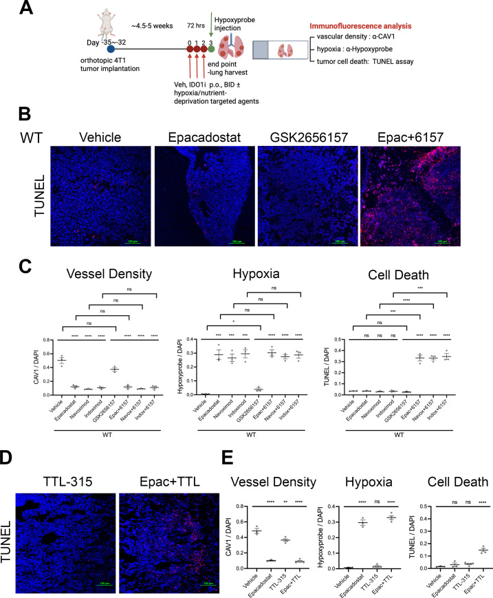
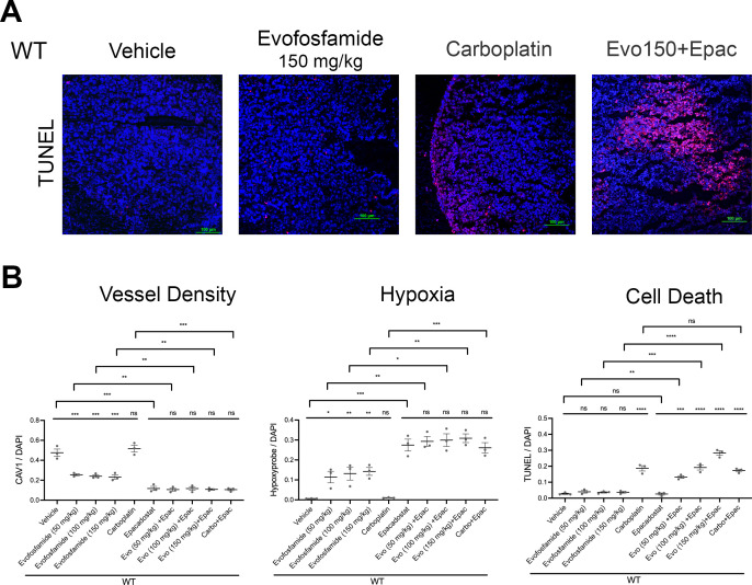
Methods: Pulmonary metastases seeded by orthotopically implanted 4T1 breast carcinoma cells were evaluated by confocal microscopy for the impact of both genetic and pharmacological IDO1 inhibition, alone or in combination with ischemia-directed cytotoxic agents, on markers of blood vessel density, hypoxia and cell death. Tumor immunogenicity and programmed death-ligand 1 (PD-L1) elevation were also evaluated. Quantitative analysis of these results was used to guide combinatorial treatment regimen development.

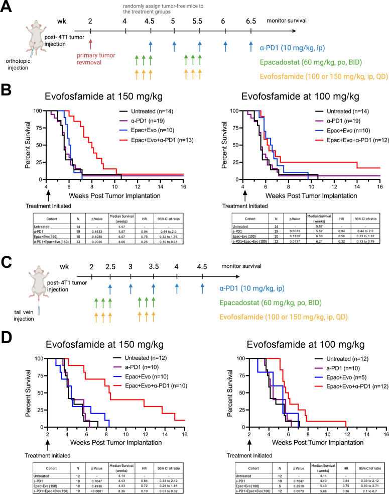
Results: Inhibiting IDO1 activity resulted in reduced neovascular density and elevated hypoxia in pulmonary metastases for which host IFN-γ was essential while adaptive immunity was dispensable. The tumors were consequently sensitized to the cytotoxic activity of ischemia-targeted agents including the protein kinase R-like endoplasmic reticulum kinase (PERK) inhibitor GSK2656157, the dithiol oxidative antimetabolite TTL-315, and the hypoxia-activated prodrug evofosfamide. Evofosfamide provoked the greatest degree of immunogenic cell death, while hypoxia, among other stressors, induced PD-L1. Based on this information, synergistic improvement in median survival was demonstrated in mice with established lung metastases through combined administration of anti-programmed cell death protein-1 (PD-1) antibody with evofosfamide and the IDO1 inhibitor epacadostat.

Conclusions: Improving therapeutic outcomes for patients with lung tumors, arising either as primary lesions or metastatic colonies, is of vital clinical importance. Building on preclinical evidence for IDO1's role in promoting inflammatory neovascularization of lung tumors, this study demonstrates how the intratumoral ischemic stress elicited by IDO1 inhibition can potentiate the immunogenic cytotoxicity of ischemia-targeted agents to effectively leverage immune checkpoint blockade responsiveness to confer a synergistic survival benefit. These findings provide a novel perspective on how IDO1 inhibitors can impact tumor biology and open up new possibilities for therapeutic applications.

Keywords: Combination therapy; Immune Checkpoint Inhibitor; Indoleamine 2, 3-dioxygenase - IDO; Lung Cancer; Tumor microenvironment - TME.


Conflict of interest statement

Competing interests: AJM is the recipient of financial support through sponsored research and consulting agreements with IO Biotech, Inc. AJM is also a co-inventor in the discovery and development of IDO1 inhibitor technologies patented by the Lankenau Institute for Medical Research and licensed to Augen Therapeutics, Inc., a private company for which he serves as a scientific advisor. SD is employed by Wuxi Advanced Therapeutics, Inc. MTH is Executive Chairman and CEO of Telesis Therapeutics, Inc. GCP is a co-inventor in the discovery and development of IDO1 inhibitor technologies patented by the Lankenau Institute for Medical Research and licensed to Augen Therapeutics, Inc., a private company for which he serves as a scientific advisor. The remaining authors declare no competing interests.

Figures

Figure 1
Figure 1. Lung metastases in mice lacking IDO1 exhibit elevated hypoxia and PERK inhibitor sensitization. (A) Representative confocal images of immunofluorescence staining for blood vessels (α-CAV1; cy3, red), hypoxia (α-Hypoxyprobe; FITC, green), apoptosis (TUNEL; Cy3, red) and nuclei (DAPI, blue) in 4T1 lung metastases from WT and Ido1−/− mice administered vehicle or the PERK inhibitor, GSK2656157. (B) Quantitative assessment of neovascular density, hypoxic area, and cell death within 4T1 lung metastases corresponding to images in (A) (n≥3 mice/group) and plotted as means±SEM of the fluorescent signal normalized to DAPI. CAV1, caveolin 1; DAPI, 4′,6-diamidino-2-phenylindole; FITC, fluorescein isothiocyanate; IDO1, indoleamine 2,3-dioxygenase 1; PERK, protein kinase R-like endoplasmic reticulum kinase; TUNEL, terminal deoxynucleotidyl transferase dUTP nick end labeling.
Figure 2
Figure 2. Coordinate neovascular regression and hypoxia elevation in lung metastases following IDO1 inhibition. (A) Representative confocal images of immunofluorescence staining for blood vessels (α-CAV1; Cy3, red), hypoxia (α-Hypoxyprobe; FITC, green), and nuclei (DAPI, blue) in 4T1 lung metastases from WT mice prior to and following epacadostat administration over 72 hours. (B) Quantitative time course of neovascular density and hypoxia levels within 4T1 lung metastases at 0, 24, 48, 72 hours following epacadostat administration (n=3 mice/group) plotted as means±SEM of the fluorescent signal normalized to DAPI. (C) Left panel: tumor volume distribution of individual lung metastases from WT mice administered vehicle or IDO1 inhibitor (epacadostat) (n≥20 nodules/group). Right panel: paired quantitative assessment of neovascular density and hypoxia levels in individual lung metastases from WT mice administered vehicle or epacadostat together with corresponding nodule volumes (n≥4 nodules/group). CAV1, caveolin 1; DAPI, 4′,6-diamidino-2-phenylindole; FITC, fluorescein isothiocyanate; IDO1, indoleamine 2,3-dioxygenase 1.
Figure 3
Figure 3. IDO1 inhibitors sensitize lung metastases to oxygen/nutrient deprivation-directed cytotoxic agents. (A) Schematic of the treatment regimen and immunofluorescence staining analysis of 4T1 lung metastasis samples. (B,D) Representative confocal images of apoptosis (TUNEL; Cy3, red) and nuclei (DAPI, blue) in 4T1 lung metastases from WT mice administered IDO1 inhibitor (epacadostat), protein kinase R-like endoplasmic reticulum kinase inhibitor (GSK2656157), or oxidative antimetabolite (TTL-315) alone or in combination. (C,E) Quantitative assessment of neovascular density, hypoxic area, and cell death within 4T1 lung metastases corresponding to the images in (B,D and online supplemental figure 2) (n≥3 mice/group) and plotted by means±SEM of the fluorescent signal normalized to DAPI. CAV1, caveolin 1; DAPI, 4′,6-diamidino-2-phenylindole; IDO1, indoleamine 2,3-dioxygenase 1; TUNEL, terminal deoxynucleotidyl transferase dUTP nick end labeling.
Figure 4
Figure 4. IDO1 inhibitor-elicited neovascular regression requires host IFN-γ but not adaptive immunity. (A) Representative confocal images of immunofluorescence staining for apoptosis (TUNEL; Cy3, red) and nuclei (DAPI, blue) in 4T1 lung metastases from WT, Ifng−/− and Rag1−/− mice administered either IDO1 inhibitor (epacadostat) or aryl hydrocarbon receptor inhibitor (CH-223191) in combination with protein kinase R-like endoplasmic reticulum kinase inhibitor (GSK2656157). (B) Quantitative assessment of neovascular density, hypoxic area, and cell death within 4T1 lung metastases corresponding to the images in (A and online supplemental figures 3–5) (n≥3 mice/group) and plotted as mean±SEM of the fluorescent signal normalized to DAPI. CAV1, caveolin 1; DAPI, 4′,6-diamidino-2-phenylindole; IDO1, indoleamine 2,3-dioxygenase 1; IFN, interferon; TUNEL, terminal deoxynucleotidyl transferase dUTP nick end labeling.
Figure 5
Figure 5. IDO1 inhibition sensitizes lung metastases to HAP (hypoxia-activated prodrug) cytotoxicity. (A) Representative confocal images of apoptosis (TUNEL; Cy3, red) and nuclei (DAPI, blue) in 4T1 lung metastases from WT mice administered epacadostat, carboplatin (non-targeted), evofosfamide (HAP) or epacadostat+evofosfamide. (B) Quantitative assessment of neovascular density, hypoxic area, and cell death within 4T1 lung metastases corresponding to the images in (A and online supplemental figure 6) (n=3 mice/group) and plotted as means±SEM of the fluorescent signal normalized to DAPI. CAV1, caveolin 1; DAPI, 4′,6-diamidino-2-phenylindole; IDO1, indoleamine 2,3-dioxygenase 1; TUNEL, terminal deoxynucleotidyl transferase dUTP nick end labeling.
Figure 6
Figure 6. IDO1 inhibition induces immunogenic cell death preferentially with the alkylating HAP but also elevates PD-L1. (A,B) Representative confocal images of immunofluorescence staining for calreticulin (α-CALR; Alexa488, green), wheat-germ agglutinin (α-WGA; Cy3, red), PD-L1 (α-PD-L1; Cy3, red) and nuclei (DAPI, blue) in 4T1 lung metastases from WT mice administered IDO1 inhibitor (epacadostat), protein kinase R-like endoplasmic reticulum kinase inhibitor (GSK2656157), oxidative antimetabolite (TTL-315) or the non-targeted/HAP alkylating agents (carboplatin/evofosfamide) alone or in combination. (C,D) Quantitative evaluation of immunogenic cell death and PD-L1 expression correlated to images shown in (A,B) (n≥3 mice/group) and plotted as means±SEM of either the co-localized fluorescent signals from WGA and CALR staining or the individual signal from PD-L1 staining normalized to DAPI. CALR, calreticulin; DAPI, 4′,6-diamidino-2-phenylindole; HAP, hypoxia-activated prodrug; IDO1, indoleamine 2,3-dioxygenase 1; PD-L1, programmed death-ligand 1; WGA, wheat germ agglutinin.
Figure 7
Figure 7. IDO1 inhibitor+hypoxia-activated prodrug combines with ICB to improve lung metastasis survival. (A,C) Schematic detailing the schedule for surgery and dosing in the orthotopic and tail vein-injected 4T1 lung metastasis models. (B,D) Kaplan-Meier survival curves for individual and combined administration of anti-PD-1 and epacadostat+evofosfamide at 150 and 100 mg/kg (n≥5 mice/group) compared with no treatment with accompanying tables listing p values, median survival, HR and 95% CI for each treatment parameter. BID, two times a day; ICB, immune checkpoint blockade; IDO1, indoleamine 2,3-dioxygenase; ip, intraperitoneal; PD-1, programmed cell death protein-1; QD, once a day.

References

    1. Stella GM, Kolling S, Benvenuti S, et al. Lung-Seeking Metastases. Cancers (Basel) 2019;11:1010. doi: 10.3390/cancers11071010. - DOI - PMC - PubMed
    1. Smith C, Chang MY, Parker KH, et al. IDO is a nodal pathogenic driver of lung cancer and metastasis development. Cancer Discov. 2012;2:722–35. doi: 10.1158/2159-8290.CD-12-0014. - DOI - PMC - PubMed
    1. Yoshida R, Imanishi J, Oku T, et al. Induction of pulmonary indoleamine 2,3-dioxygenase by interferon. Proc Natl Acad Sci U S A. 1981;78:129–32. doi: 10.1073/pnas.78.1.129. - DOI - PMC - PubMed
    1. Mondal A, Smith C, DuHadaway JB, et al. IDO1 is an Integral Mediator of Inflammatory Neovascularization. EBioMedicine. 2016;14:74–82. doi: 10.1016/j.ebiom.2016.11.013. - DOI - PMC - PubMed
    1. Shankaran V, Ikeda H, Bruce AT, et al. IFNgamma and lymphocytes prevent primary tumour development and shape tumour immunogenicity. Nature New Biol. 2001;410:1107–11. doi: 10.1038/35074122. - DOI - PubMed

MeSH terms

Substances