. 2025 Aug 15;80(9):604-615.
doi: 10.1136/thorax-2024-222242.

Effect of elexacaftor/tezacaftor/ivacaftor on systemic inflammation in cystic fibrosis

Affiliations

Effect of elexacaftor/tezacaftor/ivacaftor on systemic inflammation in cystic fibrosis

Rosemary E Maher et al. Thorax. .

Abstract

Background: Despite significant clinical improvements, there is evidence of persisting airway inflammation in people with cystic fibrosis (CF) established on elexacaftor/tezacaftor/ivacaftor (ETI) therapy. As CF is a multi-system disease, systemic immune profiles can reflect local inflammation within the lungs and other organs. Understanding systemic inflammation after ETI therapy may reveal important translational insights. This study aims to profile systemic inflammatory changes and relate these to the well-documented improvements observed with ETI therapy.

Methods: We conducted a single-centre longitudinal study with 57 CF subjects initiating ETI therapy. All participants were Phe508del homozygous or Phe508del/minimal function. Blood samples were collected pre-ETI and 3-12 months post-therapy initiation. Analyses included mass spectrometry-based proteomics, a multiplex immunoassay, and flow cytometry for peripheral immune cell counts and phenotype. Controls samples were provided by 29 age-matched healthy controls.

Results: Systemic inflammation reduced with ETI therapy; however, the immune profile remained distinct from healthy controls. ETI reduced neutrophil counts and was associated with a more mature, less inflammatory phenotype, as well as a shift towards an immune resolving state associated with increased CD206 expression. Cytokines known to influence neutrophil levels reduced with therapy. Despite ETI therapy, neutrophil and monocyte counts remained elevated compared with healthy controls. There was no obvious association between the ETI-related improvements in systemic inflammation and lung function.

Conclusions: Patients with CF showed evidence of persisting systemic inflammation despite ETI therapy, which may have long-term potentially adverse effects on respiratory and other organ systems.

Keywords: Cystic Fibrosis; Neutrophil Biology.


Conflict of interest statement

Competing interests: PB declares speaker fees (Vertex and Chiesi) and Advisory board participation (Vertex, Boehringer Ingelheim and Insmed).

Figures

Figure 1
Figure 1. Immune profiles of pre-ETI CF subjects stratified by baseline modulator status. Absolute cell counts were measured by high-dimensional flow cytometry, with CF subjects stratified according to baseline modulator status (dual therapy vs modulator naïve). For each boxplot, individual cell count, together with median and IQRs, is plotted. Differences between cohorts are calculated by t-tests: *p<0.05, **p<0.01, ***p<0.001, ****p<0.0001. CF, cystic fibrosis; ETI, elexacaftor/tezacaftor/ivacaftor.
Figure 2
Figure 2. Quantitative profiling of circulating immune cells pre-ETI and post-ETI. Absolute cell counts were measured by high-dimensional flow cytometry. (A) Stacked bar chart displaying median values of selected circulating immune cells. (B) Box plots of the selected immune cells frequently implicated in CF lung disease pathology, with CF subjects stratified according to ETI status. (C) Box plots of the monocyte subsets. For each boxplot, individual cell count, together with median and IQRs, is plotted. Differences between cohorts are calculated by t-tests: *.
Figure 3
Figure 3. Immunophenotyping of neutrophils pre-ETI and post-ETI. Median fluorescence intensity (MFI) was measured by high-dimensional flow cytometry. MFI values are plotted, with CF subjects stratified according to ETI status. (A) Represents those markers that significantly changed with ETI and were related to neutrophil maturation. (B) Represents changes in the cell marker CD206. (C) These are the other cell markers that were tested. For each group, individual median cell marker MFI, together with median and IQR, is plotted. Difference between cohorts is calculated by Mann-Whitney U test: *p<0.05, **p<0.01, ***p<0.0001. CF, cystic fibrosis; ETI, elexacaftor/tezacaftor/ivacaftor.
Figure 4
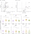
Figure 4. Quantitative profiling of systemic soluble immune mediators pre-ETI and post-ETI. (A) Volcano plot displaying pre-ETI CF versus healthy controls with log2 median fold change in soluble mediator abundance against adjusted p value calculated by Mann-Whitney U tests. Dots are coloured by thresholds based on log-fold change and adjusted p value. Horizontal dashed line indicates cut-off for adjusted p<0.05. Vertical dashed lines indicate positive and negative cut-offs for absolute log2 fold change of 1. (B) Volcano plot showing pre-ETI versus post-ETI. (C) Box plots of key altered soluble mediators. For those that were different at baseline between healthy and pre-ETI and then changed with therapy (adjusted p<0.05), the absolute concentrations are shown, with CF subjects stratified by ETI state. For each group, individual median cell marker MFI, together with median and IQRs, is plotted. Differences between cohorts are calculated by Mann-Whitney with adjustment for false discovery rate: *p<0.05, **p<0.01, ***p<0.001, ****p<0.0001. CF, cystic fibrosis; ETI, elexacaftor/tezacaftor/ivacaftor; G-CSF, granulocyte-colony stimulating factor; MFI, median fluorescence intensity.
Figure 5
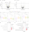
Figure 5. Quantitative profiling of cystic fibrosis (CF) plasma proteomes pre-ETI and post-ETI. (A) Volcano plot for pre-ETI CF versus healthy displaying fold change as calculated by limma against adjusted p values. Dots are coloured by thresholds based on log-fold change and adjusted p value. Horizontal dashed line indicates cut-off for adjusted p<0.05. Vertical dashed lines indicate positive and negative cut-offs for absolute log2 fold change of 1. (B) Volcano plot of pre-versus post-ETI CF. (C) Box plots of differentially expressed proteins. For those that were different at baseline between healthy and pre-ETI (adjusted p<0.05), the absolute concentrations are shown, with CF subjects stratified by ETI state. For each group, individual median cell marker MFI, together with median and IQRs, is plotted. Differences between cohorts are calculated by limma with adjustment for false discovery rate: *p<0.05. (D) The protein set enrichment analysis for CF versus healthy. X-axis indicates the Net Enrichment Scores (NES) of a pathway, with a negative score indicating that the pathway was downregulated in the CF group compared to the healthy group and a positive score indicating that the pathway was upregulated. The colour of the dot reflects p adjusted by Benjamini-Hochberg correction. The size of the dot represents the number of proteins present in the pathway that were also present in the data. (E) Protein set enrichment analysis of pre-ETI versus post-ETI therapy. ETI, elexacaftor/tezacaftor/ivacaftor; ITIH2, inter-alpha-trypsin inhibitor heavy chain H2; LRG1, leucine rich alpha-2-glycoprotein 1; PEX11B, peroxisomal membrane protein 11B; SPP2, secreted phosphoprotein 2.

References

    1. Gushue C, Eisner M, Bai S, et al. Impact of Elexacaftor-Tezacaftor-Ivacaftor on lung disease in cystic fibrosis. Pediatr Pulmonol. 2023;58:2308–16. doi: 10.1002/ppul.26485. - DOI - PubMed
    1. Heijerman HGM, McKone EF, Downey DG, et al. Efficacy and safety of the elexacaftor plus tezacaftor plus ivacaftor combination regimen in people with cystic fibrosis homozygous for the F508del mutation: a double-blind, randomised, phase 3 trial. Lancet. 2019;394:1940–8. doi: 10.1016/S0140-6736(19)32597-8. - DOI - PMC - PubMed
    1. Middleton PG, Mall MA, Dřevínek P, et al. Elexacaftor-Tezacaftor-Ivacaftor for Cystic Fibrosis with a Single Phe508del Allele. N Engl J Med. 2019;381:1809–19. doi: 10.1056/NEJMoa1908639. - DOI - PMC - PubMed
    1. Schaupp L, Addante A, Völler M, et al. Longitudinal effects of elexacaftor/tezacaftor/ivacaftor on sputum viscoelastic properties, airway infection and inflammation in patients with cystic fibrosis. Eur Respir J. 2023;62:62. doi: 10.1183/13993003.02153-2022. - DOI - PubMed
    1. Maher RE, Barry PJ, Emmott E, et al. Influence of highly effective modulator therapy on the sputum proteome in cystic fibrosis. J Cyst Fibros. 2024;23:269–77. doi: 10.1016/j.jcf.2023.10.019. - DOI - PubMed

Publication types

MeSH terms