

Automated diagnosis for extraction difficulty of maxillary and mandibular third molars and post-extraction complications using deep learning
- PMID: 40447616
- PMCID: PMC12125366
- DOI: 10.1038/s41598-025-00236-7
Automated diagnosis for extraction difficulty of maxillary and mandibular third molars and post-extraction complications using deep learning
Abstract
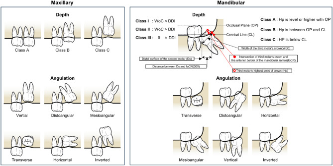
Optimal surgical methods require accurate prediction of extraction difficulty and complications. Although various automated methods related to third molar (M3) extraction have been proposed, none fully predict both extraction difficulty and post-extraction complications. This study proposes an automatic diagnosis method based on state-of-the-art semantic segmentation and classification models to predict the extraction difficulty of maxillary and mandibular M3s and possible complications (sinus perforation and inferior alveolar nerve (IAN) injury). A dataset of 4,903 orthopantomographys (OPGs), annotated by experts, was used. The proposed diagnosis method segments M3s (#18, #28, #38, #48), second molars (#17, #27, #37, #47), maxillary sinuses, and inferior alveolar canal (IAC) in OPGs using a segmentation model and extracts the region of interest (RoI). Using the RoI as input, the classification model predicts extraction difficulty and complication possibilities. The model achieved 87.97% and 88.85% accuracy in predicting maxillary and mandibular M3 extraction difficulty, with area under the receiver operating characteristic curve (AUROC) of 96.25% and 97.3%, respectively. It also predicted the possibility of sinus perforation and IAN injury with 91.45% and 88.47% accuracy, and AUROC of 91.78% and 94.13%, respectively. Our results show that the proposed method effectively predicts the extraction difficulty and complications of maxillary and mandibular M3s using OPG, and could serve as a decision support system for clinicians before surgery.
Keywords: Deep learning; Extraction difficulty; Orthopantomographys; Post-extraction complications; Third molar.
© 2025. The Author(s).
Conflict of interest statement
Declarations. Competing interests: The authors declare no competing interests. Code availability: The code for implementing this project is open-sourced at https://github.com/gist-ailab/man-max-third-molar .
Figures


References
-
- Hugoson, A. & Kugelberg, C. F. The prevalence of third molars in a Swedish population. An epidemiological study. Community Dent. Health. 5, 121–138 (1988). - PubMed
-
- Kim, H. J. et al. Anatomical risk factors of inferior alveolar nerve injury association with surgical extraction of mandibular third molar in Korean population. Appl. Sci.11, 816 (2021). - DOI
MeSH terms
LinkOut - more resources
Full Text Sources
Medical
