. 2025 May 30;15(1):18983.
doi: 10.1038/s41598-025-01736-2.

Chitosan attenuates titanium dioxide nanoparticles induced hepatic and renal toxicities

Affiliations

Chitosan attenuates titanium dioxide nanoparticles induced hepatic and renal toxicities

Amal Abdelmonem Halawa et al. Sci Rep. .

Abstract

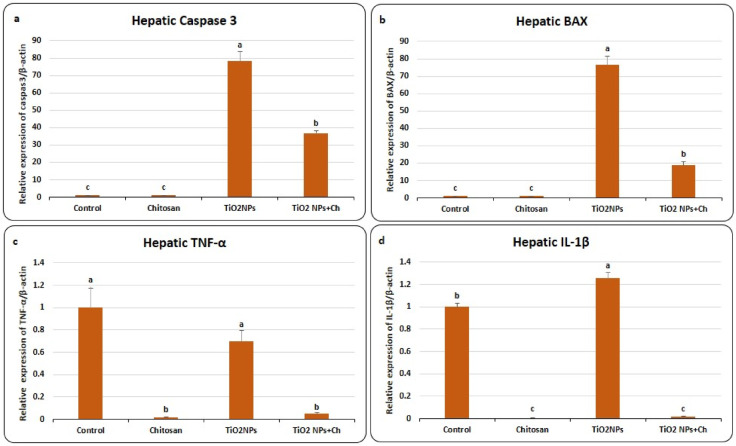
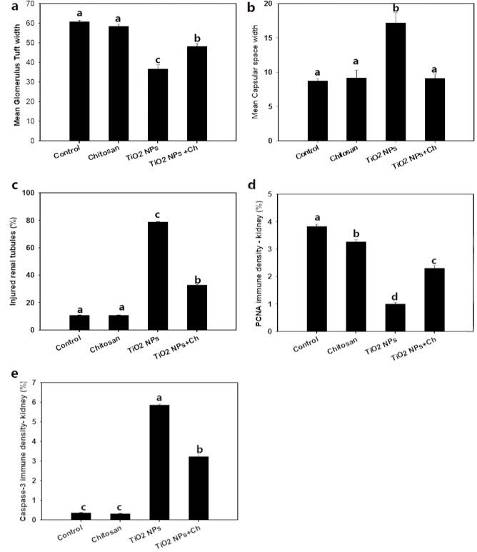
Titanium dioxide nanoparticles (TiO2 NPs) are extensively incorporated in numerous industrial products. Adult male Albino rats received oral TiO2 NPs at a dose of 150 mg/kg body weight for 14 days exhibited both hepatic and renal toxicities manifested by disruption in serum hepatic and renal biomarkers, imbalance in oxidative-antioxidant system, up-regulation of mRNA expression of genes encode inflammation (IL-1β, TNF-α) and apoptosis (Caspase-3, BAX) with down-regulation of PCNA immune-staining density and histological modifications in hepatic and renal architecture. Carboxymethyl chitosan (5 mg/kg BW) significantly improved the harmful effects of nano-titanium particles highlighting its relevance in reducing TiO2 NPs - induced hepatic and renal dysfunction.

Keywords: Chitosan; Nanotoxicology; Titanium dioxide nanoparticles.


Conflict of interest statement

Declarations. Competing interests: The authors declare no competing interests. Ethics approval: We confirmed that all experiments in this study were performed in accordance with the relevant guidelines and regulations.

Figures

Fig. 1
Fig. 1
Characterization of TiO2 nanoparticles, X-ray diffraction (XRD) peak of crystalline powder of TiO2.
Fig. 2
Fig. 2
Effects of TiO2 NPs and chitosan on mRNA expression of hepatic genes encode apoptosis; Caspase 3 (a) and BAX (b) and hepatic genes encode inflammation; TNFα (c) and IL-1β (d). Data were expressed as means ± SEM (n = 5). Bars with different superscripts means significant difference (p < 0.05).
Fig. 3
Fig. 3
Effects of TiO2 NPs and chitosan on relative expression of renal genes encode apoptosis, Caspase 3 (a) and BAX (b) and renal genes encode inflammation; TNFα (c) and IL-1β (d). Data were expressed as means ± SEM (n = 5). Bars with different superscripts means significant difference (p < 0.05).
Fig. 4
Fig. 4
Photomicrograph of rat’s hepatic tissue stained with H&E stain in various groups (1–4) showing normal lobule with normal mononucleated hepatocyte (thin black arrow), binucleated hepatocyte (thick black arrow), normal central vein (CV), normal hepatic sinusiod (curved black arrow), abnormal hepatic cord (green arrow), congested central vein (curved blue arrow), pycnotic nucleus (blue arrow), congested blood sinusiod (green arrow head). The photos (5–8) represent enzyme immunohistochemical staining of hepatic tissue for PCNA showing normal strong positive reaction in normal hepatocyte (red arrow), negative reaction in some hepatocyte (black arrowhead). The photos (9–12) represent enzyme immunohistochemical staining of hepatic tissue for Caspase-3 showing normal negative reaction of hepatocyte (black arrow head). Strong positive reaction of Caspase-3 antibody in the hepatocyte (red thin arrow).
Fig. 5
Fig. 5
Morphometric analysis comparison among the different groups including: (a) Normal hepatocyte, (b) PCNA density in liver and (c) Caspase-3 density in liver.Data are presented as means ± SD. Bars with different superscripts mean significant difference (p < 0.05).
Fig. 6
Fig. 6
Photomicrograph of rat’s renal cortex stained with H&E stain in various groups (1–4) showing normal corpuscles (curved black arrow), parietal layer (thin arrow), glomerulus tuft (G), normal proximal convoluted (thick arrow) and collecting (CT) tubules, shrunken corpuscles (green curved arrow), coagulative necrosis (green arrow), inflammatory cells (blue curved arrow), tubular cast (green arrow head). The photos (5–8) represent enzyme immunohistochemical staining of renal cortex tissues for PCNA showing normal strong positive reaction of PCNA antibody in the normal corpuscles and renal tubular cells (red arrow). Negative reaction of degenerated renal tubule (black arrowhead). The photos (9–12) represent enzyme immunohistochemical staining of renal cortex tissues for Caspase-3 showing normal negative reaction of normal corpuscle and renal tubule (red arrow). Strong positive reaction of Caspase-3 antibody in the degenerated renal tubular cells (black thin arrow) and shrunken glomerulus.
Fig. 7
Fig. 7
Morphometric analysis comparison among the different groups including: (a) glomerulus tuft width, (b) capsular space width, (c) injured renal tubule, (d) PCNA density in kidney and (e) Caspase-3 density in kidney. Data are presented as means ± SD. Bars with different superscripts mean significant difference (p < 0.05).

References

    1. Weir, A., Westerhoff, P., Fabricius, L. & Hristovski, K. Goetz, N. Titanium dioxide nanoparticles in food and personal care products. Environ. Sci. Technol.46, 2242–2250. 10.1021/es204168d (2012). von. - PMC - PubMed
    1. Leynen, N. et al. Assessing the in vivo toxicity of titanium dioxide nanoparticles in Schmidtea mediterranea: uptake pathways and (neuro)developmental outcomes. Aquat. Toxicol.270, 106895. 10.1016/j.aquatox.2024.106895 (2024). - PubMed
    1. Peters, R. J. B. et al. Characterization of titanium dioxide nanoparticles in food products: analytical methods to define nanoparticles. J. Agric. Food Chem.62, 6285–6293. 10.1021/jf5011885 (2014). - PubMed
    1. Shakeel, M. et al. Toxicity of Nano-Titanium dioxide (TiO2-NP) through various routes of exposure: a review. Biol. Trace Elem. Res.172, 1–36. 10.1007/s12011-015-0550-x (2016). - PubMed
    1. Xu, J. et al. Acute toxicity of intravenously administered titanium dioxide nanoparticles in mice. PLoS ONE. 8, e70618. 10.1371/journal.pone.0070618 (2013). - PMC - PubMed

LinkOut - more resources