. 2025 May 30;15(1):18982.
doi: 10.1038/s41598-025-02997-7.

The a subunit isoforms of V-ATPase are involved in glucose-dependent trafficking of insulin granules

Affiliations

The a subunit isoforms of V-ATPase are involved in glucose-dependent trafficking of insulin granules

Mizuki Sekiya et al. Sci Rep. .

Abstract

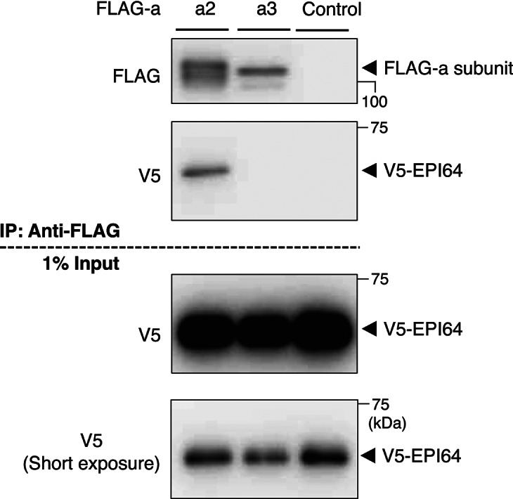
In pancreatic β cells, insulin granules move toward the plasma membrane to secrete insulin upon glucose stimulation, but the amount of secreted insulin is only a small portion of the total, and many granules do not release insulin. Here, using MIN6 cells derived from mouse pancreatic β cells, we observed that granules that moved toward the plasma membrane returned to the inner area after the stimulation was removed. This back-and-forth trafficking is likely important for strict regulation of insulin secretion in response to the blood glucose level. However, the mechanism was largely unknown. We found that "back" (inward) and "forth" (outward) trafficking was reduced in cells with knockdown of the a2 and a3 subunit isoforms of the proton pump V-ATPase, respectively. Interestingly, the amount of secreted insulin was increased in a2 knockdown cells. Both a2 and a3 interacted with GDP-bound form Rab27A, a member of the Rab small GTPase family that regulates insulin secretion. These results indicate that a2 and a3 are involved in back-and-forth trafficking of insulin granules, respectively. The a subunit isoforms of V-ATPase seem to determine the direction of insulin granule trafficking dependent on the glucose level.

Keywords: Insulin secretion; Membrane trafficking; Rab27A; V-ATPase; a subunit isoforms.


Conflict of interest statement

Declarations. Competing interests: The authors declare no competing interests.

Figures

Fig. 1
Fig. 1
Interaction of a subunit isoforms with Rab27A and A subunit. (A) Schematic illustration of the subunits of the mammalian V-ATPase. (B) Interaction of a subunit isoforms with Rab27A. FLAG-tagged a isoforms and various V5-fused forms of Rab27A were co-expressed in HEK293T cells. The cells were lysed, and lysates were immunoprecipitated with an anti-FLAG antibody. The precipitates were analyzed using antibodies against FLAG (upper panel) and V5 (middle panel). As a control, cells were co-transfected with an empty vector and a recombinant plasmid harboring V5-fused Rab27A (Control). WT, T23N, and Q78L indicate wild-type, GDP-bound form, and GTP-bound form Rab27A, respectively. In addition, 5% of the cell lysate was subjected to western blotting with an anti-V5 antibody (lower panel). Original blots are presented in Supplementary Fig. 7. The blots in (B) were cut horizontally at ~ 60 kDa. Upper and lower membranes were subjected to hybridization with anti-FLAG and anti-V5 antibodies, respectively. A full-length membrane was cut because the amount of protein in immunoprecipitation samples was insufficient to prepare multiple full-length membranes. (C) Interaction of the a2 subunit and Rab27A with the A subunit in V1. FLAG-Rab27A(T23N) (lanes 1–3) and FLAG-a2 (lanes 4–6) were co-expressed with V5-a2 and V5-Rab27A(T23N), respectively. Immunoprecipitation was performed as described in (B). Original blots are presented in Supplementary Fig. 8. D Interaction of the a3 subunit and Rab27A with the A subunit in V1. FLAG-Rab27A(T23N) (lanes 1–3) and FLAG-a3 (lanes 4–6) were co-expressed with V5-a3 and V5-Rab27A(T23N), respectively. Immunoprecipitation was performed as described in (B). Original blots are presented in Supplementary Fig. 8. (E) Presence of a2 and a3 isoforms and Rab27A in the membrane fraction. FLAG-Rab27A(T23N) and V5-a isoforms were co-expressed in HEK293T cells. The cells were lysed and centrifuged at 100,000 × g for 30 min to separate the membrane (M) and soluble (S) fractions. Immunoprecipitation was performed as described in (B). α-Tubulin was used as a control cytosolic protein. Original blots are presented in Supplementary Fig. 9.
Fig. 2
Fig. 2
Localizations of insulin and Rab27A in the MIN6 mouse pancreatic β cell line. (A) Effect of the glucose concentration on localizations of insulin and V5-Rab27A. Cells expressing V5-Rab27A were sequentially incubated with 2.2 mM glucose for 60 min, 20 mM glucose for 5 min, and 2.2 mM glucose for 5 min, and then stained with antibodies specific for insulin (magenta) and V5 (green). Merged images are also shown. (B) Peripheral localization of insulin. The cell peripheral area was defined as the region between the cell edge (yellow solid line) and 0.75 µm inside from this edge (yellow dotted line). Cells with peripheral insulin or V5-Rab27A were defined as those in which the average signal intensity of insulin or V5-Rab27A in the cell peripheral area was more than twice that in the other area. (C) Cells with peripheral insulin and V5-Rab27A according to the glucose concentration. The percentages of cells with peripheral insulin (magenta) and V5-Rab27A (green) as defined in (B) were calculated at each glucose concentration. Data are means ± S.E. from four independent experiments. n > 30 cells in each experiment. (D) Localization of Rab27A to insulin granules. Co-localization was quantified as the percentage of insulin-positive pixels that were also Rab27A-positive. More than 30 cells were randomly selected from four experiments. Data are means ± S.E.
Fig. 3
Fig. 3
Localization of insulin granules in a2KD MIN6 cells. (A) Expression of a2 in a2KD#1 cells. MIN6 cells were transduced with pSIREN-RetroQ-negative control shRNA (control) or pSIREN-RetroQ-a2-shRNA#1 (a2KD#1). a2KD#1 cells were further transduced with pMX-V5-a2 (a2KD#1 + V5-a2). Endogenous a2 and V5-a2 were detected with an antibody specific for a2 (left upper panel). β-actin was also detected using a corresponding antibody (left lower panel). Original blots are presented in Supplementary Fig. 10. The blots were cut horizontally at ~ 75 kDa. Upper and lower membranes were subjected to hybridization with anti-a2 and anti-β-actin antibodies, respectively. A full-length membrane was cut to conserve the anti-a2 antibody, which is commercially unavailable. Relative signal intensities of a2 were calculated as means ± S.E. from three independent experiments compared with control cells (set to 100%) (right panel). (B) Localizations of insulin and V5-Rab27A in control (upper panels) and a2KD#1 (lower panels) cells. Cells expressing V5-Rab27A were sequentially incubated with 2.2 mM glucose for 60 min, 20 mM glucose for 5 min, and 2.2 mM glucose for 5 min, and then stained with antibodies specific for insulin (magenta) and V5 (green). Merged images are also shown. (CE) Cells with peripheral insulin and Rab27A according to the glucose concentration. The percentage of cells with peripheral insulin (C, E) or Rab27A (D) was calculated as described in Fig. 2B. Control cells, open squares (insulin), open circles (Rab27A); a2KD#1 cells, closed squares (insulin), closed circles (Rab27A); a2KD#1 + V5-a2 cells, shaded squares (insulin). Data are means ± S.E. from more than three independent experiments. n > 30 cells in each experiment. *p < 0.05, (A) unpaired two-tailed Student’s t-test, (CE) Unpaired multiple t test (Holm-Šídák method).
Fig. 4
Fig. 4
Effects of colchicine on localizations of insulin and Rab27A in MIN6 cells. (A) Localizations of insulin and V5-Rab27A in cells treated with colchicine. Cells expressing V5-Rab27A were preincubated with 2.2 mM glucose for 60 min, and then, in the absence (colchicine (–), upper panels) or presence (colchicine ( +), lower panels) of 2 μM colchicine, sequentially incubated with 2.2 mM glucose for 15 min, 20 mM glucose for 5 min, and 2.2 mM glucose for 5 min. The treated cells were stained with antibodies specific for insulin (magenta) and V5 (green). Merged images are also shown. (B) Effects of colchicine on the glucose-dependent peripheral localization of insulin. The percentage of cells with peripheral insulin was calculated in the absence (colchicine (–), open squares) and presence (colchicine ( +), closed squares) of 2 μM colchicine. Data are means ± S.E. from more than three independent experiments. n > 30 cells in each experiment. *p < 0.05, unpaired multiple t test (Holm-Šídák method).
Fig. 5
Fig. 5
Insulin secretion, morphology of insulin granules, and expression of insulin and its related genes in control and a2KD MIN6 cells. (A) Total insulin content in control (open bars) and a2KD#1 (closed bars) cells. (B) Insulin secretion from control (open bars) and a2KD#1 (closed bars) cells. Cells were treated with KRH buffer containing 2 or 20 mM glucose for 2 h, and then the insulin content in the recovered medium and acid ethanol extracts was determined using an ELISA kit. Insulin secretion from control cells treated with 2 mM glucose was set to 1. Data are means ± S.E. from four independent experiments. (C) Electron micrographs of control (upper panels) and a2KD#1 (lower panels) cells in HEPES-buffered Krebs buffer containing 2.2 mM glucose. Magnified images of the boxed areas are shown in the right panels. Arrows indicate typical insulin granules with a dense core. (D) Expression of insulin and insulin-related genes in control (open bars) and a2KD#1 (closed bars) cells. Total mRNA was isolated from cells in HEPES-buffered Krebs buffer containing 2.2 mM glucose. The expression levels of Ins1, Ins2, PC1/3, and PC2 were quantified by real-time RT-PCR using β-actin as an internal standard, with expression in control cells set to 1. Ins1, insulin 1; Ins2, insulin 2; PC1/3, prohormone convertase 1/3; PC2, prohormone convertase 2. Data are means ± S.E. from three independent experiments. *p < 0.05, unpaired two-tailed Student’s t-test.
Fig. 6
Fig. 6
Interaction of a2 and a3 with EPI64, the GAP for Rab27A. FLAG-a2 or FLAG-a3 and V5-EPI64 were co-expressed in HEK293T cells. The cells were lysed, and lysates were immunoprecipitated with an anti-FLAG antibody. The precipitates were analyzed using antibodies against FLAG (top panel) and V5 (second panel from the top). In addition, 1% of the cell lysate was subjected to western blotting with an anti-V5 antibody (bottom two panels). Original blots are presented in Supplementary Fig. 11.
Fig. 7
Fig. 7
Localization of insulin granules in control and a3KD MIN6 cells. (A) Expression of a3 in a3KD cells. Cells were transduced with pSIREN-RetroQ-negative control shRNA (control) or pSIREN-RetroQ-a3-shRNA (a3KD). a3KD cells were further transduced with pMX-V5-a3 (a3KD + V5-a3). Endogenous a3 and V5-a3 were detected with an antibody specific for a3 (left upper panel). β-actin was also detected using a corresponding antibody (left lower panel). Original blots are presented in Supplementary Fig. 12. Relative signal intensities of a3 were calculated as means ± S.E. from three independent experiments compared with control cells (set to 100%) (right panel). (B) Localizations of insulin and V5-Rab27A in control (upper panels) and a3KD (lower panels) cells. Cells expressing V5-Rab27A were sequentially incubated with 2.2 mM glucose for 60 min, 20 mM glucose for 5 min, and 2.2 mM glucose for 5 min, and then stained with antibodies specific for insulin (magenta) and V5 (green). Merged images are also shown. (CE) Cells with peripheral insulin and Rab27A according to the glucose concentration. The percentage of cells with peripheral insulin (C, E) or Rab27A (D) was calculated as described in Fig. 2B. Control cells, open squares (insulin), open circles (Rab27A); a3KD cells, closed squares (insulin), closed circles (Rab27A); a3KD#1 + V5-a3 cells, shaded squares (insulin). Data are means ± S.E. from three independent experiments. n > 30 cells in each experiment. *p < 0.05, (A) unpaired two-tailed Student’s t-test, (CE) Unpaired multiple t test (Holm-Šídák method).
Fig. 8
Fig. 8
Localization of Rab27A to insulin granules and insulin secretion in control and a3KD MIN6 cells. (A) Effect of a3 on the localization of Rab27A to insulin granules. Control and a3KD cells expressing V5-Rab27A were incubated with 2.2 mM glucose for 60 min and stained with antibodies specific for insulin (magenta) and V5 (green). Merged images are also shown. (B) Co-localization was quantified as the percentage of insulin-positive pixels that were also Rab27A-positive. Open and closed bars indicate control and a3KD cells, respectively. More than 30 cells were randomly selected from three experiments using WT and a3KD cells. Data are means ± S.E. (C) Total insulin content in control (open bars) and a3KD (closed bars) cells. (D) Insulin secretion from control (open bars) and a3KD (closed bars) cells. Cells were treated with KRH buffer containing 2 or 20 mM glucose for 2 h, and then the insulin content in the recovered medium and acid ethanol extracts was determined using an ELISA kit. Insulin secretion from control cells treated with 2 mM glucose was set to 1. Data are means ± S.E. from four independent experiments. *p < 0.05, unpaired two-tailed Student’s t-test.

Similar articles

Cited by

References

    1. Chatterjee Bhowmick, D., Ahn, M., Oh, E., Veluthakal, R. & Thurmond, D. C. Conventional and unconventional mechanisms by which exocytosis proteins oversee beta-cell function and protection. Int. J. Mol. Sci.22, 1833 (2021). - PMC - PubMed
    1. Izumi, T. Multiple pathways and independent functional pools in insulin granule exocytosis. Genes Cells28, 471–481 (2023). - PMC - PubMed
    1. Ohara-Imaizumi, M. & Nagamatsu, S. Insulin exocytotic mechanism by imaging technique. J. Biochem.140, 1–5 (2006). - PubMed
    1. Seino, S., Shibasaki, T. & Minami, K. Dynamics of insulin secretion and the clinical implications for obesity and diabetes. J. Clin. Invest.121, 2118–2125 (2011). - PMC - PubMed
    1. Axelsson, A. S. et al. Sox5 regulates beta-cell phenotype and is reduced in type 2 diabetes. Nat. Commun.8, 15652 (2017). - PMC - PubMed

MeSH terms

LinkOut - more resources