. 2025 May 30;15(1):18984.
doi: 10.1038/s41598-025-00880-z.

Neuronal hyperexcitability in dystrophin-deficient mdx hippocampal neurons: the importance of interleukin-6 and GABAergic regulation

Affiliations

Neuronal hyperexcitability in dystrophin-deficient mdx hippocampal neurons: the importance of interleukin-6 and GABAergic regulation

Kimberley A Stephenson et al. Sci Rep. .

Abstract

Duchenne Muscular Dystrophy (DMD) is a severe neuromuscular disorder arising from loss of the structural protein, dystrophin. It also often presents with cognitive deficits and susceptibility to epilepsy. Expressed in neurons of the hippocampus, dystrophin plays an important role in synapse formation, specifically the post-synaptic organisation of γ-aminobutyric acid A receptors (GABAARs). This study explored possible interactions between interleukin (IL)-6, which is elevated in DMD, and GABAAR signalling in cultured hippocampal neurons of dystrophic mdx mice. Immunofluorescent imaging revealed altered development of network connectivity that displayed similar characteristics to dystrophin-expressing neurons cultured in elevated levels of IL-6. Mdx neurons dependably exhibited spontaneous oscillations. Calcium (Ca2+) signalling was further modulated by exposure to agonists and antagonists of GABAA and GABABRs. IL-6-evoked Ca2+ responses were enhanced by muscimol, a GABAAR agonist, in wildtype (WT) and mdx neurons, whilst bicuculline, a GABAAR antagonist, only suppressed IL-6-evoked Ca2+activity in WT neurons. The GABABR agonist, baclofen, enhanced IL-6-evoked Ca2+ responses only in mdx neurons. Our findings support dysfunctional GABAergic signalling in hippocampal neurons that lack dystrophin, resulting in aberrant neuronal network excitability. The contribution of elevated levels of IL-6 further impact upon Ca2+ dyshomeostasis in dystrophic neurons and may underpin cognitive changes reported in dystrophinopathies.

Keywords: Duchenne muscular dystrophy; Dystrophin; GABA; Hippocampus; Learning; Memory.


Conflict of interest statement

Declarations. Competing interests: The authors declare no competing interests. Ethics approval: All animal experiments were approved and performed following guidelines set out by the HPRA (Health Products Regulatory Authority), Ireland and following project authorisation (AEI9130/P088), as well as individual authorisation (AEI9130/I303). Animals were euthanized in accordance with European Directive 2010/63/EU.

Figures
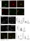
Fig. 1
Fig. 1
Elevated IL-6 increased dystrophin-expressing synaptic connections in cultured hippocampal neurons (A) The images show immunofluorescently-labelled hippocampal neurons cultured from C57BL/6 mice. Neurons have been labelled with an antibody (MANDRA-1) that identifies both Dp427 & Dp71 isoforms of dystrophin (green staining) and the Dp427 isoform alone (red staining) and (B) red MANDRA-1 staining co-expressed with the post-synaptic marker, PSD-95 (green staining). Arrows indicated co-expression of dystrophin with the post-synaptic marker (yellow labelling). (C) The representative immunofluorescent images and data plots illustrate the intensity (corrected total cell fluorescence, CTCF) of IL-6 staining in the neuronal cell bodies and processes under control culture conditions (black circles) and in culture medium supplemented with IL-6 (1 nM, > 48 h, grey open circles). Arrows indicate punctate expression of IL-6 in the cell bodies and processes. (D) The images and data plot illustrates the number of discrete clusters of dystrophin/10 µm in the processes of neurons cultured under control conditions or in culture medium supplemented with IL-6. Arrows highlight the increased expression of dystrophin clusters in treated neurons. (E) The images and data plots show clustering of IL-6 receptor (IL-6R) labelling (green staining) at synapses (indicated by arrows) labelled with the pre-synaptic marker, synaptophysin (red staining). The merged images illustrate regions where labelling is co-expressed (yellow staining). The number of IL-6R clusters and synaptophysin-labelled synapses under control and IL-6-exposed conditions are illustrated in the pooled data. * and ***p < 0.05 and p < 0.001, respectively. Scalebars: 35 µm.
Fig. 2
Fig. 2
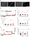
Loss of dystrophin is associated with neuronal hyperexcitability (A) The brightfield and immuno-labelled images show NMDA receptor (NMDA R) expression in wildtype (WT) and mdx cultured hippocampal neurons. Scalebar: 35 µm. (B) The dot plots show pooled data of the (i) frequency, (ii) amplitude and (iii) duration (full width, half max: FWHM) of spontaneous Ca2+ peaks in WT (black circles) and mdx (red triangles) cultured neurons under basal conditions. (C) The representative Ca2+ traces illustrate increased spontaneous Ca2+ activity in mdx hippocampal neuronal cultures (red traces) in comparison to WT neurons (black/grey traces) and traces and (D) The data plot illustrates suppression of spontaneous neuronal activity in dystrophic mdx hippocampal neurons following incubation with the neurotoxin, tetrodotoxin (TTX). (E) The data plot illustrates enhanced Ca2+ responses induced by high (50 mM) K+ in mdx neurons. ** and ***p < 0.01 and p < 0.001, respectively.
Fig. 3
Fig. 3
Dystrophic mdx neurons have increased numbers of interleukin (IL)-6 receptor-expressing synaptic contacts but similar Ca2+ responses (A) The immunofluorescent images and data plots illustrate the intensity of IL-6 staining (corrected total cell fluorescence, CTCF) in the cell bodies and processes of hippocampal neurons from wildtype (WT, black circles) and mdx (red triangles) neurons. Scalebar: 35 µm. (B) The immunofluorescent images and data plot show the number of IL-6 receptor (green staining) clusters located at synapses identified using anti-synaptophysin (red staining) /10 µm of neuronal processes in WT and mdx neurons. Yellow staining in the merged images indicates co-localisation. Scalebar: 35 µm. (C) (i) The representative traces from WT (black trace) and mdx (red trace) and the dot plots of pooled data illustrating (ii) the changes in amplitude and (iii) frequency of neuronal intracellular Ca2+ oscillations following application of IL-6. (D) (i) The representative traces and (ii) dot plot from WT and mdx neurons show suppression of the IL-6-evoked response in mdx neurons. *, ** and ***p < 0.05, p < 0.01 and p < 0.001, respectively.
Fig. 4
Fig. 4
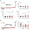
IL-6-evoked Ca2+ responses are enhanced in cultured WT and mdx hippocampal neurons when exposed to the GABAA receptor agonist, muscimol (A)The representative brightfield and immuno-labelled images of cultured hippocampal neurons from wildtype (WT) and dystrophin-deficient, mdx mice illustrate expression of GABAA receptors in the cell bodies and processes of these neurons. Scalebar: 35 µm. (B) (i) The representative Ca2+ traces illustrate spontaneous activity in WT (black trace) and mdx (red trace) cultured neurons in the absence, presence, and following washout of the GABAA receptor agonist, muscimol (1 µM, 20 min). The data plots of pooled data show the relative change (from baseline) of the (ii) frequency, (iii) amplitude and (iv) duration (time between 1/2 amplitude on ascent and descent of the transient; full width, half max (FWHM)) of Ca2+ oscillations in mdx hippocampal neurons (red triangles) as compared to WT (black circles) neurons. (C) (i) The representative traces from WT and mdx neurons and (ii) dot plot illustrate the enhanced response to IL-6 (1 nM, 3 min) in the presence of muscimol. *, ** and ***p < 0.05, p < 0.01 and p < 0.001, respectively.
Fig. 5
Fig. 5
IL-6-evoked Ca2+ responses in the presence of the GABAA receptor antagonist, bicuculline, is suppressed in WT hippocampal neurons (A) (i) The representative Ca2+ traces from WT (black trace) and mdx (red trace) hippocampal neurons and the data plots show the effects of the GABAA receptor antagonist, bicuculline (100 µM, 20 min exposure) on the relative change in (ii) frequency, (iii) amplitude and (iv) duration (time between 1/2 amplitude on ascent and descent of the transient; full width, half max (FWHM)) of Ca2+ oscillations. (B) (i) The representative traces and (ii) dot plot illustrate suppression of the Ca2+ responses to IL-6 (1 nM, 3 min) in the presence of bicuculline, only in WT neurons. *, ** and ***p < 0.05, p < 0.01 and p < 0.001, respectively.
Fig. 6
Fig. 6
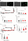
The GABAB receptor agonist, baclofen, potentiated IL-6-evoked Ca2+ responses in mdx hippocampal neurons (A) The representative brightfield and immuno-labelled images of cultured hippocampal neurons from wildtype (WT) and dystrophin-deficient mdx mice illustrate expression of GABAB receptors in the cell bodies and processes of these neurons. Scalebar: 35 µm. (B) (i) The representative Ca2+ traces from wildtype (WT, black traces) and mdx (red traces) cultured hippocampal neurons illustrate neuronal activity in the absence, presence, and following washout, of the GABAB receptor agonist, baclofen (100 µM, 20 min). The data plots of pooled data show the relative change (from baseline) of the (ii) frequency, (iii) amplitude and (iv) duration (time between 1/2 amplitude on ascent and descent of the transient; full width, half max (FWHM)) of Ca2+ oscillations in WT (black circles) and mdx hippocampal (red triangles) neurons. (C) (i) The representative traces and (ii) dot plot illustrate the enhanced response to IL-6 (1 nM, 3 min) in the presence of baclofen in mdx neurons. (D) The dot plot illustrates suppression of the amplitude of Ca2+ response evoked by high K+ in mdx neurons after exposure to baclofen. *, ** and ***p < 0.05, p < 0.01 and p < 0.001, respectively.
Fig. 7
Fig. 7
The GABAB receptor antagonist, phaclofen, had no effect on IL-6-evoked Ca2+ responses in WT and mdx hippocampal neurons (A) (i) The Ca2+ traces and the data plots show the effects of the GABAB receptor antagonist, phaclofen (100 µM, 20 min exposure), on the relative change in (ii) frequency, (iii) amplitude and (iv) duration (time between 1/2 amplitude on ascent and descent of the transient; full width, half max (FWHM)) of Ca2+ oscillations in WT and mdx hippocampal neurons. (B) (i) The representative traces and ii dot plot show that Ca2+ responses to IL-6 (1 nM, 3 min) were not altered in the presence of phaclofen in WT or mdx neurons. *p < 0.05.
Fig. 8
Fig. 8
Mdx mice exhibit increased freezing behaviours (A) The frequency of freezing (mouse did not move for more than 2 s) in the open field was increased in mdx mice as compared to wildtype (WT) controls. Other anxiety-related behaviours such as (B) the frequency of exploration of the exposed inner zone of the open field (number of times the mouse had four paws in the central area) and (C) stress-induced faecal excretion was similar in WT and mdx comparators. Exploration and escape behaviours such as the frequency of (D) rearing (number of times the mouse reared its front paws, against the wall or mid-air) and (E) jumping (number of times the mouse jumped with all four paws in the air) was similar in both strains. (F) Compulsive behaviours, such as repetitive grooming was similar in both strains. (G) The discrimination index, calculated from a novel object recognition trial, was similar in WT and mdx mice *p < 0.05.

Similar articles

References

    1. Lynch, G. S. Role of contraction-induced injury in the mechanisms of muscle damage in muscular dystrophy. Clin. Exp. Pharmacol. Physiol.31, 557–561 (2004). - PubMed
    1. Tulangekar, A. & Sztal, T. E. Inflammation in duchenne muscular dystrophy-exploring the role of neutrophils in muscle damage and regeneration. Biomedicines10.3390/biomedicines9101366 (2021). - PMC - PubMed
    1. McDonald, C. M. et al. Long-term effects of glucocorticoids on function, quality of life, and survival in patients with Duchenne muscular dystrophy: A prospective cohort study. Lancet391, 451–461 (2018). - PubMed
    1. Rae, M. G. & O’Malley, D. Cognitive dysfunction in Duchenne muscular dystrophy: A possible role for neuromodulatory immune molecules. J. Neurophysiol.116, 1304–1315 (2016). - PMC - PubMed
    1. Felisari, G. et al. Loss of Dp140 dystrophin isoform and intellectual impairment in Duchenne dystrophy. Neurology55, 559–564 (2000). - PubMed

MeSH terms

LinkOut - more resources