. 2025 May 30;15(1):18981.
doi: 10.1038/s41598-025-03879-8.

Role of galectin-9 in the development of gestational diabetes mellitus

Affiliations

Role of galectin-9 in the development of gestational diabetes mellitus

Haya Hamed Hassan Albuayjan et al. Sci Rep. .

Abstract

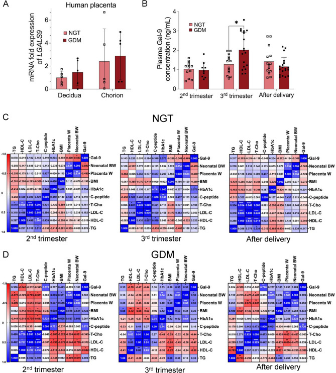
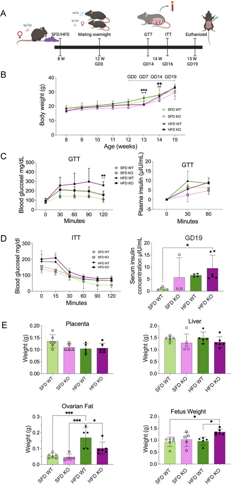
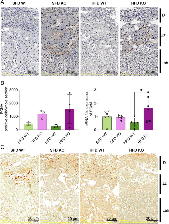
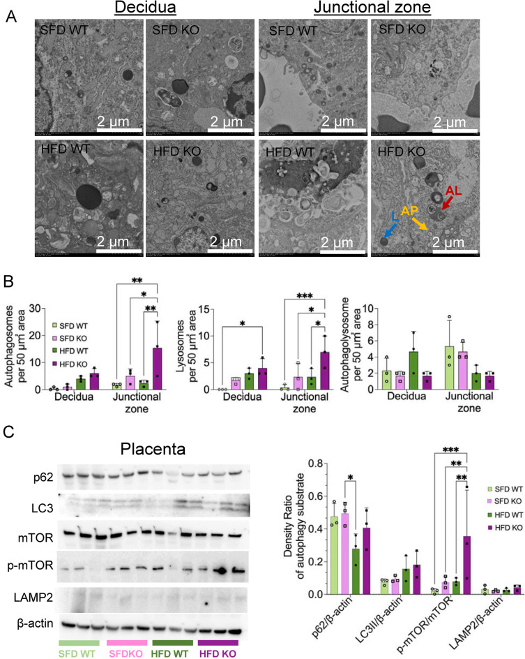
Galectin-9 (Gal-9) is highly expressed in trophoblasts in placenta. Interaction between Gal-9 and T-cell immunoglobulin and mucin-domain containing-3 (Tim-3) is important for the differentiation of tissue resident natural killer (trNK) cells in placenta and maintenance of normal pregnancy. Furthermore, the enhanced maternal systemic inflammation associated with increased proinflammatory cytokines in preeclampsia is mediated by enhanced interaction between Gal-9 and Tim-3. However, the role of Gal-9 in gestational diabetes (GDM) remains unexplored. Plasma Gal-9 levels were elevated at 3rd trimester in pregnant women with GDM and positively correlated with placenta and newborn weight. Lgals9 knockout pregnant mice fed with high fat diet (HFD KO) demonstrated maternal glucose intolerance and fetus macrosomia compared with controls (HFD WT). In HFD KO, increased proliferating cells, reduced apoptosis, and autophagy impairment were observed in junctional zones. The number of trNK cells and percentage of Tim-3 + trNK increased, while early apoptosis percentage in Tim-3 + trNK was reduced in placenta of HFD KO. The elevation of plasma Gal-9 may be a biomarker for prediction of maternal glucose intolerance and fetal macrosomia in pregnant women with GDM and Gal-9 functions as a compensation factor for GDM by inducing apoptosis in Tim-3 + trNK cells.


Conflict of interest statement

Declarations. Competing interests: Jun Wada receives speaker honoraria from Astra Zeneca, Bayer, Boehringer Ingelheim, Daiichi Sankyo, Kyowa Kirin, Novo Nordisk, and Mitsubishi Tanabe, and receives grant support from Bayer, Chugai, Kyowa Kirin, Otsuka, Shionogi, Sumitomo, and Mitsubishi Tanabe. All other authors have nothing to disclose.

Figures

Fig. 1
Fig. 1
Galectin-9 in pregnant women with normal glucose tolerance (NGT) and gestational diabetes mellitus (GDM). (A) LGALS9 mRNA expression in decidua and chorion tissues of human placenta in NGT (n = 5) and GDM (n = 5). (B) Plasma Gal-9 concentrations in NGT (n = 14) and GDM (n = 19) at 2nd trimester, 3rd trimester, and after delivery. Data are shown as mean ± SD and analyzed by unpaired t-tests. C. Pearson correlation coefficient of plasma Gal-9 with various clinical parameters in NGT (n = 14) and GDM (n = 19) at 2nd trimester, 3rd trimester, and after delivery. (*p < 0.05, **p < 0.01, ***p < 0.001)
Fig. 2
Fig. 2
Glucose metabolism in B6-Lgals9tm1glp (KO) and wild type (WT) pregnant mice. (A) Experimental design. WT (w/w) and KO (-/-) female mice were fed with standard fat diet (SFD, 10% fat) or high fat diet (HFD, 60% fat) and mated with WT male mice. GD, gestational day; W, weeks of age; GTT, glucose tolerance test; ITT, insulin tolerance test. Created in BioRender. H, H. (2025) (B) Body weight of SDF WT (n = 14), SDF KO (n = 10), HFD WT (n = 15), and HFD KO (n = 9) pregnant mice. Body weight in HFD KO mice at GD7 (21.18 ± 0.69 g) was significantly lower than HFD WT mice (25.71 ± 2.43) (P = 0.0005). The body weight in SFD KO mice at GD14 (24.23 ± 1.09) was lower than SFD WT (28.41 ± 4.68) (P = 0.0025). (C) Glucose levels [SFD WT (n = 7), SFD KO (n = 5), HFD WT (n = 8), and HFD KO (n = 6)] and plasma insulin levels [SFD WT (n = 6), SFD KO (n = 5), HFD WT (n = 5), and HFD KO (n = 6)] in GTT at GD14. (D) Glucose levels [SFD WT (n = 7), SFD HFD (n = 8), SFD KO (n = 5), and HFD KO (n = 7)] in ITT at GD16 and serum fasting insulin levels [SFD WT (n = 6), SFD KO (n = 4), HFD WT (n = 4), and HFD KO (n = 6)] at GD19. (E) Placenta, liver, ovarian fat and fetus weight of WT and KO pregnant mice fed with SFD or HFD at GD19. Data are shown as mean ± SD and analyzed by one-way ANOVA with Tukey test (*p < 0.05, **p < 0.01, ***p < 0.001).
Fig. 3
Fig. 3
Expression of Gal-9 in placenta derived from B6-Lgals9tm1glp (KO) and wild type (WT) pregnant mice. (A) Immunohistochemical staining of Gal-9 in placenta at gestational day (GD19). Gal-9 is observed in the cytoplasm and cell surface of trophoblast giant cells (TGC, red arrows) and spongiotrophoblast cells (SP, yellow arrows) in SFD WT, SFD KO, HFD WT, and HFD KO mice. D, decidua; JZ, junctional zone; Lab, labyrinth. B. Western blot analysis of Gal-9 in placenta at GD19 and densitometric analyses (n = 3 in each group). C. mRNA expression of Lgals9 normalized by Rplp0 and Gapdh in placenta, liver, and ovarian fat derived from SFD WT (n = 6), SFD KO (n = 5), HFD WT (n = 5), and HFD KO (n = 6). Data are shown as mean ± SD and analyzed by one-way ANOVA with Tukey test (*p < 0.05; **p < 0.01). SFD, standard fat diet, 10% fat; HFD, high fat diet, 60% fat.
Fig. 4
Fig. 4
Proliferating cell nuclear antigen (PCNA) and apoptosis assay in placenta derived from B6-Lgals9tm1glp (KO) and wild type (WT) pregnant mice. (A) Immunohistochemical staining of PCNA in placenta at gestational day 19 (GD19). (B) The counts of PCNA positive cells in whole placenta sections in SFD WT (n = 3), SFD KO (n = 3), HFD WT (n = 3), and HFD KO (n = 4), and mRNA expression of Pcna normalized by Rplp0 in SFD WT (n = 5), SFD KO (n = 5), HFD WT (n = 5), and HFD KO (n = 6). (C) Terminal deoxynucleotidyl transferase dUTP nick-end labeling (TUNEL assay) of placenta at GD19. The apoptotic cells are labeled by 3,3′-Diaminobenzidine (DAB). SFD, standard fat diet, 10% fat; HFD, high fat diet, 60% fat.
Fig. 5
Fig. 5
Autophagy in placenta derived from B6-Lgals9tm1glp (KO) and wild type (WT) pregnant mice. (A) Electron micrographs of placenta at gestational day 19 (GD19) showing decidua and junctional zone. AP, autophagosome (yellow arrow); L, lysosomes (blue arrow); AL, autophagolysosomes (red arrow). (B) The number of autophagosome, lysosomes and autolysosomes per 50 µm2 area of decidua and junctional zone. (C) Western blots and densitometric analyses of p62, microtubule-associated protein 1 A/1B-light chain 3 (LC3), mammalian target of rapamycin (mTOR), phosphorylated mTOR (p-mTOR) and β-actin. Data shown as mean ± SD and analyzed by one-way ANOVA with Tukey test (*p < 0.05, **p < 0.01 and ***p < 0.001). SFD, standard fat diet, 10% fat; HFD, high fat diet, 60% fat.
Fig. 6
Fig. 6
Flow cytometry analysis in placenta derived from B6-Lgals9tm1glp (KO) and wild type (WT) pregnant mice. (A) Percentage of CD45+ CD3 CD49b+ conventional NK (cNK) cells in total leukocytes. (B) Percentage of CD45+ CD3 CD49b CD49a+ trNK (tissue-resident NK) cells in total leukocytes. (C) Percentage of CD45+ CD3 CD49b CD49a+ C366+ (Tim-3+) trNK cells. Tim-3, T-cell immunoglobulin domain and mucin domain-containing molecule-3. (D) Percentage of Zombie Annexin+ early apoptotic trNK cells in Tim-3 + trNK cells. (E) mRNA expression of Cd244, Havcr2 (Tim-3), Eomes, Tnfa, Il6, and Il10. Data shown as mean ± SD and analyzed by one-way ANOVA with Tukey test (*p < 0.05). N = 4–6 in each group. SFD, standard fat diet, 10% fat; HFD, high fat diet, 60% fat.

References

    1. McElwain, C. J., McCarthy, F. P. & McCarthy, C. M. Gestational diabetes mellitus and maternal immune dysregulation: what we know so Far. Int. J. Mol. Sci.22 (2021). - PMC - PubMed
    1. Sweeting, A., Wong, J., Murphy, H. R. & Ross, G. P. A clinical update on gestational diabetes mellitus. Endocr. Rev.43, 763–793 (2022). - PMC - PubMed
    1. Chen, M. et al. Galectins: important regulators in normal and pathologic pregnancies. Int. J. Mol. Sci23 (2022). - PMC - PubMed
    1. Wada, J., Ota, K., Kumar, A., Wallner, E. I. & Kanwar, Y. S. Developmental regulation, expression, and apoptotic potential of galectin-9, a beta-galactoside binding lectin. J. Clin. Invest.99, 2452–2461 (1997). - PMC - PubMed
    1. Menkhorst, E. et al. Medawar’s postera: galectins emerged as key players during Fetal-Maternal glycoimmune adaptation. Front. Immunol.12, 784473 (2021). - PMC - PubMed

MeSH terms

LinkOut - more resources