. 2025 May 30;15(1):19094.
doi: 10.1038/s41598-025-04603-2.

Broadly reactive monoclonal antibodies against beta-lactamases for immunodetection of bacterial resistance to antibiotics

Affiliations

Broadly reactive monoclonal antibodies against beta-lactamases for immunodetection of bacterial resistance to antibiotics

Karolina Bielskė et al. Sci Rep. .

Abstract

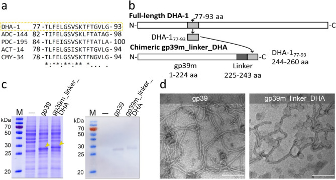
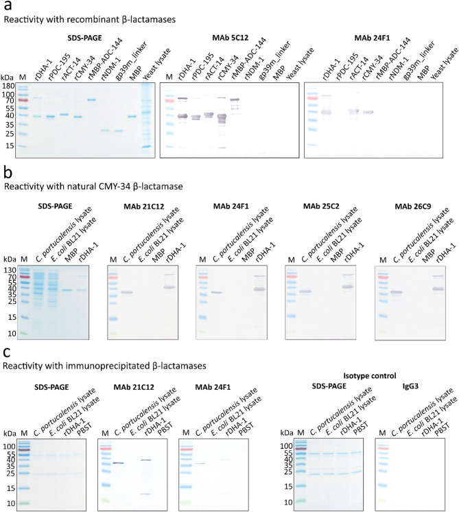
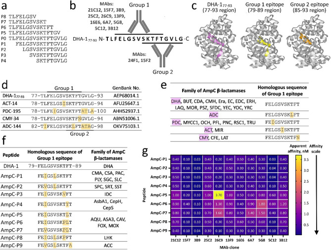
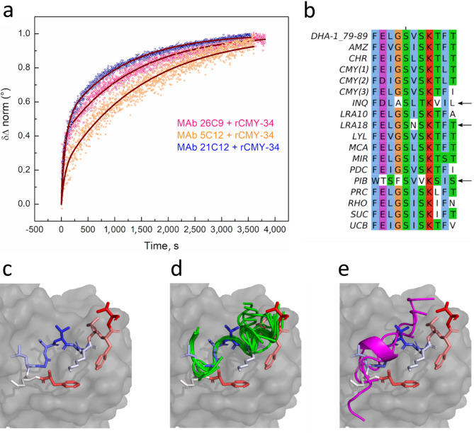
With antibiotic resistance reaching alarming levels globally, rapid detection of resistance determinants is crucial for administering appropriate antimicrobial therapies. This study aimed to develop monoclonal antibodies (MAbs) against bacterial β-lactamases, which are key enzymes in antibiotic resistance, for potential diagnostic use. To generate MAbs capable of recognising a broad range of β-lactamases in bacterial isolates, the bacteriophage vB_EcoS_NBD2 tail tube protein gp39-derived nanotubes, as a scaffold displaying a highly conserved 17-amino acid peptide of AmpC β-lactamases, were produced in yeast and used as an immunogen for generation of MAbs by hybridoma technology. Thirteen hybridoma clones producing peptide-specific MAbs were developed. To assess MAb reactivity with AmpC enzymes, recombinant DHA-1, PDC-195, ACT-14, CMY-34, and ADC-144 β-lactamases were generated. Eleven of thirteen MAbs demonstrated cross-reactivity with all tested β-lactamases in ELISA and Western blot. Immunoprecipitation and Western blot analyses confirmed MAb reactivity with natural CMY-34 in the Citrobacter portucalensis isolate. Epitope analysis revealed that most MAbs recognise a highly conserved epitope of 11 amino acids. The MAbs were comprehensively characterised using different immunoassays, total internal reflection ellipsometry and computational modelling. These novel MAbs, which recognise a wide range of AmpC enzymes, represent a promising tool for immunodetection of antibiotic resistance determinants.

Keywords: AmpC β-lactamases; Antibiotic resistance; Class C β-lactamases; Monoclonal antibodies; β-lactamase detection.


Conflict of interest statement

Competing interests: The authors declare no competing interests. Ethics approval: Animal maintenance and experimental procedures were performed in accordance with the ARRIVE and FELASA guidelines according to Lithuanian and European legislation. Ethical approval was granted by State Food and Veterinary Agency (Vilnius, Lithuania), permission No. G2–117, issued 11 June 2019.

Figures

Fig. 1
Fig. 1
Production and analysis of the chimeric gp39m_linker_DHA protein. (a) Alignment of the conserved aa region of DHA-1 (GenBank no. AEP68014.1) (squared in yellow), CMY-34 (GenBank no. ABN51006.1), ACT-14 (GenBank no. AFU25647.1), PDC-195 (GenBank no. AHH52937.1) and ADC-144 (GenBank no. OVK75103.1) β-lactamases. An asterisk “*” denotes positions that have a single, fully conserved residue. A period “.” indicates conservation between groups of weakly similar properties. A colon “:” indicates conservation between groups of strongly similar properties. (b) Schematic representation of chimeric protein construction. A 77–93 aa region of DHA-1 (DHA-177−93) was fused with the tail tube protein gp39 of the bacteriophage, introducing a linker sequence (indicated in Supplementary information file 2, Fig. S1). (c) SDS‒PAGE and WB analysis of yeast-expressed gp39 protein variants (indicated by yellow arrows). The lysates of yeast expressing gp39 and gp39m_linker_DHA were analysed. The dash “–” indicates the lysate of yeast transformed with the vector pFX7 without the DHA-177−93 coding sequence. For WB analysis, gp39 protein-specific in-house generated mouse polyclonal antibodies were used. M molecular weight marker Page Ruler Prestained protein ladder (Thermo Scientific, 26616). The original blot and gel data are presented in Supplementary information file 2, Fig. S2. (d) Electron micrographs of chimeric nanotubes. The scale bars represent 200 nm. The original micrographs are presented in Supplementary information file 2, Fig. S3.
Fig. 2
Fig. 2
Representative reactivity patterns of the MAbs raised against the DHA-177−93 sequence with different β-lactamases. (a) MAb reactivity with recombinant β-lactamases in WB. Irrelevant rNDM-1 β-lactamase, gp39m_linker, maltose binding protein (MBP) and yeast lysate were used as negative controls. Original blots and gels are presented in Supplementary information file 2, Fig. S6. (b) MAb reactivity with the natural CMY-34 β-lactamase in C. portucalensis lysate tested by WB. E. coli BL21 lysate and MBP were used as negative controls. Original blots and gels are presented in Supplementary information file 2, Fig. S8. (c) Immunoprecipitation of rDHA-1 and natural CMY-34. C. portucalensis lysate was used as a source of natural CMY-34. E. coli BL21 lysate and protein dilution buffer (PBST) were used as negative controls. In-house produced irrelevant IgG3 MAb 20D1 against the SARS-CoV-2 spike protein was tested as an isotype control. The WB result was developed using horseradish peroxidase-conjugated MAb 26C9 against DHA-177−93. SDS‒PAGE of immunoprecipitated proteins with MAbs 24F1, 21C12 and the isotype control are shown. Images of the original blots and gels are presented in Supplementary information file 2, Fig. S10. M – molecular weight marker PageRuler Prestained Protein Ladder (Thermo Scientific, 26616).
Fig. 3
Fig. 3
Determined epitopes of the MAbs against DHA-177−93 and the results of cross-reactivity testing. (a) Representation of synthetic peptides (P0–P1) used for fine mapping of MAb epitopes. P0 peptide corresponds to DHA-177−93 aa sequence. (b) Schematic representation of MAb recognition sites within DHA-177−93. Based on the determined recognition sites, two groups of MAbs (Group 1 and Group 2) are identified. The epitope recognised by Group 1 MAbs is marked with an upper line. The epitope of Group 2 MAbs is underlined. (c) Visualisation of the determined MAb epitopes in the DHA-1 protein. DHA-177−93, corresponding to the 77–93 aa region in the DHA-1 protein, is coloured violet, the epitope of Group 1 MAbs (79–89 aa region) is coloured yellow, and the epitope of Group 2 MAbs (85–93 aa region) is marked in orange. The image of the DHA-1 protein (23–379 aa) without a signal sequence (AlphaFold Protein Structure Database, No. AF-G5DDZ0-F1-v4) is shown. (d) Alignment of aa sequences of DHA-177−93 and homologous regions of β-lactamases used for MAb characterisation. The positions of mismatched aa are highlighted in yellow. (e) Alignment of the 79–89 aa region of DHA-1, recognised by Group 1 MAbs, with homologous regions of ADC, PDC, ACT, CMY (highlighted in purple) and other families of AmpC β-lactamases sharing the same region. The differences in the aa sequences are coloured yellow. (f) Synthetic peptides (AmpC-P1–AmpC-P9) used for MAb cross-reactivity testing and their alignment with the 79–89 aa recognition site of Group 1 MAbs. The mismatched aa are coloured yellow. Each peptide corresponds to a group of AmpC β-lactamases sharing the same homologous region. (g) The reactivity of Group 1 MAbs with synthetic peptides (AmpC-P1–AmpC-P9) tested by ELISA. The results are presented as apparent affinity values (nM).
Fig. 4
Fig. 4
Results of Group 1 MAbs characterisation. (a) Normalised δΔ kinetics fitted by a two-step kinetic binding model for MAbs 26C9, 5C12 and 21C12 binding to rCMY-34. The scattered curves correspond to the experimental data, and the solid lines correspond to the model. (b) Multiple sequence alignment of the experimentally untested peptides corresponding to various families of AmpC β-lactamases, and DHA-1 peptide (79–89 aa) corresponding to Group 1 MAb epitope. The aa residues are coloured according to the Clustal X colour scheme. Peptides that demonstrated unreliable binding to MAb 21C12 in computational modelling are indicated with arrows. (c) DHA-1 peptide (79–89 aa) binding to the MAb 21C12 VH and VL domains, as predicted by AlphaFold (the antibody is shown as a gray surface, and the peptide is coloured according to the AlphaFold pLDDT values: solid red represents pLDDT < 50 (unreliable prediction), solid blue represents pLDDT > 90 (reliable prediction), light colours and white correspond to pLDDT values between these numbers. (d) Prediction of experimentally untested peptides binding to the MAb 21C12. Reliably predicted peptides (green) are bound in the same orientation as the DHA-1 peptide, and peptides predicted with low reliability (magenta) (e) have different binding orientations.

Similar articles

References

    1. Barriere, S. L. Clinical, economic and societal impact of antibiotic resistance. Expert Opin. Pharmacother. 16, 151–153 (2015). - PubMed
    1. Bush, K. & Jacoby, G. A. Updated functional classification of β-lactamases. Antimicrob. Agents Chemother.54, 969–976 (2010). - PMC - PubMed
    1. Ambler, R. P. The structure of β-lactamases. Philosophical Trans. Royal Soc. Lond. B Biol. Sci.289, 321–331 (1980). - PubMed
    1. Jacoby, G. A. AmpC β-Lactamases. Clin. Microbiol. Rev.22, 161–182 (2009). - PMC - PubMed
    1. Naas, T. et al. Beta-lactamase database (BLDB) – structure and function. J. Enzyme Inhib. Med. Chem.32, 917–919 (2017). - PMC - PubMed

MeSH terms

LinkOut - more resources